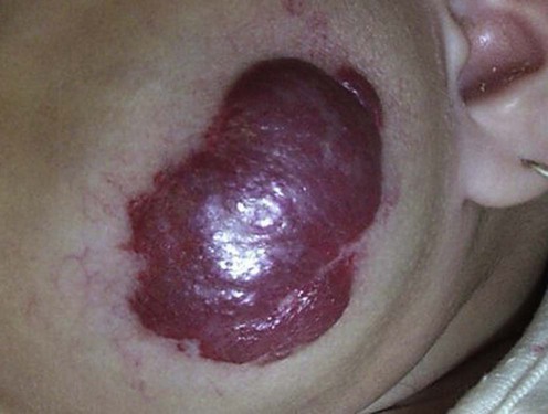
Published on 18/03/2015 by admin
Filed under Dermatology
Last modified 22/04/2025
This article have been viewed 1508 times
Omar Pacha and Adelaide A. Hebert
Evidence Levels: A Double-blind study B Clinical trial ≥ 20 subjects C Clinical trial < 20 subjects D Series ≥ 5 subjects E Anecdotal case reports
A hemangioma is a benign neoplastic proliferation of endothelial cells and is the most common soft tissue tumor of infancy. The incidence of hemangiomas in a general newborn nursery is between 1% and 2.6%, but may be as high as 10% in the Caucasian population. Hemangiomas occur four times more frequently in female infants with a predilection for premature infants.
Approximately 55% of hemangiomas present at birth, with the remainder arising in the first weeks of life. Initially, mature cutaneous hemangiomas grow rapidly for the first 3 to 9 months. After this characteristic proliferative phase, the lesions typically cease growing at 18 months of age, and subsequent spontaneous involution is the rule. Most will involute slowly over a period of 2 to 6 years, usually completing the process by age of 7–10 years. About half of children with hemangiomas will have normal skin after involution, but the rest may have residual changes, including telangiectasias, atrophy, fibrofatty residuum, and scarring. Although for uncomplicated hemangiomas treatment typically consisted of active non-intervention, the advent of use of oral and systemic steroids and/or beta-blocking agents changed the therapeutic paradigm for these vascular tumors.
Differentiating benign, common hemangiomas from other vascular anomalies is essential as the pathophysiology, treatment modalities, and prognoses are significantly different. Vascular malformations and tumors such as the kaposiform hemangioendothelioma (KE) and tufted angioma (TA) differ from hemangioma in both clinical and histological appearance as well as growth rate and involutional tendencies. In particular, KE and TA are associated with the Kasabach–Merritt syndrome and its accompanying coagulopathy, whereas common hemangiomas are not. The majority of common hemangiomas occur as solitary lesions, but organ system involvement or, rarely, syndromes such as diffuse neonatal hemangiomatosis and the PHACES syndrome (posterior fossa malformation, hemangiomas, cardiac anomalies, eye abnormalities, and sternal cleft/supraumbilical raphe) can also occur.
Although the natural course of hemangiomas is self-limited, treatment is probably indicated for approximately 25%, including the 5% that ulcerate and the 20% that may compress, obstruct, or distort vital structures, such as the larynx, eyes, ears, and nose. Medical management of low-risk hemangiomas is generally centered on the administration of topical and systemic beta-blockers, corticosteroids, either topically or intralesionally. One group of investigators used clobetasol propionate cream 0.05% to treat periocular hemangiomas. Other less frequently used management options include cryosurgery, surgical excision, and laser therapy.
Topical and systemic beta-blockers are quickly becoming the preferred first-line therapy in hemangiomas that will require treatment. Starting in 2008, publication of successful treatment of large morbid hemangiomas with propranolol was described. Since that time, other iterations of beta-blocker therapy such as topical timolol have also been used. Current available recommendations for oral propranolol range from 0.5 mg/kg/day to 2 mg/kg/day divided into two to three daily doses. Timolol, available as solution or gel, has reported success in treating periocular and facial infantile hemangiomas with twice daily topical application.
Systemic corticosteroids may be added for larger, deforming, or life-threatening lesions, and are usually indicated during the growth phase. Prednisone or prednisolone can be given at doses from 2–4 mg/kg daily from 2 to 6 months, and then gradually tapered over several months. Stopping treatment before adequate therapeutic response may result in rebound growth. Approximately one-third of patients will show an accelerated rate of involution, but another one-third may have no response to this treatment modality. Reported risks include hypothalamopituitary–adrenal axis suppression, growth delays, pseudotumor cerebri, infections, and avascular bone necrosis. Surgical excision, laser treatment (especially flashlamp-pumped pulsed-dye laser), and cryosurgery, either alone or in combination with corticosteroids, may also be employed in certain cases.
For endangering or life-threatening lesions refractory to systemic corticosteroids, interferon-α2a or -α2b may be used. The typical delivery route is subcutaneously at a dose of 3 million units/m2 daily for 6 to 12 months. Side effects include transient neutropenia, fever, elevated liver enzymes, and flu-like symptoms. In addition, there are rare reports of neurotoxicity, specifically spastic diplegia, which may develop in 5–10% of patients. For the exceptional recalcitrant hemangioma, other treatments include cyclophosphamide, vincristine, bleomycin, propranolol, and embolization.
Ultrasound with Doppler
MRI
The diagnosis of hemangioma is usually made clinically, and the above investigations may only be needed in atypical cases to monitor the progress of treatment, establish the extent of the vascular lesion, or screen for other complications.
Paltiel HJ, Burrows PE, Kozakewich HPW, Zurakowski D, Mulliken JB. Radiology 2000; 214: 747–54.
Doppler ultrasonography is an available, low-cost, non-invasive method to confirm the diagnosis of a vascular anomaly, monitor therapeutic response, or preclude the involvement of visceral organs. Hemangiomas can be differentiated from vascular malformations on ultrasonography by distinguishing features such as the presence of a solid tissue mass.
Vilanova JC, Barcelo J, Smirniotopoulos JG, Pérez-Andrés R, Villalón M, Miró J, et al. Radiographics 2004; 24: 367–85.
MRI is a useful non-invasive imaging technique to diagnose, characterize, and determine the extent of vascular lesions. On T2-weighted images hemangiomas have the characteristic appearance of multiple lobules, similar to a bunch of grapes.
Although not specifically mentioned in this article, infants with large, segmental, plaque-like facial hemangiomas should have MRI head evaluation for possible posterior fossa brain abnormalities as a component of the PHACES syndrome.
Systemic corticosteroids are the mainstay of therapy for life-threatening or endangering hemangiomas. In contrast, topical and intralesional administration remains the first-line therapy for relatively uncomplicated cases.
Garzon MC, Lucky AW, Hawrot A, Frieden IJ. J Am Acad Dermatol 2005; 52: 281–6.
Retrospective review of 34 charts of patients with hemangiomas treated with class I topical steroids showed good response in 35%, partial response in 38%, and no response in 27%. Treatment protocols varied between patients.
Elsas F, Lewis A. J Pediatr Ophthalmol Strabismus 1994; 31: 153–6.
Five patients with sight-threatening periocular hemangiomas were treated with the ultrapotent topical corticosteroid clobetasol propionate 0.05% cream. Therapy was well tolerated, with involution proceeding at a slower rate than with intralesional therapy, but eliminating the risk of central retinal artery occlusion (Shorr N, Seiff SR. Central retinal artery occlusion associated with periocular corticosteroid injection for juvenile hemangioma. Ophthalm Surg 1986; 17: 229–31).
This may be a useful initial treatment for lesions that are not amenable to injection.
Chen MT, Yeong EK, Horng SY. J Pediatr Surg 2000; 35: 420–3.
In this retrospective study, 155 hemangiomas of the head and neck region treated with intralesional corticosteroid injections (three to six injections of triamcinolone acetonide 10 mg/mL at monthly intervals, with an average of four injections per lesion) were analyzed. At the 1-month visit, 85% of hemangiomas showed greater than 50% reduction, with superficial hemangiomas showing the most improvement. Perioral hemangiomas appeared the most recalcitrant to intralesional corticosteroid treatment.
Bennett ML, Fleischer AB, Chamlin SL, Frieden IJ, Arch Dermatol 2001; 137: 1208–13.
This meta-analysis of 10 case series with 184 patients analyzed the efficacy of systemic corticosteroids in the treatment of cutaneous hemangiomas. The mean age of infants at the initiation of therapy was 4.5 months at an average prednisone dose equivalent to 2.9 mg/kg. The mean response rate was 84%, and the mean rate of rebound growth after treatment cessation was 36%. Treatment with higher doses of corticosteroids resulted in a higher response rate but greater adverse effects. The average incidence of side effects was 35% (behavior changes, irritability, cushingoid appearance, and transient growth delay).
Chakkittakandiyil A, Phillips R, Frieden IJ, Siegfried E, Lara-Corrales I, Lam J, et al. Pediatr Dermatol 2011; 29: 28–31.
This retrospective cohort study reviewed 73 patients with infantile hemangioma. The mean age at initiation of therapy was 8 months with 85% of patients dosed at 0.5% strength and the remainder being treated with 0.1%. All patients were treated twice daily and without occlusion. Mean treatment period was 3.4 months and photos were evaluated by investigators to correspond to a 0–100 visual analog scale. Mean visual analog scale improvement was 45 units at the last visit. Longer treatment period was associated with greater improvement. The incidence of adverse events was extremely low, only one patient, and no skin-related adverse events were noted in any subjects. No rebound growth was observed after discontinuation at 3–6 months.
Schupp CJ, Kleber J-B, Günther P, Holland-Cunz S. Pediatr Dermatol 2011 28: 640–4.
This prospective study started 55 patients with a mean age of 6 months on an escalating dose of propranolol. The monitored doses were administered during a 2-day in-hospital evaluation starting with 1 mg/kg/day that was increased to 2 mg/kg/day if no adverse effects were noted. Therapy was continued for 4 to 6 months and then response was measured as ‘total regression without residual skin changes, partial regression, and no regression.’ Results revealed 14.5% with complete regression without visible residual changes, 83.4% with partial regression, and one without regression. Minimal adverse effects were noted with just one patient being discontinued secondary to asthma exacerbation and another due to parental concerns about fatigue.
Ezekowitz RA, Mulliken JB, Folkman J. N Engl J Med 1992; 326: 1456–63.
Twenty infants with life-threatening or vision-threatening hemangiomas who failed corticosteroid therapy were treated with up to 3 million units/m2 daily of interferon-α2a; 90% experienced a 50% or greater regression in their lesions by 7.8 months of treatment. Side effects were transient, including fever, neutropenia, and skin necrosis. No long-term effects were documented.
Jiminez-Hernandez E, Duenas-Gonzalez MT, Quintero-Curiel JL, Velasquez-Ortega J, Magana-Perez JA, Berges-Garcia A, et al. Dermatol Surg 2008; 34: 640–7.
Of 20 patients with deforming or life-threatening hemangiomas who failed earlier treatment with interferon-α2b (3 million IU/m2 daily for 5 days/week) for 6 months, 85% demonstrated an excellent response, 5% a moderate response, and 10% a poor response. Toxicity was short-lived, including fever, fatigue, nausea, and hair loss. No transaminase or neurologic symptoms were reported with 7- to 10-year follow-up.
Although not observed in this study, spastic diplegia is a documented and potentially irreversible complication of this treatment and should be considered prior to initiation of therapy.
Poetke M, Philipp C, Berlien HP. Arch Dermatol 2000; 136: 628–32.
A prospective study of 165 children with 225 separate hemangiomas treated with flashlamp-pumped pulsed-dye laser demonstrated that this therapy produced excellent results in superficial hemangiomas. However, this method was less successful in treating deeper lesions because the efficacy of the laser was limited by the depth of the vascular proliferation.
Pulsed-dye laser therapy promotes epithelialization and is also a safe and effective means of treating ulcerated hemangiomas. Adverse effects more recently reported include scarring, ulceration, and pain (Witman PM, Wagner AM, Scherer K, Waner M, Frieden IJ. Complications following pulsed dye laser treatment of superficial hemangiomas. Lasers Surg Med 2006; 38: 112–15).
Mcheik JN, Renauld V, Duport G, Vergnes P, Levard G. Br J Plast Surg 2005; 58: 1067–72.
Retrospective study of 31 patients (average age 30 months) showed very good results in 20%, good in 66%, and fair in 14%. Recommendations included consideration of surgery in lesions that had not changed in 2 years or that interfered with the child’s social integration.
Metz BJ, Rubenstein MC, Levy ML, Metry DW. Arch Dermatol 2004; 140: 867–70.
Eight infants were treated with becaplermin gel 0.01% for ulcerated perineal hemangiomas of infancy. Rapid ulcer healing occurred in all patients within three to 21 days (average, 10.25 days).
The rapid healing achieved with 0.01% becaplermin gel allows a reduction in the risk of secondary infection, pain, and the need for hospitalization, as well as in the costs that often accumulate from multiple follow-up visits and long-term therapy.
Fawcett SL, Grant I, Hall PN, Kelsall AW, Nicholson JC. Br J Plast Surg 2004; 57: 168–71.
A 21-month-old infant with a large, deep hemangioma in the beard distribution, necessitating multiple hospital admissions for respiratory and feeding difficulties, was treated with vincristine after failing to respond to systemic corticosteroid therapy. Vincristine (1.5 mg) was administered in weekly doses for 1 month, with improvement in feeding, speech, and external appearance of the lesion, with no adverse effects.
Although there are literature reports of success achieved with vincristine, the data are anecdotal at best.
Ho NTC, Lansang P, Pope E. J Am Acad Dermatol 2007; 56: 63–8.
Eighteen children (median age 18 weeks) with imiquimod 5% cream applied three times weekly or five times weekly for a mean of 17 weeks showed improvement in superficial hemangiomas and accelerated ulcer healing; no change was seen in deep hemangiomas. A 31% clearance rate was reported. Adverse events included crusting and inflammation. Mechanism presumably related to cytokine (such as interferon) release.
Subsequent criticisms reported control arms of earlier studies (using only observation) showing similar clearance rates to those treated in this study without the use of any intervention.
Omidvari S, Nezakatgoo N, Ahmadloo N, Mohammadianpanah M, Mosalaei A. Dermatol Surg 2005; 31: 499–501.
Thirty-two patients with complicated hemangiomas (median age 45 months) were treated with intralesional bleomycin (1–2 mg/cm2 of lesion every 2 weeks for four to six courses). Following the initial swelling and erythema, 56% of patients had 70–100% regression; 21.9% had 50–70% regression. No severe adverse effects were reported.
Treatment of Skin Disease Comprehensive Therapeutic Strategies 4e
WhatsApp us