Published on 18/03/2015 by admin
Filed under Dermatology
Last modified 22/04/2025
This article have been viewed 2113 times
Rajani Nalluri and Ian Coulson
Evidence Levels: A Double-blind study B Clinical trial ≥ 20 subjects C Clinical trial < 20 subjects D Series ≥ 5 subjects E Anecdotal case reports
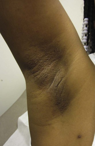
Obliteration of the follicular infundibulum with keratin in the apocrine gland-bearing skin is the cause of this rare, paroxysmally intensely itchy condition. Apocrine sweat retention and rupture of the gland duct under periods of apocrine sudomotor stimulation, particularly emotional stress, results in the development of an itchy, spongiotic intraepidermal vesicle. It mainly affects women between the ages of 13 and 35 years, but has rarely been reported prior to puberty, after the menopause, and in men. Itchy, dome-shaped, flesh-colored or keratotic papules that develop peripubertally in the apocrine areas of the axillae, pubic, periumbilical and periareolar skin characterize this condition. Sparsity of axillary hair and hypohidrosis is usual, although rarely it can be exacerbated by hyperhidrosis. Improvement in pregnancy and during the administration of the oral contraceptive pill has led to speculation regarding an endocrine etiology, but this has been unsubstantiated by blood sex hormone investigations. Very few reports in twins and within families suggest a possible genetic component. It can occur following laser hair removal and has been reported in Turner syndrome.
There are no controlled trials of any agents in Fox–Fordyce disease.
Topical and intralesional corticosteroids are frequently tried and may be of limited benefit, but atrophy in the axillary area will limit their potency and duration of use. Topical tretinoin has been reported to reduce itch, but its alternation with a mild corticosteroid may be needed to reduce retinoid irritancy. Clindamycin lotion may be of help. The oral contraceptive pill (OCP) may bring relief to some women. Oral isotretinoin may give temporary help. Electrocautery and excision of the periareolar skin may offer permanent solutions. A recent report advocates an ingenious method of removal of the apocrine glands using a microliposuction cannula.
Biopsy
Stashower ME, Krivda SJ, Turiansky GW. J Am Acad Dermatol 2000; 42: 89–91.
Transverse sectioning demonstrates the follicular plugging and infundibular spongiosis more readily than conventional sections.
Böer A. Am J Dermatopathol 2004; 26: 482–92.
An exhaustive review of the subtleties of the dermatopathology of Fox–Fordyce disease.
Macarenco RS, Garces SJC. Am J Dermatopathol 2009; 31: 93–7.
Apocrine gland dilation may be used as a low-power magnification clue which should be followed by a search for further histological changes to confirm or rule out the diagnosis.
Kao PH, Hsu CK, Lee JY. J Dermatol 2009; 36: 485–90.
Focal spongiosis in the upper infundibulum with perifollicular fibrosis and lymphohistiocytic infiltrate were consistent features in their case series.
Kossard S, Dwyer P. Australas J Dermatol 2004; 45: 146–8.
Mataix J, Silvestre JF, Niveiro M, Lucas A, Pérez-Crespo M. Actas Dermosifiliogr 2008; 99: 145–8.
There are occasional conditions to consider in the differential diagnosis. There is even controversy as to whether perifollicular xanthomatosis is part of the spectrum of this disorder.
Helfamn RJ. South Med J 1962; 55: 681–4.
A single report of successful symptom relief of axillary lesions with 10 mg/mL triamcinolone diluted with an equal volume of 1% lidocaine to four sites on nine occasions over 3 months.
Feldmann R, Masouye I, Chavaz P, Saurat JH. Dermatology 1992; 184: 310–13.
A single report of Fox–Fordyce disease in the axillary, pubic, and inguinal areas responding to 1% clindamycin in an alcoholic propylene glycol solution within 1 month (clindamycin 10 mg/mL, propylene glycol 50 mg/mL, isopropyl alcohol 0.5 mg/mL, water). Nine months later the treatment was stopped and no recurrence was observed. The authors speculate that the keratolytic effect of propylene glycol may have been responsible for the therapeutic effect.
Kronthal HI, Pomeranz JR, Sitomer. G. Arch Dermatol 1965; 91: 243–5.
Two female patients responded to a high estrogen dose combined OCP, norethynodrel, and mestranol.
Giacobetti R, Caro WA, Roenigk HH Jr. Arch Dermatol 1979; 115: 1365–6.
A single report of 0.1% tretinoin cream applied to the axillae on alternate nights resulting in reduction of itch and regrowth of hair. Local retinoid irritation was controlled with 1% hydrocortisone cream.
Pinkus H. JAMA 1973; 223: 924.
Erythemogenic doses of UVB (once weekly for 4 to 6 weeks) produced long-lasting relief to several patients.
Pock L, Svrcková M, Machácková R, Hercogová J. Int J Dermatol 2006; 45: 1134–5.
A series of three patients who benefited from topical pimecrolimus.
Shelley WB. JAMA 1972; 222: 1069.
A concise review of the therapies available to that date. The author admits that sometimes all fails and that relief may only come at the menopause.
Effendy I, Ossowski B, Happle R. Clin Exp Dermatol 1994; 19: 67–9.
Oral treatment with isotretinoin (30 mg daily for 8 weeks then 15 mg daily for 2 months) resulted in temporary relief. Relapse occurred 3 months after discontinuation.
Pasricha JS, Nayyar KC. Dermatologica 1973; 147: 271–3.
Electrocoagulation to a level of 3–4 mm under local anesthetic produced a permanent resolution of symptoms in the axillae of two patients.
Chavoin J-P, Charasson T, Barnard J-D. Ann Chir Plast Esthet 1994; 39: 233–8.
A simple technique involving dermal detachment of the areola, excision of the underlying apocrine glands, and reattachment of the areola with good cosmetic results.
This treatment has not proved beneficial long term.
Chae KM, Marschall MA, Marschall SF. Arch Dermatol 2002; 138: 452–4.
A novel technique of curettage removal of the apocrine glands using a small liposuction cannula with symptom relief, great cosmesis, and a follow-up at publication of 8 months. A liposuction cannula was introduced through a stab incision in the axilla and, with the aperture of the cannula turned up towards the underside of the dermis, the deeper dermis was curetted to create inflammation and subsequent fibrosis. The same technique can be used to treat axillary hyperhidrosis.
Treatment of Skin Disease Comprehensive Therapeutic Strategies 4e
WhatsApp us

 Topical and intralesional corticosteroids
 Topical clindamycin
 Oral contraceptive pill
 Topical retinoids
 UVB
UVB Topical pimecrolimus
 Oral isotretinoin
 Electrocautery
 Excision
 Apocrine gland removal using microliposuction