Examination of the Eye

Published on 25/03/2015 by admin

Filed under Pediatrics

Last modified 22/04/2025

Print this page

rate 1 star rate 2 star rate 3 star rate 4 star rate 5 star
Your rating: none, Average: 0 (0 votes)

This article have been viewed 1314 times

Chapter 611 Examination of the Eye

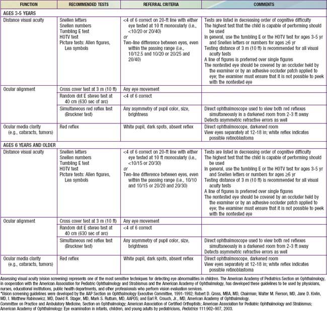
Examination of the eyes is a routine part of the periodic pediatric assessment beginning in the newborn period. The primary care physician is very important in detecting both obvious and insidious asymptomatic eye diseases. Screening by lay persons in schools and community programs can also be effective in detecting problems early. The best method of screening (ages 3-5 yrs) is currently being investigated. The American Academy of Ophthalmology recommend preschool vision screening as a means of reducing preventable visual loss (Table 611-1). This testing should also be done by pediatricians during well child visits. Children should be examined by an ophthalmologist whenever a significant ocular abnormality or vision defect is noted or suspected. Children who are at high risk of ophthalmologic problems, such as genetically inherited ocular conditions and various systemic disorders, should also be examined by an ophthalmologist.

Basic examination, whether done by a pediatrician or an ophthalmologist, must include evaluation of visual acuity and the visual fields, assessment of the pupils, ocular motility and alignment, a general external examination, and an ophthalmoscopic examination of the media and fundi. When indicated, biomicroscopy (slit-lamp examination), cycloplegic refraction, and tonometry are performed by an ophthalmologist. Special diagnostic procedures, such as ultrasonic examination, fluorescein angiography, electroretinography, or visual evoked response (VER) testing, are also indicated for specific conditions.

Visual Acuity

There are many tests of visual acuity. Which test is used depends on a child’s age and ability to cooperate, as well as a clinician’s preference and experience with each test. The most common visual acuity test in infants is an assessment of their ability to fixate and follow a target. If appropriate targets are used, this reflex can be demonstrated by about 6 weeks of age. The test is performed by seating the child comfortably in the caretaker’s lap. The object of visual interest, usually a bright-colored toy, is slowly moved to the right and to the left. The examiner observes whether the infant’s eyes turn toward the object and follow its movements. The examiner can use a thumb to occlude one of the infant’s eyes and test each eye separately. Although a sound-producing object might compromise the purity of the visual stimulus, in practice, toys that squeak or rattle heighten an infant’s awareness and interest in the test.

The human face is a better target than test objects. The examiner can exploit this by moving his or her face slowly in front of the infant’s face. If the appropriate following movements are not elicited, the test should be repeated with the caretaker’s face as the test stimulus. It should be remembered that even children with poor vision can follow a large object without apparent difficulty, especially if only one eye is affected.

An objective measurement of visual acuity is usually possible when children reach 2.5-3 years of age. Children this age are tested using a schematic picture or other illiterate eye chart. Each eye should be tested separately. It is essential to prevent peeking. The examiner should hold the occluder in place and observe the child throughout the test. The child should be reassured and encouraged throughout the test because many children are intimidated by the procedure and fear a “bad grade” or punishment for errors.

The E test, in which a child points in the direction of the letter, is the most widely used visual acuity test for preschool children. Right-left presentations are more confusing than up-down presentations. With pretest practice, this test can be performed by most children 3-4 years of age.

An adult-type Snellen acuity chart can be used at about 5 or 6 years of age if the child knows letters. An acuity of 20/40 is generally accepted as normal for 3 yr old children. At 4 yr of age, 20/30 is typical. By 5 or 6 years of age, most children attain 20/20 vision.

Optokinetic nystagmus (the response to a sequence of moving targets; “railroad” nystagmus) can also be used to assess vision; this can be calibrated by targets of various sizes (stripes or dots) or by a rotating drum at specified distances. The VER, an electrophysiologic method of evaluating the response to light and special visual stimuli, such as calibrated stripes or a checkerboard pattern, can also be used to study visual function in selected cases. Preferential looking tests are also used for evaluating vision in infants and children who cannot respond to standard acuity tests. This is a behavioral technique based on the observation that given a choice, an infant prefers to look at patterned rather than unpatterned stimuli. Because these tests require the presence of a skilled examiner, their use is often limited to research protocols involving preverbal children.

External Examination

The external examination begins with general inspection in good illumination noting size, shape, and symmetry of the orbits; position and movement of the lids; and position and symmetry of the globes. Viewing the eyes and lids from above aids in detecting orbital asymmetry, lid masses, proptosis (exophthalmos), and abnormal pulsations. Palpation is also important in detecting orbital and lid masses.

The lacrimal apparatus is assessed by looking for evidence of tear deficiency, overflow of tears (epiphora), erythema, and swelling in the region of the tear sac or gland. The sac is massaged to check for reflux when obstruction is suspected. The presence and position of the puncta are also checked.

The lids and conjunctivae are specifically examined for focal lesions, foreign bodies, and inflammatory signs; loss and maldirection of lashes should also be noted. When necessary, the lids can be everted in the following manner: (1) instruct the patient to look down; (2) grasp the lashes of the patient’s upper lid between the thumb and index finger of one hand; (3) place a probe, a cotton-tipped applicator, or the thumb of the other hand at the upper margin of the tarsal plate; and (4) pull the lid down and outward, everting it over the probe, using the instrument as a fulcrum. Foreign bodies commonly lodge in the concavity just above the lid margin and are exposed only by fully everting the lid.

The anterior segment of the eye is then evaluated with oblique focal illumination, noting the luster and clarity of the cornea, the depth and clarity of the anterior chamber, and the features of the iris. Transillumination of the anterior segment aids in detecting opacities and in demonstrating atrophy or hypopigmentation of the iris; these latter signs are important when ocular albinism is suspected. When necessary, fluorescein dye can be used to aid in diagnosing abrasions, ulcerations, and foreign bodies.

Bibliography

Bennett DM, Gordon G, Dutton GN. The useful field of view test, normative data in children of school age. Optom Vis Sci. 2009;86:717-721.

Committee on Practice and Ambulatory Medicine, Section on Ophthalmology; American Association of Certified Orthoptists; American Association for Pediatric Ophthalmology and Strabismus; American Academy of Ophthalmology. Eye examination in infants, children, and young adults by pediatricians. Pediatrics. 2003;111:902-907.

Donahue SP, Johnson TM, Ottar W, et al. Sensitivity of photoscreening to detect high-magnitude amblyogenic factors. J AAPOS. 2002;6:86-91.

Fulton A. Screening preschool children to detect visual and ocular disorders. Arch Ophthalmol. 1992;110:1553-1554.

Isenberg SJ. Clinical application of the pupil examination in neonates. J Pediatr. 1991;118:650-652.

Powell C, Hatt SR: Vision screening for amblyopia in childhood. Cochrane Database Syst Rev (3):CD005020, 2009.

Reinecke RD. Screening 3-year olds for visual problems: are we gaining or falling behind? Arch Ophthalmol. 1986;104:245-248.

Salcido AA, Bradley J, Donahue SP. Predictive value of photoscreening and traditional screening of preschool children. J AAPOS. 2005;9:114-120.

Simons K. Preschool vision screening: rationale, methodology and outcome. Surv Ophthalmol. 1996;41:3-30.

Solouki AM, Verhoeven VJM, van Duijn CM, et al. A genome-wide association study identifies a susceptibility locus for refractive errors and myopia at 15q14. Nat Genet. 2010;42(10):897-901.

US Preventive Services Task Force. Vision screening for children 1 to 5 years of age: US preventive services task force recommendation statement. Pediatrics. 2011;127:340-346.