[level-membership-for-basic-science-category]
Eukaryotic Microorganisms
After reading this chapter, the student will be able to:
• Describe, compare, and differentiate the characteristics of fungi, algae, protozoans, and helminths
• Describe the defining characteristics of fungi and explain the difference between their sexual and asexual reproduction
• Identify the medically important fungi
• Name the defining characteristics of algae, classify them, and describe the difficulties in their classification
• Describe the general characteristics of asexual and sexual reproduction of algae
• Identify the three defining and the common characteristics of protozoans
• Describe the general life cycle of protozoans and explain the importance of the macro- and micronucleus
• Discuss the fundamental differences between cellular and plasmodial slime molds
• Describe the main characteristics of parasitic helminths
• Describe the life cycles of flukes, tapeworms, and roundworms, and classify them into the appropriate categories
Introduction
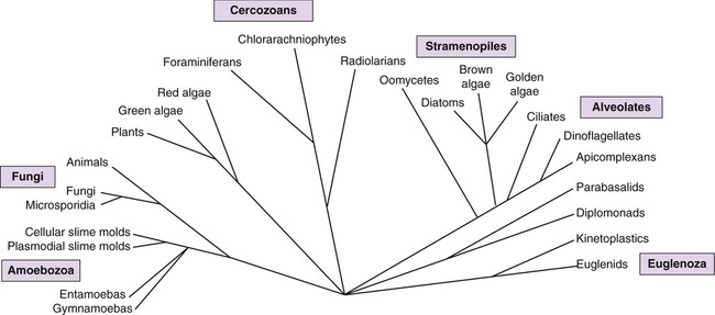
Eukaryotic organisms differ from bacteria and archaea in many ways including cell size, internal structure, and genetic properties (see Chapter 3, Cell Structure and Function). The classification of eukaryotes has changed over the centuries starting in the late eighteenth century with Linnaeus, who classified all organisms as either plants or animals. At present, instead of the traditional classification schemes, many taxonomists favor classification schemes that are based on distinguishing characteristics at the molecular level. Modern taxonomy attempts to explain the genetic relationships of microbes and place them in groups based on similarities in their biochemical and metabolic characteristics, their nucleotide sequences, as well as their cellular ultrastructure as revealed by electron microscopy. Although universal support for a particular classification scheme has not been reached, many taxonomists favor a scheme similar to the one shown in Figure 8.1.
Fungi
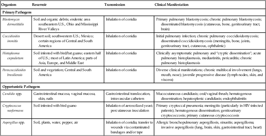
The study of fungi is called mycology, and although more than 100,000 species of fungi are known, only about 200 are pathogenic to humans and animals, and relatively few fungi are virulent enough to be considered primary pathogens (Table 8.1). Moreover, the incidence of fungal infections has been increasing, especially those acquired in hospitals (nosocomial), other institutional settings such as nursing homes, and in people with a compromised immune system.
TABLE 8.1
Primary and Opportunistic Fungal Pathogens
| Organism | Reservoir | Transmission | Clinical Manifestation |
| Primary Pathogens | |||
| Blastomyces dermatitidis | Soil and organic debris; endemic area southeastern U.S., Ohio and Mississippi River Valleys | Inhalation of conidia | Primary pulmonary blastomycosis; chronic pulmonary blastomycosis; disseminated blastomycosis (cutaneous, bone, genitourinary tract, brain) |
| Coccidioides immitis | Desert soil; southwestern U.S.; Mexico; certain regions of Central and South America | Inhalation of conidia | Initial pulmonary infection; chronic pulmonary coccidioidomycosis; disseminated coccidioidomycosis (meningitis, bone, joints, genitourinary tract, cutaneous, ophthalmic) |
| Histoplasma capsulatum | Soil infested with bird/bat guano; eastern half of U.S.; most of Latin America; parts of Asia, Europe, and Middle East | Inhalation of conidia | Clinically asymptomatic pulmonary and “cryptic dissemination”; acute pulmonary histoplasmosis, mediastinitis, pericarditis; chronic pulmonary histoplasmosis |
| Paracoccidioides brasiliensis | Soil and vegetation; Central and South America | Inhalation of conidia | Diverse clinical manifestations; chronic multifocal involvement (lungs, mouth, nose); juvenile progressive disease (lymph nodes, skin, and viscera) |
| Opportunistic Pathogens | |||
| Candida spp. | Gastrointestinal mucosa, vaginal mucosa, skin, nails | Gastrointestinal translocation, intravascular catheters | Mucocutaneous candidiasis; oral/vaginal thrush; hematogenous dissemination; hepatosplenic candidiasis; endophthalmitis |
| Cryptococcus neoformans | Soil infested with bird guano | Inhalation of aerosolized yeast; percutaneous inoculation | Primary cryptoccal pneumonia; meningitis (particularly in HIV-infected patients); hematogenous dissemination; genitourinary cryptococcosis; primary cutaneous cryptococcosis |
| Aspergillus spp. | Soil, plants, water, pepper, air | Inhalation of conidia; transfer to wounds via contaminated bandages and/or tape | Allergic bronchopulmonary aspergillosis, sinusitis; aspergilloma; invasive aspergillosis (lung, brain, skin, gastrointestinal tract, heart) |
Fungi have important symbiotic relationships with other organisms and therefore can be beneficial to life in general. For example, they play a major role in the decomposition of dead plant material, and they are the primary decomposers of material such as cellulose, which cannot be digested by animals. The majority of plants are dependent on their symbiotic relationship with mycorrhizae (fungi that help the roots of plants to absorb minerals and water from the soil). Fungi provide food for humans in the form of mushrooms and truffles, and they are used in the manufacture of foods and beverages, including breads, alcoholic beverages, citric acid, soy sauce, and some cheeses. Fungi also produce pharmaceuticals such as the antibiotics penicillin and cephalosporin (see Chapter 22, Antimicrobial Drugs, for details). In addition, fungi are of use in the making of the immunosuppressive drug cyclosporine for organ transplant patients, and mevinic acids, which are used in the manufacture of cholesterol-reducing drugs. Fungi are also important research tools in genetics and biotechnology (see Chapter 25, Biotechnology).
Characteristics of Fungi
Fungi are eukaryotes but are quite different from plants and animals. Fungal colonies are vegetative structures because they are made up of cells involved in catabolism and growth. Whether microscopic or macroscopic, they generally grow as filamentous, multinucleated organisms that form threadlike filaments called hyphae, or as unicellular fungal organisms (Figure 8.2, A). Morphologically, fungi are often divided into three groups.
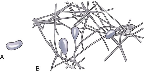
Fungi can exist as (A) unicellular organisms, or (B) form a mycelium, a mass of hyphae that continues to grow.
Most fungi that are identified as human pathogens are microscopic. Hyphae are long, branching filamentous cells of a fungus and they represent its main vegetative growth. A collective mass of the threadlike hyphae is called the mycelium (Figure 8.2, B). A mycelium can be small, forming colonies too small to see with the naked eye, or they can be extensive, such as Armillaria gallica, which was described as extending through 37 acres of forest soil and was estimated to weigh at least 9700 kg.
• Fungi tend toward maximal growth in a somewhat acidic environment, at about pH 5, which is too acidic for the growth of most bacteria (with such exceptions as Helicobacter pylori, which likes to grow at the pH of the human stomach [see Chapter 12, Infections of the Gastrointestinal System]).
• Most fungi are more resistant to osmotic pressure than are bacteria and can grow in relatively high concentrations of sugar or salt.
• Fungi can grow on substances with low moisture content, too low to support bacterial growth.
• Fungi metabolize more complex carbohydrates than bacteria.
• Chitin synthesis and other compounds for use in the cell wall, materials that induce hypersensitivity (see Chapter 20, The Immune System)
• Ergosterol synthesis for the plasma membrane, making the plasma membrane susceptible to antimicrobial agents targeting ergosterol synthesis or ergosterol incorporation into the plasma membrane (see Chapter 22, Antimicrobial Drugs)
Fungal pathogens can be classified on the basis of their growth forms—filamentous, yeast, or dimorphic—and on the type of infection they can cause. For example, in superficial mycoses, the fungus grows on the surface of the skin or hair. In cutaneous and subcutaneous mycoses, nails and deeper layers of the skin are involved, and in systemic (deep) mycoses, the fungus spreads to internal organs (Box 8.1). Patients with a compromised immune system are especially susceptible to fungal infections. Whereas superficial and cutaneous mycoses are generally mild, systemic mycoses are serious and difficult to treat.
Yeasts
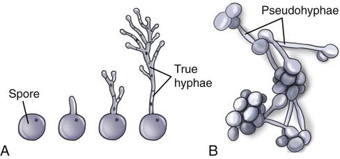
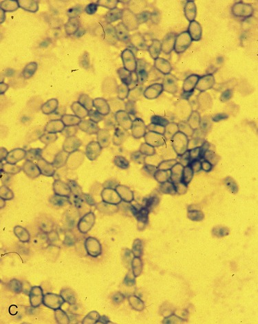
Yeasts exist as single cells that reproduce by budding. However, some species may become multicellular through the formation of connected budding cells referred to as pseudohyphae, or true hyphae (Figure 8.3). They can be classified according to the presence or absence of capsules, the size and shape of the cells, the mechanism of daughter cell formation, formation of hyphae, presence of sexual spores, and other physiological/genetic data. Yeasts may require oxygen for their metabolic activities as obligate aerobes or may be able to continue their metabolic activities in the absence of oxygen as facultative anaerobes. Yeasts do not require light for their growth.
Molds
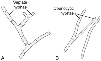
Molds are rapidly growing, asexually reproducing fungi that grow on a variety of substances. They are characterized by the development of hyphae (long filaments of cells joined together), which results in colony characteristics that can be used for identification purposes. Hyphae can grow to immense proportions (see Armillaria gallica, earlier this chapter). In general, the hyphae in molds contain cross-walls called septa, which divide the hyphae into distinct uninuclear, cell-like units called septate hyphae. Some classes of fungi have coenocytic hyphae, which do not contain septa and appear as long, continuous cells with many nuclei (Figure 8.4). In an appropriate environment, the hyphae grow to form a mycelium that is visible to the naked eye.
Dimorphic Fungi
• As a mold with septate hyphae in their natural reservoir (i.e., soil) and in the laboratory when incubated at 25° C on conventional fungal media
• As yeast in tissues of a human or other animals or in the laboratory when incubated at 37° C on enriched media (i.e., brain heart infusion agar)
Several dimorphic fungi can cause systemic mycoses (see Table 8.1) that usually start by the inhalation of spores from the mold form and then germinate in the lungs, where the fungus grows as yeast. These fungi include, but are not limited to, Sporothrix schenckii (sporotrichosis), Histoplasma capsulatum (histoplasmosis), Blastomyces dermatitidis (blastomycosis), Paracoccidioides brasiliensis (paracoccidioidomycosis), and Coccidioides spp. (coccidioidomycosis).
Life Cycle of Fungi
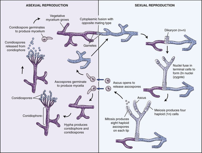
Asexual Reproduction
Yeast typically bud in a manner similar to the binary fission of prokaryotic organisms (see Chapter 3, Cell Structure and Function; and Chapter 6, Bacteria and Archaea). Filamentous fungi can reproduce asexually by fragmentation of their hyphae, and by spore formation either sexually or asexually. Asexual reproduction results in the formation of sporangia, which ultimately release spores into the environment. Spores, after entering the respiratory tract, are common causes of infection (see Chapter 11, Infections of the Respiratory System). These asexual spores are often categorized on the basis of the nature and manner of spore development:
• Sporangiospores form inside a sac, the sporangium, which is attached to a stalk, the sporangiophore, located on the tips or sides of hyphae. The spores are released when the sporangium ruptures.
• Conidia (conidiospores) are produced at the tips or sides of hyphae but are not enclosed by a saclike structure. Their development involves the pinching off of the tip of a fertile hypha or the segmentation of a preexisting vegetative hypha. They are the most common of the asexual spores and exist in different forms (Figure 8.5):
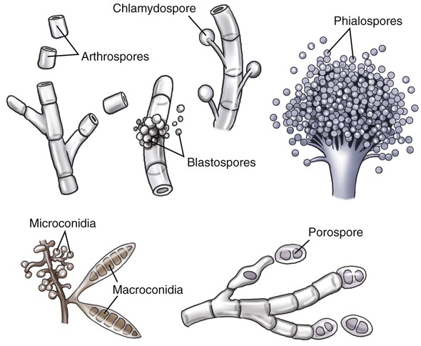
• Arthrospore: A rectangular spore, formed when a septate hypha breaks at the cross wall
• Chlamydospore: A spherical conidium produced by the thickening of a hyphal cell that is released when the surrounding hyphae crack
• Blastospore: A spore that buds from a parent cell
• Phialospore: A spore that buds from the mouth of a vase-shaped, spore-bearing cell
• Microconidium and macroconidium: Spores formed by the same fungus under different conditions, either as one cell (microconidium) or as two or more cells (macroconidium)
• Porospore: A spore that grows out through small pores in a spore-bearing cell
Sexual Reproduction
• Haploid (n) cells from a + mycelium and a − mycelium fuse and form a dikaryon. This stage is not considered to be diploid or haploid, but is designated as (n + n).
• After a period of time, which ranges from hours to years and maybe even centuries, a pair of nuclei within the dikaryon finally fuses, forming a diploid (2n) nucleus.
• The meiotic cell division that follows restores the haploid state of the organism.
• The haploid nuclei are then divided into + and − spores, reestablishing the + and − mycelia.
Classification of Fungi
• Zygomycota are coenocytic (multinucleate) molds, most of which are saprobes (fungi that derive their nutrition from nonliving organic material); the remaining are obligate parasites of insects or other fungi.
• Ascomycota are the primary fungi causing food spoilage. This group also includes plant pathogens, both producing a negative impact on the economy. However, many of the Ascomycota are beneficial and probably the best known fungus in this category is Penicillium (Figure 8.6, A), the mold responsible for the production of the antibiotic penicillin (see Chapter 22, Antimicrobial Drugs). Another fungus in this group, Saccharomyces (Figure 8.6, B), ferments sugar to produce alcohol and carbon dioxide, the basic processes necessary in the baking and brewing industries. Furthermore, Neurospora, the pink bread mold, is an important contributor in genetic and biochemical research.
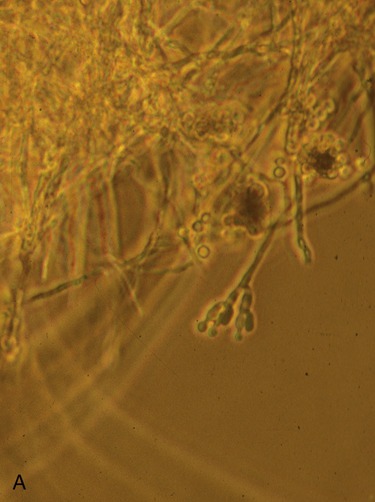
A, This is a phase-contrast micrograph of the fruiting structure of the fungus Penicillium, showing spore formation. B, This micrograph illustrates a dark-field image of the yeast Saccharomyces.
• Basidiomycota include mushrooms (fleshy fungi) and other fruiting bodies of fungi (Figure 8.7). Many of the mushrooms are edible and some produce toxins and/or hallucinatory chemicals. In addition, the fungus Cryptococcus neoformans grows in the form of yeast in humans and is the leading cause of fungal meningitis. Besides the edible and poisonous forms, Basidiomycota are important decomposers, digesting cellulose and lignin of dead plants, which returns essential nutrients back to the soil.
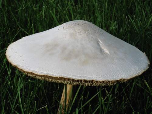
Fungi display a wide variety of structures. This photograph shows a typical mushroom, a fleshy fungus with the cap and stem formed by a thick mat of hyphae that is collectively called a mycelium. The overall vegetative structure is called a thallus. Courtesy Andrew W. Van Meter.
• Deuteromycota are somewhat different than the other subgroups, and sometimes are referred to as “imperfect fungi.” Whereas stages of sexual reproduction in the previously described groups have been well established, the sexual stages of Deuteromycota are unknown. Either this group of fungi does not produce sexual spores, or their sexual spores have not been identified at this time (recall that sexual spores may not form diploid nuclei for centuries).
• Lichens (Figure 8.8) consist of hyphae (mold) of a fungus and cyanobacteria or green algae. The mold, often an ascomycete, surrounds the photosynthetic cell and provides nutrients, water, and protection from drying out and excessive light. Conversely, the algae or cyanobacterium supplies carbohydrates and oxygen, the by-products of photosynthesis, to the fungus.
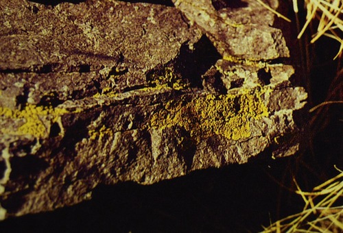
This photo of a lichen shows the yellow fungal hyphae, which are visible to the naked eye. With a microscope one can see that the hyphae of the fungus form a matrix woven around algal cells; together the fungus and algae form a mutualistic association with physiological properties different from those of either of the individual species. From Atlas RM: Principles of microbiology, St. Louis, 1995, Mosby-Year Book.
Algae
Life Cycle of Algae
All algae are capable of asexual reproduction. In unicellular algae the nucleus divides by mitosis and when the newly formed nuclei move to the opposite poles of the cell, the cell divides into two new cells by cytokinesis (see Chapter 3, Cell Structure and Function). Multicellular algae also may reproduce asexually by fragmentation, and each fragment is capable of forming a new thallus (the entire vegetative structure) or filaments.
Unicellular algae can also reproduce sexually and each algal cell then serves as a gamete, which can fuse with another gamete to form a zygote. This zygote then undergoes meiosis to return to the haploid state. When multicellular algae reproduce sexually, every cell in the reproductive structures of the algae becomes a gamete. The sexual reproduction of many algae can occur by alternation of haploid and diploid generations (Figure 8.9). In these life cycles, the diploid organism undergoes meiosis to produce male and female haploid spores, which then develop into haploid male and female thalli. In some algae, each of these will produce gametes that fuse to form a zygote, which then creates a new diploid thallus.
Classification of Algae
As mentioned earlier, the classification of algae is problematic and a summary of the algal groups described in this section and their characteristics is shown in Table 8.2.
TABLE 8.2
| Group (Common Name) | Organization | Pigments | Storage Product(s) | Cell Wall | Habitat |
| Chlorophyta (green algae) | Varies from unicellular, colonial, filamentous, to multicellular | Chlorophylls a and b, carotene, xanthophylls | Sugar, starch | Cellulose or protein, absent in some | Fresh, brackish, and salt water; terrestrial |
| Rhodophyta (red algae) | Multicellular | Chlorophyll a, phycoerythrin, phycocyanin, xanthophylls | Glycogen (floridean starch) | Agar or carrageenan, some with calcium carbonate | Mostly salt water |
| Phaeophyta (brown algae) | Multicellular, vascular system, holdfasts | Chlorophylls a and b, xanthophylls | Laminarin, oils | Cellulose and alginic acid | Brackish and salt water |
| Chrysophyta (golden algae, yellow-green algae, diatoms) | Mainly unicellular, some filamentous forms, usually some form of motility | Chlorophylls, phycoerythrin, phycocyanin, xanthophylls | Chrysolaminarin | Cellulose, silica, calcium carbonate | Fresh, brackish, and salt water; terrestrial; ice |
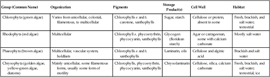
• Chlorophyta are green algae whose pigments are chlorophylls a and b. They use sugar and starch as food reserves. Because of these and other similarities with plants, green algae are often considered to be progenitors of plants. Most green algae are unicellular or filamentous, living in freshwater ponds, lakes, and pools (Figure 8.10). They form a characteristic green to yellow scum.
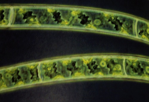
This is a micrograph of Spirogyra, a filamentous green algae. From Atlas RM: Principles of microbiology, St. Louis, 1995, Mosby-Year Book.
• Rhodophyta (red algae) contain a red accessory pigment (thus their name) and use glycogen as their energy-storing molecule. Red algae have cell walls containing agar, a gelatinous substance used as a solidifying substance in microbiological culture media. This polysaccharide has many more applications, including its use as a thickener for soups, jellies, and ice cream, to name a few.
• Phaeophyta are commonly referred to as brown algae because of the presence of brown pigments (xanthophylls) in addition to chlorophylls and carotene. Depending on the amount of brown pigments, these algae may appear brown, tan, yellow-brown, greenish brown, or green. Most brown algae are marine organisms.
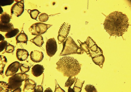
• Chrysophyta is a diverse group of algae and some taxonomists lately have grouped them with brown algae because of similarities in the nucleotide sequences and flagellar structure. Diatoms, comprising one taxon in this group (Figure 8.11), have a unique cell wall composed of silica, and are a major component of marine phytoplankton. Phytoplankton include photosynthetic microorganisms that form the basis of food chains in the oceans. Significantly, because of their massive numbers, diatoms are the major source of the world’s oxygen.
Protozoans
Characteristics of Protozoans
Protozoans are unicellular organisms that vary in size and are defined by the three characteristics described in the previous section. Protozoa consist of a diverse group of microbes and with the exception of one subgroup they are motile due to cilia, flagella, and/or pseudopodia (see Chapter 3, Cell Structure and Function). Protozoans may vary in size, but all require a moist environment to survive. Most species live in ponds, streams, lakes, and oceans, whereas others live in moist soil, beach sand, and decaying organic matter. Aquatic protozoans are important components of plankton, the basis of the food chain of aquatic animals.
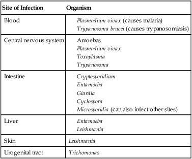
A few protozoans are pathogens that infect the human body as intracellular or extracellular parasites. Intracellular parasites infect a wide variety of cells including erythrocytes, macrophages, epithelial cells, muscle cells, and cells of the nervous system. Extracellular parasites reside in the blood, intestine, or urogenital system (Table 8.3).
HEALTHCARE APPLICATION
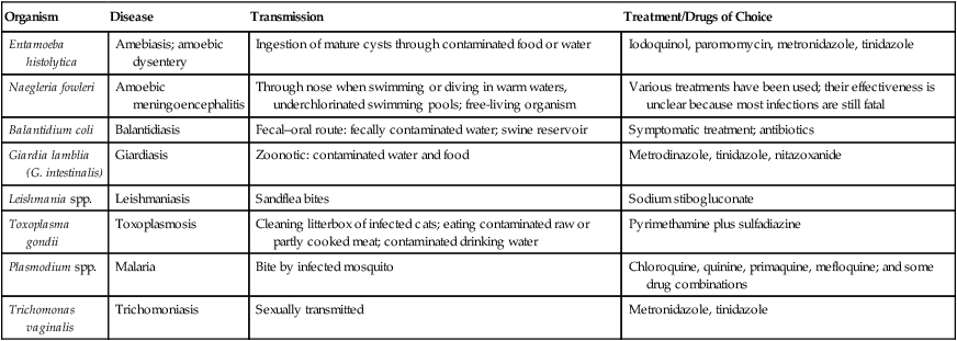
Examples of Protozoal Infections
| Organism | Disease | Transmission | Treatment/Drugs of Choice |
| Entamoeba histolytica | Amebiasis; amoebic dysentery | Ingestion of mature cysts through contaminated food or water | Iodoquinol, paromomycin, metronidazole, tinidazole |
| Naegleria fowleri | Amoebic meningoencephalitis | Through nose when swimming or diving in warm waters, underchlorinated swimming pools; free-living organism | Various treatments have been used; their effectiveness is unclear because most infections are still fatal |
| Balantidium coli | Balantidiasis | Fecal–oral route: fecally contaminated water; swine reservoir | Symptomatic treatment; antibiotics |
| Giardia lamblia (G. intestinalis) | Giardiasis | Zoonotic: contaminated water and food | Metrodinazole, tinidazole, nitazoxanide |
| Leishmania spp. | Leishmaniasis | Sandflea bites | Sodium stibogluconate |
| Toxoplasma gondii | Toxoplasmosis | Cleaning litterbox of infected cats; eating contaminated raw or partly cooked meat; contaminated drinking water | Pyrimethamine plus sulfadiazine |
| Plasmodium spp. | Malaria | Bite by infected mosquito | Chloroquine, quinine, primaquine, mefloquine; and some drug combinations |
| Trichomonas vaginalis | Trichomoniasis | Sexually transmitted | Metronidazole, tinidazole |
Life Cycle of Protozoans
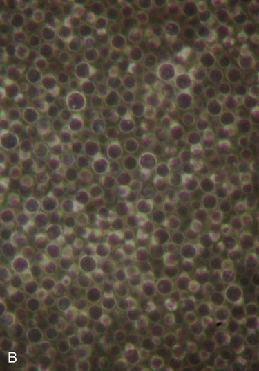
A few protozoa reproduce sexually by conjugation as seen in the ciliate Paramecium (see the next section, Classification of Protozoans). Conjugation is a form of genetic exchange by which members of two different mating types temporarily fuse and exchange nuclei. Each cell has two nuclei (Figure 8.12): a macronucleus, responsible for growth, and a haploid micronucleus, specialized for carrying out the process of conjugation. When two cells fuse, a micronucleus from each cell migrates to the other cell and fuses with a micronucleus within that cell. After this process is completed the parent cells separate; each is now a fertilized cell, and with later cell division they will produce daughter cells with recombinant DNA. The conjugation of protozoans is completely different from the bacterial process of the same name, discussed in Chapter 6 (Bacteria and Archaea).
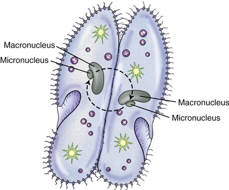
Each cell has a macronucleus, which is responsible for growth, and a haploid micronucleus. During conjugation the two cells fuse and the micronuclei of the two cells fuse. The parent cells, now fertilized, will separate and produce daughter cells with recombined DNA.
Classification of Protozoans
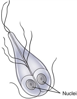
Archaezoa
Many Archaezoa are spindle shaped and have two or more flagella, located mostly on the anterior (front) end, that are used for whiplike motility and allow the cells to move through their environment (Figure 8.13). Human parasites include Trichomonas vaginalis (see Chapter 17, Sexually Transmitted Infections/Diseases) and Giardia spp. (see Chapter 12, Infections of the Gastrointestinal System). Trichomonas vaginalis lacks a cyst stage and must be transferred from host to host rather quickly before it dries out. Giardia lamblia, on the other hand, is a cyst-forming organism that is excreted in the feces and can survive in the environment before ingested by its next host.
Ciliophora
In general, ciliates are harmless, and Balantidium coli is the only ciliate pathogenic to humans, causing a disease called balantidiasis. It is the largest protozoan parasite of humans and is known to be able to exist as a trophozoite as well as a cyst. It produces proteolytic enzymes that break down and digest the intestinal epithelium (see Chapter 12, Infections of the Gastrointestinal System).
Euglenozoa
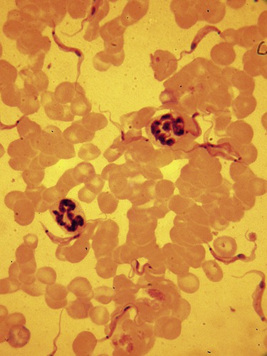
Hemoflagellates are blood parasites, transmitted by the bite of blood-feeding insects. They have long, slender bodies and an undulating membrane, giving them mobility in the circulatory system of the host. The genus Trypanosoma (Figure 8.14) includes T. brucei, which is transmitted by the tsetse fly and is the causative agent of sleeping sickness (see Chapter 14, Infections of the Circulatory System).
Amoebozoa
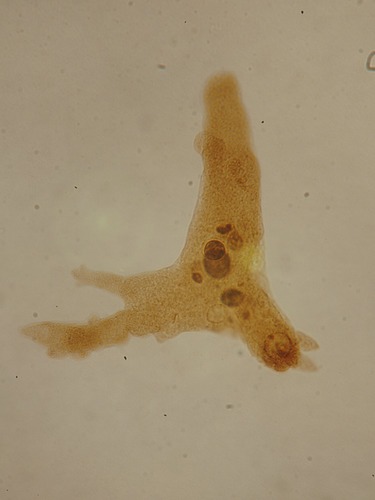
Amoebozoa, or amoebas, are a major group of amoeboid protozoans. Most of them are unicellular and common habitants in soil and water. They move by means of cytoplasmic flow, by extending blunt, lobelike projections of the cytoplasm, called pseudopods (Figure 8.15). The primary way in which these protozoans obtain nutrition is by phagocytosis, whereby the cell surrounds potential food particles and seals them into vacuoles, where digestion followed by absorption occurs. The only organism pathogenic to humans is Entamoeba histolytica, a parasite in the human intestine causing amoebic dysentery (see Chapter 12, Infections of the Gastrointestinal System). Entamoeba is transmitted between humans through ingestion of cysts that are present in the feces of infected persons. Another amoeba that can infect humans, typically immunocompromised people, is Balamuthia, which has been reported to cause brain abscesses referred to as primary amoebic meningoencephalitis (see Chapter 13, Infections of the Nervous System and Senses). Balamuthia is a free-living organism found in water and can enter the body through the lower respiratory tract or through open wounds. The organism cannot be transmitted by person-to-person contact.
Apicomplexa
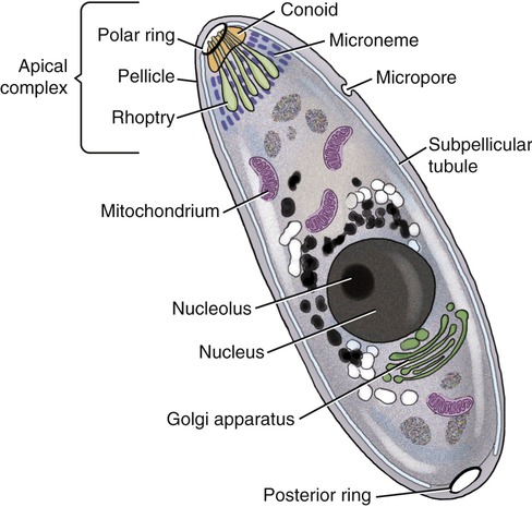
Apicomplexa are a large group of protozoans characterized by the presence of a unique, complex organelle called the apical complex (Figure 8.16). This complex can be visualized only by electron microscopy and it has been shown to consist of an apical conoid into which the ducts of saclike organelles called rhoptries lead. Furthermore, microtubules extend backward from this complex, apparently to support the surface of the organism. These rhoptries are believed to secrete an adhesive substance that facilitates attachment to the host cell, followed by entry of the parasite through the membrane.
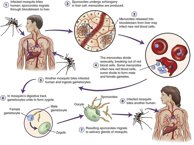
Apicomplexans have a complex life cycle requiring transmission between several hosts. An example is Plasmodium, the causative agent of malaria. Malaria is a devastating parasitic disease, endemic to tropical and subtropical areas of Asia, North and South America, the Middle East, North Africa, and the South Pacific. Plasmodium vivax is the most common among the four human malaria species: P. falciparum, malariae, ovale, and vivax. The organism is transmitted through the bite of infected Anopheles mosquitoes. The infective stage of Plasmodium carried by the mosquito is called the sporozoite. Once injected into the human the sporozoites are carried in the bloodstream to the liver, where they undergo schizogony to produce thousands of progeny called merozoites. Leaving the liver, the merozoites then invade red blood cells and reproduce. The young trophocytes (cells that provide nourishment to other cells) form ringlike structures referred to as a ring stage, which enlarges and divides repeatedly. Eventually the red blood cells rupture and release more merozoites, which then can infect more red blood cells to perpetuate their asexual reproduction. The waste products of the merozoites are responsible for the resulting fever and chills, characteristics of malaria. Some merozoites develop into male and female sexual forms (gametocytes), which can be picked up by a bite of another Anopheles mosquito. The gametocytes then enter the mosquito’s intestine and begin their sexual cycle by uniting to form a zygote. The zygote produces an oocyst in which cell division occurs and asexual sporozoites are formed. After rupturing of the oocytes the sporozoites migrate to the salivary glands of the mosquito and the cycle can start again (Figure 8.17).
Other apicomplexans are Babesia microti, Toxoplasma gondii, and Cryptosporidium.
Babesia microti is a parasite that infects erythrocytes and causes fever and anemia in humans. Before entering the human host, the life cycle of Babesia involves a rodent, primarily the white-footed mouse Peromyscus leucopus, and a Babesia-infected tick that introduces sporozoites into the mouse host. Here the sporozoites undergo asexual reproduction and some parasites differentiate into male and female gametes. Once ingested by an appropriate tick, usually the deer tick Ixodes dammini, they undergo a sporogonic cycle resulting in the production of sporozoites (Figure 8.18). Humans enter the cycle when bitten by infected ticks. The sporozoites enter erythrocytes and undergo asexual reproduction, which is responsible for the clinical symptoms of the disease—babesiosis. Humans are dead-end hosts and there is little chance that subsequent transmission occurs from ticks feeding on an infected person. However, human-to-human transmission can occur through blood transfusions.
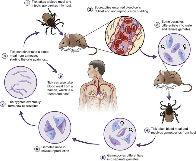
Note that the cycle consists of both sexual and asexual forms of reproduction.
Toxoplasma gondii is another intracellular parasite in humans and its life cycle includes domestic cats. The organism is the causative agent of toxoplasmosis, which is discussed in Chapter 16 (Infections of the Reproductive System).
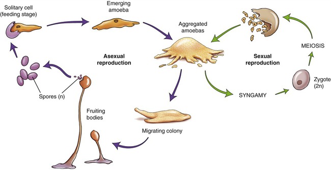
Slime Molds
Cellular Slime Molds
Cellular slime molds are typical eukaryotic cells that resemble amoebas and spend most of their lives as single amoeboid cells feeding on fungi and bacteria by phagocytosis. Under unfavorable conditions large numbers of these cells aggregate to form a single structure. This aggregation occurs in response to the release of chemicals by some individual amoebas and others respond by migrating toward the released chemical (cyclic adenosine monophosphate, or cAMP). The aggregated amoebas become enclosed in a slimy sheath called a slug, which migrates toward light. After migration, the slug begins to form differentiated structures; some form a stalk whereas others go up the stalk and form a spore cap. Most of these differentiate into spores, are released under appropriate conditions, and germinate to become single amoebas (Figure 8.19). Cellular slime molds are of great interest to biologists because they provide a relatively simple and easily manipulated system for understanding cellular migration and aggregation.
Helminths
Classification of Helminths
Platyhelminths
• Trematodes (flukes) are generally flat and leaf shaped, with oral and ventral suckers holding the organism in place (Figure 8.20). They have complex life cycles and their common names are given according to the tissue of the definitive host in which the adults live (e.g., lung fluke, liver fluke, blood fluke).
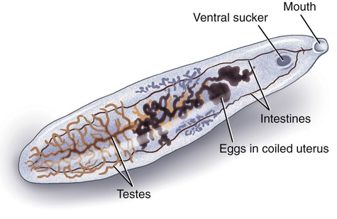
Trematodes (flukes) are generally flat and leaf shaped, with oral and ventral suckers holding the organism in place.
• Cestodes or tapeworms are intestinal parasites (see Chapter 12, Infections of the Gastrointestinal System). Adult tapeworms share a basic body structure: a scolex (head), a neck, and one or more proglottids (segments; see Figure 8.21). The scolex of the worm attaches to the intestine of the definite host, and the neck is a relatively undifferentiated mass of cells forming new segments—this is where all growth in the adult tapeworm occurs. The body of the worm is composed of successive units of proglottids collectively called strobila. It makes the worm look like a strip of tape and this is the source of its common name. Mature proglottids are released from the mature tapeworm and leave the host in its feces. The most mature proglottids are at the tail of the tapeworm and the most immature are closest to the neck.
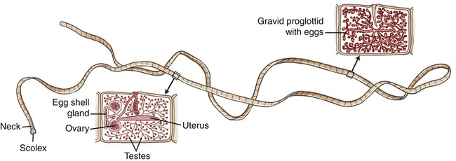
The scolex attaches the worm to the host tissue. Behind the scolex is the neck region, followed by the proglottids (body segments), which grow from the neck region as long as the worm is attached to the host. The proglottids closest to the neck are immature; those in the middle portion have matured, and the proglottids nearest the end become gravid (full of fertilized eggs). At this point the gravid proglottids break off and leave the host along with the feces.
Nematodes
• Enterobius vermicularis, the pinworm or seatworm, spends its life in a human host. The adult worms reside in the large intestine, from where it migrates to the anal area to deposit its eggs (hence the name, seatworm). The eggs can then be transmitted to another host by ingestion or via a fomite (contaminated clothing or bedding). Ascaris lumbricoides (a large roundworm that infects humans) is a parasite without an intermediate host. It spends its adult life in the human small intestine and feeds primarily on predigested food. Eggs can be excreted with feces and are able to survive in soil for long periods of time until accidentally ingested by another host. The eggs hatch in the small intestine and the larvae dig out of the intestine into the bloodstream. They are transported to the lungs, where they grow and eventually are coughed up, swallowed, and returned to the intestine for maturation into adults.
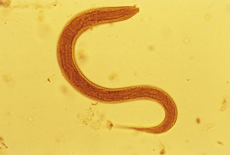
• Necator americanus and Ancylostoma duodenale are also called hookworms (Figure 8.22), and live in the small intestine of humans. Their eggs are excreted in feces, and their larvae hatch in the soil. The larvae feed on bacteria and can enter their human host by penetrating the host’s skin, after which they enter a blood or lymphatic vessel to be carried to the lungs. The larvae will be coughed up in sputum, swallowed, and carried to the small intestine, where the worms mature.
Summary
• Many eukaryotic organisms are pathogenic to humans and other animals. These pathogens include fungi, algae, protozoans, and helminths.
• Fungi are vegetative structures that generally grow as filamentous, multinucleate organisms (a mass of hyphae forms a mycelium), or they grow as unicellular fungi. Fungi are free-living, heterotrophic organisms that can appear as yeasts, molds, or fleshy fungi.
• Fungal reproduction can be asexual (mitosis) or sexual. Asexual reproduction results in the formation of sporangia and can occur at any time, whereas sexual reproduction generally occurs under conditions that are unfavorable for growth. Sexual reproduction provides variability of characteristics, making survival of the species more likely.
• Algae are aquatic, photosynthetic organisms, but they differ in distribution, morphology, reproduction, and biochemical preferences. Historically, algae were classified according to their photosynthetic pigments, but advancements in genetic analysis have indicated different relationships. Therefore, the current classification of algae is unsettled and complicated.
• All algae are capable of asexual reproduction and most can reproduce sexually as well. In some species sexual and asexual reproduction can alternate, depending on the given growth conditions.
• Protozoans are unicellular eukaryotes, lacking cell walls, and most are motile due to cilia, flagella, and/or pseudopodia. Most are chemoheterotrophs, obtaining their nutrients by phagocytosis of bacteria, organic matter, other protozoans, and host tissue.
• Protozoans exist in a motile, active feeding state called the trophozoite, or in a dormant state called a cyst. Many are human parasites and their life cycle ranges from simple to complex.
• Slime molds have both fungal and amoebal characteristics, but they are more closely related to amoeba than to fungi. Slime molds can be classified as cellular slime molds or plasmodial slime molds. They are not human pathogens.
• Helminths, although not microorganisms, exhibit microscopic infective stages in their life cycles in the form of eggs and/or larvae. Many of the parasitic helminths spend much or all of their lives in a mammalian host, including humans.
• The life cycle of parasitic helminths includes a fertilized egg, larval stage, and the adult organism. In general, their life cycles are complex, involving intermediate hosts as well as a definitive (final) host.
Review Questions
1. The antibiotics penicillin and cephalosporin are produced by:
2. Fungi are free-living __________ organisms.
3. Algae that contain agar in their cell walls belong to the:
4. Diatoms, major components of marine phytoplankton, belong to:
5. The process by which the nucleus of protozoans undergoes multiple divisions before the cell divides is called:
6. The eukaryotes known for the presence of a macronucleus and a micronucleus are:
8. Toxoplasma gondii belongs to which group of eukaryotic organisms?
9. A scolex is a structure found in:
10. Which of the following is commonly referred to as a pinworm?
11. The study of fungi is called __________.
12. The vegetative structure of algae is referred to as a(n) __________.
13. The unique cell organelle found among the Archaezoa, which appears to be a remnant of mitochondria, is called a(n) __________.
14. Masses of protoplasm containing thousands of nuclei are a characteristic of __________.
15. The common name for nematodes is __________.
16. Describe the classification of fungi with emphasis on the medically important species.
17. Describe the general characteristics of algae and their possible life cycles.
18. Name and describe three protozoans that are human parasites.
19. Describe the fundamental difference between cellular and plasmodial slime molds.
20. Describe the basic structure of cestodes (tapeworms) and their life cycle.
[/level-membership-for-basic-science-category][not-level-membership-for-basic-science-category]
Eukaryotic Microorganisms
After reading this chapter, the student will be able to:
• Describe, compare, and differentiate the characteristics of fungi, algae, protozoans, and helminths
• Describe the defining characteristics of fungi and explain the difference between their sexual and asexual reproduction
• Identify the medically important fungi
• Name the defining characteristics of algae, classify them, and describe the difficulties in their classification
• Describe the general characteristics of asexual and sexual reproduction of algae
• Identify the three defining and the common characteristics of protozoans
• Describe the general life cycle of protozoans and explain the importance of the macro- and micronucleus
• Discuss the fundamental differences between cellular and plasmodial slime molds
• Describe the main characteristics of parasitic helminths
• Describe the life cycles of flukes, tapeworms, and roundworms, and classify them into the appropriate categories
Introduction
Eukaryotic organisms differ from bacteria and archaea in many ways including cell size, internal structure, and genetic properties (see Chapter 3, Cell Structure and Function). The classification of eukaryotes has changed over the centuries starting in the late eighteenth century with Linnaeus, who classified all organisms as either plants or animals. At present, instead of the traditional classification schemes, many taxonomists favor classification schemes that are based on distinguishing characteristics at the molecular level. Modern taxonomy attempts to explain the genetic relationships of microbes and place them in groups based on similarities in their biochemical and metabolic characteristics, their nucleotide sequences, as well as their cellular ultrastructure as revealed by electron microscopy. Although universal support for a particular classification scheme has not been reached, many taxonomists favor a scheme similar to the one shown in Figure 8.1.
Fungi
The study of fungi is called mycology, and although more than 100,000 species of fungi are known, only about 200 are pathogenic to humans and animals, and relatively few fungi are virulent enough to be considered primary pathogens (Table 8.1). Moreover, the incidence of fungal infections has been increasing, especially those acquired in hospitals (nosocomial), other institutional settings such as nursing homes, and in people with a compromised immune system.
TABLE 8.1
Primary and Opportunistic Fungal Pathogens
| Organism | Reservoir | Transmission | Clinical Manifestation |
| Primary Pathogens | |||
| Blastomyces dermatitidis | Soil and organic debris; endemic area southeastern U.S., Ohio and Mississippi River Valleys | Inhalation of conidia | Primary pulmonary blastomycosis; chronic pulmonary blastomycosis; disseminated blastomycosis (cutaneous, bone, genitourinary tract, brain) |
| Coccidioides immitis | Desert soil; southwestern U.S.; Mexico; certain regions of Central and South America | Inhalation of conidia | Initial pulmonary infection; chronic pulmonary coccidioidomycosis; disseminated coccidioidomycosis (meningitis, bone, joints, genitourinary tract, cutaneous, ophthalmic) |
| Histoplasma capsulatum | Soil infested with bird/bat guano; eastern half of U.S.; most of Latin America; parts of Asia, Europe, and Middle East | Inhalation of conidia | Clinically asymptomatic pulmonary and “cryptic dissemination”; acute pulmonary histoplasmosis, mediastinitis, pericarditis; chronic pulmonary histoplasmosis |
| Paracoccidioides brasiliensis | Soil and vegetation; Central and South America | Inhalation of conidia | Diverse clinical manifestations; chronic multifocal involvement (lungs, mouth, nose); juvenile progressive disease (lymph nodes, skin, and viscera) |
| Opportunistic Pathogens | |||
| Candida spp. | Gastrointestinal mucosa, vaginal mucosa, skin, nails | Gastrointestinal translocation, intravascular catheters | Mucocutaneous candidiasis; oral/vaginal thrush; hematogenous dissemination; hepatosplenic candidiasis; endophthalmitis |
| Cryptococcus neoformans | Soil infested with bird guano | Inhalation of aerosolized yeast; percutaneous inoculation | Primary cryptoccal pneumonia; meningitis (particularly in HIV-infected patients); hematogenous dissemination; genitourinary cryptococcosis; primary cutaneous cryptococcosis |
| Aspergillus spp. | Soil, plants, water, pepper, air | Inhalation of conidia; transfer to wounds via contaminated bandages and/or tape | Allergic bronchopulmonary aspergillosis, sinusitis; aspergilloma; invasive aspergillosis (lung, brain, skin, gastrointestinal tract, heart) |
Fungi have important symbiotic relationships with other organisms and therefore can be beneficial to life in general. For example, they play a major role in the decomposition of dead plant material, and they are the primary decomposers of material such as cellulose, which cannot be digested by animals. The majority of plants are dependent on their symbiotic relationship with mycorrhizae (fungi that help the roots of plants to absorb minerals and water from the soil). Fungi provide food for humans in the form of mushrooms and truffles, and they are used in the manufacture of foods and beverages, including breads, alcoholic beverages, citric acid, soy sauce, and some cheeses. Fungi also produce pharmaceuticals such as the antibiotics penicillin and cephalosporin (see Chapter 22, Antimicrobial Drugs, for details). In addition, fungi are of use in the making of the immunosuppressive drug cyclosporine for organ transplant patients, and mevinic acids, which are used in the manufacture of cholesterol-reducing drugs. Fungi are also important research tools in genetics and biotechnology (see Chapter 25, Biotechnology).
Characteristics of Fungi
Fungi are eukaryotes but are quite different from plants and animals. Fungal colonies are vegetative structures because they are made up of cells involved in catabolism and growth. Whether microscopic or macroscopic, they generally grow as filamentous, multinucleated organisms that form threadlike filaments called hyphae, or as unicellular fungal organisms (Figure 8.2, A). Morphologically, fungi are often divided into three groups.
Fungi can exist as (A) unicellular organisms, or (B) form a mycelium, a mass of hyphae that continues to grow.
Most fungi that are identified as human pathogens are microscopic. Hyphae are long, branching filamentous cells of a fungus and they represent its main vegetative growth. A collective mass of the threadlike hyphae is called the mycelium (Figure 8.2, B). A mycelium can be small, forming colonies too small to see with the naked eye, or they can be extensive, such as Armillaria gallica, which was described as extending through 37 acres of forest soil and was estimated to weigh at least 9700 kg.
• Fungi tend toward maximal growth in a somewhat acidic environment, at about pH 5, which is too acidic for the growth of most bacteria (with such exceptions as Helicobacter pylori, which likes to grow at the pH of the human stomach [see Chapter 12, Infections of the Gastrointestinal System]).
• Most fungi are more resistant to osmotic pressure than are bacteria and can grow in relatively high concentrations of sugar or salt.
• Fungi can grow on substances with low moisture content, too low to support bacterial growth.
• Fungi metabolize more complex carbohydrates than bacteria.
• Chitin synthesis and other compounds for use in the cell wall, materials that induce hypersensitivity (see Chapter 20, The Immune System)
• Ergosterol synthesis for the plasma membrane, making the plasma membrane susceptible to antimicrobial agents targeting ergosterol synthesis or ergosterol incorporation into the plasma membrane (see Chapter 22, Antimicrobial Drugs)
Fungal pathogens can be classified on the basis of their growth forms—filamentous, yeast, or dimorphic—and on the type of infection they can cause. For example, in superficial mycoses, the fungus grows on the surface of the skin or hair. In cutaneous and subcutaneous mycoses, nails and deeper layers of the skin are involved, and in systemic (deep) mycoses, the fungus spreads to internal organs (Box 8.1). Patients with a compromised immune system are especially susceptible to fungal infections. Whereas superficial and cutaneous mycoses are generally mild, systemic mycoses are serious and difficult to treat.
Yeasts
Yeasts exist as single cells that reproduce by budding. However, some species may become multicellular through the formation of connected budding cells referred to as pseudohyphae, or true hyphae (Figure 8.3). They can be classified according to the presence or absence of capsules, the size and shape of the cells, the mechanism of daughter cell formation, formation of hyphae, presence of sexual spores, and other physiological/genetic data. Yeasts may require oxygen for their metabolic activities as obligate aerobes or may be able to continue their metabolic activities in the absence of oxygen as facultative anaerobes. Yeasts do not require light for their growth.
Molds
Molds are rapidly growing, asexually reproducing fungi that grow on a variety of substances. They are characterized by the development of hyphae (long filaments of cells joined together), which results in colony characteristics that can be used for identification purposes. Hyphae can grow to immense proportions (see Armillaria gallica, earlier this chapter). In general, the hyphae in molds contain cross-walls called septa, which divide the hyphae into distinct uninuclear, cell-like units called septate hyphae. Some classes of fungi have coenocytic hyphae, which do not contain septa and appear as long, continuous cells with many nuclei (Figure 8.4). In an appropriate environment, the hyphae grow to form a mycelium that is visible to the naked eye.
Dimorphic Fungi
• As a mold with septate hyphae in their natural reservoir (i.e., soil) and in the laboratory when incubated at 25° C on conventional fungal media
• As yeast in tissues of a human or other animals or in the laboratory when incubated at 37° C on enriched media (i.e., brain heart infusion agar)
Several dimorphic fungi can cause systemic mycoses (see Table 8.1) that usually start by the inhalation of spores from the mold form and then germinate in the lungs, where the fungus grows as yeast. These fungi include, but are not limited to, Sporothrix schenckii (sporotrichosis), Histoplasma capsulatum (histoplasmosis), Blastomyces dermatitidis (blastomycosis), Paracoccidioides brasiliensis (paracoccidioidomycosis), and Coccidioides spp. (coccidioidomycosis).
Life Cycle of Fungi
Asexual Reproduction
Yeast typically bud in a manner similar to the binary fission of prokaryotic organisms (see Chapter 3, Cell Structure and Function; and Chapter 6, Bacteria and Archaea). Filamentous fungi can reproduce asexually by fragmentation of their hyphae, and by spore formation either sexually or asexually. Asexual reproduction results in the formation of sporangia, which ultimately release spores into the environment. Spores, after entering the respiratory tract, are common causes of infection (see Chapter 11, Infections of the Respiratory System). These asexual spores are often categorized on the basis of the nature and manner of spore development:
• Sporangiospores form inside a sac, the sporangium, which is attached to a stalk, the sporangiophore, located on the tips or sides of hyphae. The spores are released when the sporangium ruptures.
• Conidia (conidiospores) are produced at the tips or sides of hyphae but are not enclosed by a saclike structure. Their development involves the pinching off of the tip of a fertile hypha or the segmentation of a preexisting vegetative hypha. They are the most common of the asexual spores and exist in different forms (Figure 8.5):
• Arthrospore: A rectangular spore, formed when a septate hypha breaks at the cross wall
• Chlamydospore: A spherical conidium produced by the thickening of a hyphal cell that is released when the surrounding hyphae crack
• Blastospore: A spore that buds from a parent cell
• Phialospore: A spore that buds from the mouth of a vase-shaped, spore-bearing cell
• Microconidium and macroconidium: Spores formed by the same fungus under different conditions, either as one cell (microconidium) or as two or more cells (macroconidium)
• Porospore: A spore that grows out through small pores in a spore-bearing cell
Sexual Reproduction
• Haploid (n) cells from a + mycelium and a − mycelium fuse and form a dikaryon. This stage is not considered to be diploid or haploid, but is designated as (n + n).
[/not-level-membership-for-basic-science-category]
