Disorders of Malabsorption

Published on 27/03/2015 by admin

Filed under Pediatrics

Last modified 22/04/2025

Print this page

rate 1 star rate 2 star rate 3 star rate 4 star rate 5 star
Your rating: none, Average: 0 (0 votes)

This article have been viewed 5006 times

Chapter 330 Disorders of Malabsorption

All disorders of malabsorption are associated with diminished intestinal absorption of one or more dietary nutrients. Malabsorption can result from a defect in the nutrient digestion in the intestinal lumen or from defective mucosal absorption. Malabsorption disorders can be categorized into generalized mucosal abnormalities usually resulting in malabsorption of multiple nutrients (Table 330-1) or malabsorption of specific nutrients (carbohydrate, fat, protein, vitamins, minerals, and trace elements) (Table 330-2). Almost all the malabsorption disorders are accompanied by chronic diarrhea (Chapter 333).

Table 330-2 CLASSIFICATION OF MALABSORPTION DISORDERS AND CHRONIC DIARRHEA BASED ON THE PREDOMINANT NUTRIENT MALABSORBED

CARBOHYDRATE MALABSORPTION

FAT MALABSORPTION

AMINO ACID MALABSORPTION

MINERAL AND VITAMIN MALABSORPTION

DRUG INDUCED

Clinical Approach

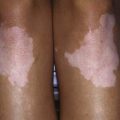
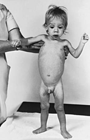
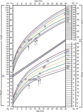
The clinical features depend on the extent and type of the malabsorbed nutrient. The common presenting features, especially in toddlers with malabsorption, are diarrhea, abdominal distention, and failure to gain weight, with a fall in growth chart percentiles. Physical findings include muscle wasting and the disappearance of the subcutaneous fat, with subsequent loose skinfolds (Fig. 330-1). The nutritional consequences of malabsorption are more dramatic in toddlers because of the limited energy reserves and higher proportion of calorie intake being used for weight gain and linear growth. In older children, malnutrition can result in growth retardation, as is commonly seen in children with late diagnosis of celiac disease. If malabsorption is untreated, linear growth slows, and with prolonged malnutrition, death can follow (Chapter 43). This extreme outcome is usually restricted to children living in the developing world, where resources to provide enteral and parenteral nutrition support may be limited. Specific findings on examination can guide toward a specific disorder; edema is usually associated with protein-losing enteropathy, digital clubbing with cystic fibrosis and celiac disease, perianal excoriation and gaseous abdominal distention with carbohydrate malabsorption, perianal and circumoral rash with acrodermatitis enteropathica, abnormal hair with Menkes syndrome, and the typical facial features diagnostic of the Johanson-Blizzard syndrome.

Many children with malabsorption disorders have very good appetites as they try to compensate for the fecal protein and energy losses. In exocrine pancreatic insufficiency, fecal losses of up to 40% of ingested protein and energy do not lead to malnutrition, as long as they are compensated by an increased appetite. In conditions associated with villous atrophy or inflammation (celiac disease, postinfectious enteropathy), fecal protein and energy losses are usually modest, but associated anorexia and reduced food intake results in malnutrition.

The nutritional assessment is an important part of clinical evaluation in children with malabsorptive disorders (Chapter 41). Long-term calcium and vitamin D malabsorption can lead to reduced bone mineral density and metabolic bone disease, with increased risk of bone fractures. Vitamin K malabsorption, irrespective of the underlying mechanism (fat malabsorption, mucosal atrophy), can result in coagulopathy. Severe protein-losing enteropathy is often associated with malabsorption syndromes (celiac disease, intestinal lymphangiectasia) and causes hypoalbuminemia and edema. Other nutrient deficiencies include iron malabsorption causing microcytic anemia and low reticulocyte count, low serum folate levels in conditions associated with mucosal atrophy, and low serum vitamin A and vitamin E concentrations in fat malabsorption.

Clinical history alone might not be sufficient to make a specific diagnosis, but it can direct the pediatrician toward a more structured and rational investigative approach. Diarrhea is the main clinical expression of malabsorption. Onset of diarrhea in early infancy suggests a congenital defect (Table 330-3). In secretory diarrhea due to disorders such as congenital chloride diarrhea and microvillus inclusion disease, the stool is watery and voluminous and can be mistaken for urine. Onset of symptoms after introduction of a particular food into a child’s diet can provide diagnostic clues, such as with sucrose in sucrase-isomaltase deficiency. The nature of the diarrhea may be helpful: explosive watery diarrhea suggests carbohydrate malabsorption; loose, bulky stools are associated with celiac disease; and pasty and yellowish offensive stools suggest an exocrine pancreatic insufficiency. Stool color is usually not helpful; green stool with undigested “peas and carrots” can suggest rapid intestinal transit in toddler’s diarrhea, which is a self-limiting condition unassociated with failure to thrive.

Table 330-3 DIARRHEAL DISEASES APPEARING IN THE NEONATAL PERIOD

CONDITION CLINICAL FEATURES
Microvillus inclusion disease Secretory watery diarrhea
Tufting enteropathy Secretory watery diarrhea
Congenital glucose-galactose malabsorption Acidic diarrhea
Congenital lactase deficiency Acidic diarrhea
Congenital chloride diarrhea Hydramnion, secretory watery diarrhea
Metabolic alkalosis
Congenital defective jejunal Na+/H+ exchange Hydramnion, secretory watery diarrhea
Congenital bile acid malabsorption Steatorrhea
Congenital enterokinase deficiency Failure to thrive, edema
Congenital trypsinogen deficiency Failure to thrive, edema
Congenital lipase and/or co-lipase deficiency Failure to thrive, oily stool
Enteric anendocrinosis (NEUROG 3 mutation) Hyperchloremic acidosis, failure to thrive

Adapted from Schmitz J: Maldigestion and malabsorption. In Walker WA, Durie PR, Hamilton JR, et al, editors: Pediatric gastrointestinal disease, ed 3, Hamilton, Ontario, 2000, BC Decker, p 55.

330.1 Evaluation of Children with Suspected Intestinal Malabsorption

Michael J. Lentze and David Branski

The investigation is guided by the history and physical examination. In a child presenting with chronic or recurrent diarrhea, the initial work-up should include stool cultures and antibody tests for parasites, stool microscopy for ova and parasites such as Giardia, and stool occult blood and leukocytes to exclude inflammatory disorders. Stool pH and reducing substances for carbohydrate malabsorption, and quantitative stool fat examination and α1-antitrypsin to demonstrate fat and protein malabsorption, respectively, should also be determined. Fecal stool elastase-1 can determine exocrine pancreatic insufficiency.

A complete blood count including peripheral smear for microcytic anemia, lymphopenia (lymphangiectasia), neutropenia (Shwachman syndrome), and acanthocytosis (abetalipoproteinemia) is useful. If celiac disease is suspected, serum immunoglobulin A (IgA) and tissue transglutaminase (TG2) antibody levels should be determined. Depending on the initial test results, more-specific investigations can be planned.

Investigations for Carbohydrate Malabsorption

Measurement of carbohydrate in the stool, using a Clinitest reagent that identifies reducing substances, is a simple screening test. An acidic stool with >2+ reducing substance suggests carbohydrate malabsorption. Sucrose or starch in the stool is not recognized as a reducing sugar until after hydrolysis with hydrochloric acid, which converts them to reducing sugars.

Breath hydrogen test is used to identify the specific carbohydrate that is malabsorbed. After an overnight fast, the suspected sugar (lactose, sucrose, fructose, or glucose) is administered as an oral solution (carbohydrate load 1-2 g/kg, maximum 50 g). In malabsorption, the sugar is not digested or absorbed in the small bowel, passes on to the colon, and is metabolized by the normal bacteria flora. One of the products of this process is hydrogen gas, which is absorbed through the colon mucosa and excreted in the breath. Increased hydrogen concentration in the breath samples suggests carbohydrate malabsorption. A rise in breath hydrogen of 20 ppm above the baseline is considered a positive test. The child should not be on antibiotics at the time of the test, because colonic flora is essential for fermenting the sugar.

Small bowel mucosal biopsies can measure mucosal disaccharidase (lactase, sucrase, maltase, palatinase) concentrations directly. In primary enzyme deficiencies the mucosal enzyme levels are low and small bowel mucosal morphology is normal. Partial or total villous atrophy due to disorders such as celiac disease, or following rotavirus gastroenteritis can result in secondary disaccharidase deficiency and transient lactose intolerance. The disaccharidase levels revert to normal after mucosal healing.

Investigations for Exocrine Pancreatic Function (Fig. 330-2)

Cystic fibrosis is the most common cause of exocrine pancreatic insufficiency in children; therefore, a sweat chloride test must be performed before embarking on invasive tests to investigate possible exocrine pancreatic insufficiency. Many cases of cystic fibrosis are detected by neonatal genetic screening programs; occasional rare mutations are undetected.

Fecal elastase-1 estimation is a sensitive test to assess exocrine pancreatic function in chronic cystic fibrosis and pancreatitis. Elastase-1 is a stable endoprotease unaffected by exogenous pancreatic enzymes. One disadvantage of the fecal elastase-1 test is the lack of full differentiation between primary exocrine pancreatic insufficiency and exocrine pancreatic dysfunction secondary to intestinal villous atrophy. The proximal small bowel is the site for pancreozymin/cholecystokinin production; the latter is the hormone that stimulates enzyme secretion from the exocrine pancreas. Mucosal atrophy can lead to diminished pancreozymin/cholecystokinin secretion and subsequently to exocrine pancreatic insufficiency. Fecal elastase-1 can also give a false-positive result during acute episodes of diarrhea.

Serum trypsinogen concentration can also be used as a screening test for exocrine pancreatic insufficiency. In cystic fibrosis, the levels are greatly elevated early in life, and then they gradually fall, so that by 5-7 yr of age, most patients with cystic fibrosis with pancreatic insufficiency have subnormal levels. Patients with cystic fibrosis and adequate exocrine pancreatic function tend to have normal or elevated levels. In such patients, observing the trend in serial serum trypsinogen estimation may be useful in monitoring exocrine pancreatic function. In Shwachman syndrome, another condition associated with exocrine pancreatic insufficiency, the serum trypsinogen level is low.

Other tests for pancreatic insufficiency (NBT-PABA test and pancreolauryl test) measure urine or breath concentrations of substances released and absorbed across the mucosal surface following pancreatic digestion. These tests lack specificity and are rarely used in clinical practice.

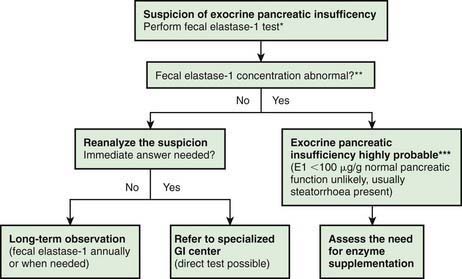
The gold standard test for exocrine pancreatic function is direct analysis of duodenal aspirate for volume, bicarbonate, trypsin and lipase upon secretin and pancreozymin/cholecysto-kinin stimulation. This involves duodenal intubation, and only a few centers perform this test (Chapter 340).

330.2 Gluten-Sensitive Enteropathy (Celiac Disease)

Etiology and Epidemiology

Celiac disease is an immune-mediated disorder elicited by the ingestion of gluten in genetically susceptible persons and characterized by chronic inflammation of the small intestine. It is considered an autoimmune condition because of the presence of anti–TG2 antibodies and the association with other autoimmune diseases (thyroid, liver, diabetes, adrenal).

Celiac disease is triggered by the ingestion of wheat gluten and related prolamines from rye and barley. In most studies oats proved to be safe; however, a few celiac patients have oats prolamine–reactive mucosal T cells that can cause mucosal inflammation.

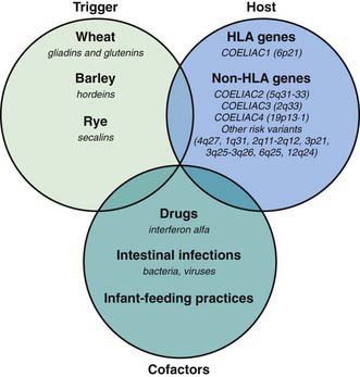
Celiac disease is a common disorder (1% prevalence of biopsy-proven disease). It is thought to be rare in Central Africa and East Asia. Environmental factors might affect the risk of developing celiac disease or the timing of its presentation. Prolonged breastfeeding has been associated with a reduced incidence of symptomatic disease. Less clear is the effect of the time of gluten introduction in the infant diet; the ingestion of increased amounts of gluten in the 1st year of life can increase the incidence. Infectious agents have been hypothesized to play a role because frequent rotavirus infections are associated with an increased risk. It is plausible that the contact with gliadin at a time when there is ongoing intestinal inflammation, altered intestinal permeability, and enhanced antigen presentation can increase the risk of developing the disease, at least in a subset of persons (Fig. 330-3).

image

Figure 330-3 Causative factors in celiac disease. HLA, human leukocyte antigen.

(From Di Sabatino A, Corazza GR: Coeliac disease, Lancet 373:1480-1490, 2009.)

Genetics and Pathogenesis

A genetic predisposition is suggested by the family aggregation and the concordance in monozygotic twins, which approaches 100%. It is suggested that the primary association of CD is with the DQ αβ heterodimer encoded by the DQA1*05 and the DQB1*02 genes. Such a DQ molecule is present in ≥95% of celiac patients compared with 20-30% of controls. DQ2-negative celiac patients are invariably HLA DQ8 positive (DQA1*0301/DQB1*0302). A gene dosage effect has been suggested, and a molecular hypothesis for such a phenomenon has been proposed, based on the impact of the number and quality of the HLA DQ2 molecules on gluten peptide presentation to T cells. Other non-HLA genes confer susceptibility to celiac disease. Genome-wide association studies have shown risk variants in genes controlling the immune response, some being shared with type 1 diabetes.

Celiac disease is a T cell–mediated chronic inflammatory disorder with an autoimmune component. Altered processing by intraluminal enzymes, changes in intestinal permeability, and activation of innate immunity mechanisms may be involved and precede the activation of the adaptive immune response. Immunodominant epitopes from gliadin are highly resistant to intraluminal and mucosal digestion; incomplete degradation favors the immunostimulatory and toxic effects. Some gliadin peptides (p31-43) can activate innate immunity, and in particular they induce interleukin 15 (IL-15). Others activate lamina propria T cells in the context of HLA-DQ2 or DQ8 molecules. Gliadin-specific T-cell responses are enhanced by the action of TG2; the enzyme converts particular glutamine residues into glutamic acid, which results in higher affinity of these gliadin peptides for HLA-DQ2 or HLA-DQ8. The pattern of cytokines produced following gliadin activation is dominated by interferon-γ (IFN-γ) (Th1 skewed); IFN-α, IL-18, and IL-21 are also upregulated. A complex remodeling of the mucosa then takes place, involving increased levels of metalloproteinases and growth factors, which leads to the classic flat mucosa. Increased density of CD8+ cytotoxic intraepithelial lymphocytes are a hallmark of celiac disease. IL-15 is implicated in the expression of natural killer receptors CD94 and NKG2D, as well as in epithelial expression of stress molecules, thus enhancing cytotoxicity, cell apoptosis, and villous atrophy.

The most evident expression of autoimmunity is the presence of serum antibodies to TG2. However, the mechanisms leading to autoimmunity are largely unknown. The finding of IgA deposits on extracellular TG2 in the liver, lymph nodes, and muscles indicates that TG2 is accessible to the gut-derived autoantibodies. Several extraintestinal clinical manifestations of celiac disease (e.g., liver, heart, nervous system) are possibly related to the presence of autoantibodies.

Clinical Presentation and Associated Disorders

Clinical features of celiac disease vary considerably (Table 330-4). Intestinal symptoms are common in children whose disease is diagnosed within the 1st 2 years of life; failure to thrive, chronic diarrhea, vomiting, abdominal distention, muscle wasting, anorexia, and irritability are present in most cases (see Fig. 330-1). Occasionally there is constipation, rectal prolapse, or intussusception. As the age at presentation of the disease shifts to later in childhood, and with the more liberal use of serologic screening tests, extraintestinal manifestations and associated disorders, without any accompanying digestive symptoms, have increasingly become recognized, affecting almost all organs (Table 330-5).

Table 330-4 SOME CLINICAL MANIFESTATIONS OF CELIAC DISEASE IN CHILDREN AND ADOLESCENTS

SYSTEM MANIFESTATION (POSSIBLE) CAUSE
Gastrointestinal Diarrhea
Distended abdomen
Vomiting
Anorexia
Weight loss
Failure to thrive
Aphthous stomatitis
Atrophy of the small bowel mucosa
Malabsorption
Hematologic Anemia Iron malabsorption
Skeletal Rickets
Osteoporosis
Enamel hypoplasia of the teeth
Calcium/vitamin D malabsorption
Muscular Atrophy Malnutrition
Neurologic Peripheral neuropathy
Epilepsy
Irritability
Thiamine/vitamin B12 deficiency
Endocrinologic Short stature
Pubertas tarda
Secondary hyperparathyroidism
Malnutrition
Calcium/vitamin D malabsorption
Dermatologic Dermatitis herpetiformis
Alopecia areata
Erythema nodosum
Autoimmunity
Respiratory Idiopathic pulmonary hemosiderosis  

Adapted from Mearin ML: Celiac disease among children and adolescents, Curr Prob Pediatr Adolesc Health Care 37:81–112, 2007.

The most common extraintestinal manifestation of celiac disease is iron-deficiency anemia, unresponsive to iron therapy. Osteoporosis may be present; in contrast to the situation in adults, it can be reversed by a gluten-free diet, with restoration of normal peak bone densitometric values. Other extraintestinal manifestations include short stature, endocrinopathies, arthritis and arthralgia, epilepsy with bilateral occipital calcifications, peripheral neuropathies, cardiomyopathy, chronic lung disease, isolated hypertransaminasemia, dental enamel hypoplasia, aphthous stomatitis, and alopecia. The mechanisms responsible for the severity and the variety of clinical presentations remain obscure. Nutritional deficiencies or abnormal immune responses have been advocated.

Silent celiac disease is being increasingly recognized, mainly in asymptomatic 1st-degree relatives of celiac patients investigated during screening studies. However, small bowel biopsy in these people reveals severe mucosal damage consistent with celiac disease. Potential celiac disease is defined when patients are identified by positive screening studies but without documented celiac disease on small bowel biopsy. It is important to follow these patients because they can develop established celiac disease in the future (Table 330-6).

Table 330-6 CLINICAL SPECTRUM OF CELIAC DISEASE

SYMPTOMATIC

Frank malabsorption symptoms: chronic diarrhea, failure to thrive, weight loss

Extraintestinal manifestations: anemia, fatigue, hypertransaminasemia, neurologic disorders, short stature, dental enamel defects, arthralgia, aphthous stomatitis

SILENT

No apparent symptoms in spite of histologic evidence of villous atrophy

In most cases identified by serologic screening in at-risk groups (see Table 330-1)

LATENT

Subjects who have a normal histology, but at some other time, before or after, have shown a gluten-dependent enteropathy

POTENTIAL

Subjects with positive celiac disease serology but without evidence of altered jejunal histology

It might or might not be symptomatic

Some diseases, many with an autoimmune pathogenesis, are found with a higher than normal incidence in celiac patients. Among these are type 1 diabetes, autoimmune thyroid disease, Addison disease, Sjögren syndrome, autoimmune cholangitis, autoimmune hepatitis, primary biliary cirrhosis, IgA nephropathy, alopecia, and dilated cardiomyopathy. Such associations have been interpreted as a consequence of the sharing of identical HLA haplotypes. The relation between celiac disease and other autoimmune diseases is poorly defined; once those diseases are established, they are not influenced by a gluten-free diet. Other associated conditions include selective IgA deficiency, Down syndrome, Turner syndrome, and Williams syndrome.

Patients with celiac disease show increased long-term mortality, the risk rising with delayed diagnosis and/or poor dietary compliance. Non-Hodgkin lymphoma is the main cause of death. Adult patients can develop complications such a refractory celiac disease, ulcerative jejunoileitis, or enteropathy-associated T-cell lymphoma.

Diagnosis

Serologic tests have a crucial role in the diagnosis of celiac disease; sensitivity of the IgA anti-TG2 is 61-100% (mean, 87%), and specificity is 86-100% (mean, 95%). Some 10% of patients whose disease is diagnosed earlier than 2 yr of age show absence of IgA anti-TG2. For them, the measurement of serum antigliadin antibodies is generally advised. Antibodies against gliadin-derived deamidated peptides (D-AGA) have been assessed. Compared with conventional AGA, the peptide antibodies (IgG and IgA) have a greater sensitivity and specificity. A problem with serology is represented by the association of celiac disease with IgA deficiency (10-fold increase compared to the general population). Serum IgA should always be checked, and in the case of IgA deficiency, D-AGA, IgG anti-endomysium, or TG2 should be sought. Negative serology should not preclude a biopsy examination when the clinical suspicion is strong.

Genetic tests have an increasing role in the diagnosis. Less than 2% of celiac patients lack both HLA specificities; at the same time, approximately one third of the “normal” population has one or the other marker; that means that the measurement of HLA DQ2 and/or DQ8 has a strong negative predictive value but a very weak positive predictive value for the diagnosis of celiac disease. With these limitations the test can prove useful to exclude celiac disease when the genetic studies are negative in subjects on a gluten-free diet or in subjects belonging to an at-risk group (e.g., 1st-degree relatives, insulin-dependent diabetics, patients with Down syndrome) to avoid long-term follow-up.

The ultimate diagnosis of celiac disease relies on the demonstration of specific, though not pathognomonic, histopathologic abnormalities in the small bowel mucosa (Table 330-7). According to The European Society for Pediatric Gastroenterology, Hepatology and Nutrition (ESPGHAN) current criteria, the 2 requirements mandatory for the diagnosis of celiac disease are the finding of villous atrophy with hyperplasia of the crypts and abnormal surface epithelium, while the patient is eating adequate amounts of gluten, and a full clinical remission after withdrawal of gluten from the diet. The finding of circulating IgA celiac disease–associated antibodies at the time of diagnosis and their disappearance on a gluten-free diet adds weight to the diagnosis. A control biopsy to verify the consequences of the gluten-free diet on the mucosal architecture is considered mandatory only in patients with an equivocal clinical response to the diet. Gluten challenge is not considered mandatory except in situations where there is doubt about the initial diagnosis, for example, when an initial biopsy was not performed or when the biopsy specimen was inadequate or atypical of celiac disease.

It is now accepted that the spectrum of histologic abnormalities in the celiac small intestine is wider than previously recognized. In some celiac disease patients, only subtle changes of crypt elongation with an increase in intraepithelial lymphocytes may be present. In those cases, it is very important to also evaluate the serology and the HLA typing so as to reach the correct diagnosis. Analysis of multiple biopsies is also very important.

Nevertheless, many cases of celiac disease are undiagnosed, and the ratio between patients with diagnosed and with undiagnosed disease may be as high as 1 : 7. Case finding by liberal use of anti-endomysium or anti-TG2 antibodies, followed by confirmatory jejunal biopsy, is more cost effective in primary care than mass screening is. Patients with symptoms or diseases known to be associated with celiac disease should undergo serologic evaluation.

Treatment

The only treatment for celiac disease is lifelong strict adherence to a gluten-free diet (Fig. 330-4). This requires a wheat-, barley-, and rye-free diet. Despite evidence that oats are safe for most patients with celiac disease, there is concern regarding the possibility of contamination of oats with gluten during harvesting, milling, and shipping. Nevertheless, it seems wise to add oats to the gluten-free diet only when the latter is well established, so that possible adverse reactions can be readily identified. There is a consensus that all celiac disease patients should be treated with a gluten-free diet regardless of the presence of symptoms. However, whereas it is relatively easy to assess the health improvement after treatment of celiac disease in patients with clinical symptoms of the disease, it proves difficult in persons with asymptomatic celiac disease. The nutritional risks, particularly osteopenia, are those mainly feared for subjects who have silent celiac disease and continue on a gluten-containing diet. Little is known about the health risks in untreated patients with minor enteropathy, which may be clinically silent. There are no guidelines concerning the need for a gluten-free diet in subjects with “potential” celiac disease (patients with positive celiac disease–associated serology but without enteropathy).

The Codex Alimentarius Guidelines define gluten-free as <20 ppm, but, although analytical methods for gluten detection have already reached a satisfactory degree of sensitivity, more information is needed on the daily gluten amount that may be tolerated by celiac disease patients. The data available so far seem to suggest that the threshold should be set to <50 mg/day, although individual variability makes it difficult to set a universal threshold.

It is important that an experienced dietician with specific expertise in celiac disease counseling educates the family and the child about dietary restriction. Compliance with a gluten-free diet can be difficult, especially in adolescents. It is recommended that children with celiac disease be monitored with periodic visits for assessment of symptoms, growth, physical examination, and adherence to the gluten-free diet. Periodic measurements of TG2 antibody levels to document reduction in antibody titers can be helpful as indirect evidence of adherence to a gluten-free diet, although they are inaccurate in detecting slight dietary transgressions.

Bibliography

Branski D, Fasano A, Troncone R. Latest development in the pathogenesis and treatment of celiac disease. J Pediatr. 2006;149:295-300.

Branski D, Troncone R. Celiac disease: a reappraisal. J Pediatr. 1998;133:181-187.

Collin P, Huhtala H, Virta L, et al. Diagnosis of celiac disease in clinical practice: physician’s alertness to the condition essential. J Clin Gastroenterol. 2007;41:152-156.

Di Sabatino A, Corazza RG. Coeliac disease. Lancet. 2009;373:1480-1493.

Dubois PC, Van Heel DA. Translational mini-review series on the immunogenetics of gut disease: immunogenetics of celiac disease. Clin Exp Immunol. 2008;153:162-173.

Ford AC, Chey WD, Talley NJ, et al. Yield of diagnostic tests for celiac disease in individuals with symptoms suggestive of irritable bowel syndrome. Arch Intern Med. 2009;169:651-658.

Green PHR, Cellier C. Celiac disease. N Engl J Med. 2007;357:1731-1743.

Jones R, Sleet S. Coeliac disease. BMJ. 2009;338:539-540.

Kline RM, Neudorf SML, Baron HI. Correction of celiac disease after allogeneic hematopoietic stem cell transplantation for acute myelogenous leukemia. Pediatrics. 2007;120:e1120-e1122.

Kurppa K, Ashorn M, Iltanen S, et al. Celiac disease without villous atrophy in children: a prospective study. J Pediatr. 2010;157:373-380.

Ludvigsson JF, Montgomery SM, Ekbom A, et al. Small-intestinal histopathology and mortality risk in celiac disease. JAMA. 2009;302:1171-1178.

McGowan KE, Castiglione DA, Butzner JD. The changing face of childhood celiac disease in North America: Impact of serological testing. Pediatrics. 2009;124:1572-1578.

Mearin ML. Celiac disease among children and adolescents. Curr Prob Pediatr Adolesc Health Care. 2007;37:81-112.

Meresse B, Ripoche J, Heyman M, et al. Celiac disease: from oral tolerance to intestinal inflammation, autoimmunity and lymphomagenesis. Mucosal Immunol. 2009;2:8-23.

Olsson C, Hernell O, Hornell A, et al. Difference in celiac disease risk between Swedish birth cohorts suggests an opportunity for primary prevention. Pediatrics. 2008;122:528-534.

Rashtak S, Murray JA. Tailored testing for celiac disease. Ann Intern Med. 2007;147:339-340.

Richey R, Howdle P, Shaw E, et al. Recognition and assessment of coeliac disease in children and adults: summary of NICE guidance. BMJ. 2009;338:1386-1388.

Rodrigues AF, Jenkins HR. Investigation and management of coeliac disease. Arch Dis Child. 2008;93:251-254.

Sattar N, Lazare F, Kacer M, et al. Celiac disease in children, adolescents, and young adults with autoimmune disease. J Pediatr. 2011;158:272-275.

Simmons JH, Klingensmith GJ, McFann K, et al. Celiac autoimmunity in children with type 1 diabetes: a two-year follow-up. J Pediatr. 2011;158:276-281.

Smyth DJ, Plagnol V, Walker NM, et al. Shared and distinct genetic variants in type 1 diabetes and celiac disease. N Engl J Med. 2008;359:2767-2776.

Sollid LM, Lundin KEA. Diagnosis and treatment of celiac disease. Mucosal Immunol. 2009;2:3-7.

Telega G, Bennet TR, Welin S. Emerging new clinical patterns in the presentation of celiac disease. Arch Pediatr Adolesc Med. 2008;162:164-168.

Troncone R, Ivarsson A, Szajewska H, et al. Review article: future research on celiac disease—a position report from the European multistakeholder platform on celiac disease (CDEUSSA). Aliment Pharmacol Ther. 2008;27:1030-1043.

van Koppen EJ, Schweizer JJ, Csizmadia CGDS, et al. Long-tern health and quality-of-life consequences of mass screening for childhood celiac disease: a 10-year follow-up study. Pediatrics. 2009;123:e582-e588.

Zanchi C, Di Leo G, Ronfani L, et al. Bone metabolism in celiac disease. J Pediatr. 2008;153:262-265.

330.3 Other Malabsorptive Syndromes

Congenital Intestinal Mucosal Defects

Microvillus Inclusion Disease (Congenital Microvillus Atrophy)

Microvillus inclusion disease is an autosomal recessive disorder, which manifests at birth with profuse watery secretory diarrhea. It is the most commonly recognized cause of congenital diarrhea. Light microscopy of the small bowel mucosa demonstrates diffuse thinning of the mucosa, with hypoplastic villus atrophy and no inflammatory infiltrate. Diagnosis may be easily performed with light microscopy using PAS and CD10 staining, which shows a very thin or absent brush border, together with positive PAS and CD10 intracellular inclusions. Electron microscopy shows enterocytes with absent or sparse microvilli. The apical cytoplasm of the enterocytes contains electron-dense secretory granules; the hallmark is presence of microvilli within involutions of the apical membrane. Polyhydramnios is not a classic presentation of MID. Neonates usually present with dehydration and failure to thrive. Despite parenteral nutrition, diarrhea continues and initial fluid management is difficult. The disease is fatal without long-term parenteral nutrition support. Some infants present with rapid onset of liver disease, which is associated with pruritus. Most children die in infancy or early childhood. The long-acting somatostatin analog octreotide has been used as treatment and can reduce the volume of stool in some infants (Chapter 331). Intestinal transplantation is the only definitive treatment for this rare disease. Rarely, in milder forms of the disease, the patient can reach young adulthood and enjoy partially oral feeding. The underlying gene defect is a mutation in MYO5B, which encodes a protein involved in subcellular protein trafficking. Several types of mutations are involved.

Tufting Enteropathy

Tufting enteropathy (intestinal epithelial dysplasia) manifests in the 1st weeks of life with persistent watery diarrhea and accounts for a small fraction of infants with protracted diarrhea of infancy. Symptoms typically do not begin immediately after birth but occur in early infancy. The distinctive feature on small intestinal mucosal biopsy is focal epithelial “tufts” (teardrop-shaped groups of closely packed enterocytes with apical rounding of the plasma membrane) involving 80-90% of the epithelial surface. However, the typical pathology does not appear immediately after birth, and in other known enteropathies tufts are seen on ≤15% of the epithelial surface. Colonic epithelium shows abnormalities that are more difficult to identify. Electron microscopy does not help in the diagnosis.

The pathogenesis of this disorder may be due to a disorder of cell-cell and cell-matrix interactions, because there is an abnormal distribution of α2β1-integrin along the crypt-villus axis, increased expression of desmoglein, and ultrastructural changes of desmosomes. Tufting enteropathy is often associated with punctiform keratitis and conjunctival dysplasia resembling typical pictures of tufts. The genetic basis of tufting enteropathy supports this speculation, because a single amino acid substitution in exon 4 of the EPCAM gene encoding an epithelial cell adhesion molecule protein has been described.

No treatment has been effective, so management requires permanent parenteral nutrition with possible intestinal transplantation (Chapter 331). Several types of mutations are involved, opening the door to genotype-phenotype analysis.

Carbohydrate-Deficient Glycoprotein Syndrome and Enterocyte Heparan Sulfate Deficiency

Congenital disorders of glycosylation (also carbohydrate-deficient glycoprotein, CDG) are genetic disorders of assembly of N-glycans in the cytosol and endoplasmic reticulum, resulting in a variety of manifestations (Chapter 81.6). The subtypes of CDG I are all associated with protein-losing enteropathy. Diagnosis can be established by isoelectric focusing of serum transferrin, enzyme analysis, and/or DNA analysis. Oral mannose can provide effective therapy in CDG Ib, so early identification of children presenting with hypoglycemia, hypothyroidism, and/or thyroid binding globulin deficiency is beneficial.

Congenital enterocyte heparan deficiency (CEHD) is a rare cause of intractable diarrhea with protein-losing enteropathy, which may be an unusual presentation of the carbohydrate-deficient glycoprotein syndrome (CDGS) type 1 (also known as Jaeken syndrome) (Chapter 81.6). Heparan sulfate is a glycosaminoglycan with multiple roles in the intestine, including restriction of charged macromolecules, such as albumin, in the vascular lumen.

Intestinal Lymphangiectasia

Obstruction of the lymphatic drainage of the intestine can be due to either congenital defects in lymphatic duct formation or to secondary causes (Table 330-8). The congenital form is often associated with lymphatic abnormalities elsewhere in the body, as occur with Turner, Noonan, and Klippel-Trenaunay-Weber syndromes. Causes of secondary lymphangiectasia include constrictive pericarditis, heart failure, retroperitoneal fibrosis, abdominal tuberculosis, and retroperitoneal malignancies. Lymph rich in proteins, lipids, and lymphocytes leaks into the bowel lumen, resulting in protein-losing enteropathy, steatorrhea, and lymphocyte depletion. Hypoalbuminemia, hypogammaglobulinemia, edema, lymphopenia, malabsorption of fat and fat-soluble vitamins, and chylous ascites often occur. Intestinal lymphangiectasia can also manifest with ascites, peripheral edema, and a low serum albumin.

The diagnosis is suggested by the typical findings in association with an elevated fecal α1-antitrypsin clearance. Radiologic findings of uniform, symmetric thickening of mucosal folds throughout the small intestine are characteristic but nonspecific. Small bowel mucosal biopsy can show dilated lacteals with distortion of villi and no inflammatory infiltrate. A patchy distribution and deeper mucosal involvement on occasion causes false-negative results on small bowel histology. Treatment of lymphangiectasia includes restricting the amount of long-chain fat ingested and administering a formula containing protein and medium-chain triglycerides (MCTs). Supplementing a low-fat diet with MCT oil in cooking is used in the management of older children with lymphangiectasia. Rarely, parenteral nutrition is required. If only a portion of the intestine is involved, surgical resection may be considered.

Syndromic Diarrhea

Syndromic diarrhea (SD), also known as phenotypic diarrhea (PD) or tricho-hepato-enteric (THE) syndrome, is a congenital enteropathy manifesting with early onset of severe diarrhea requiring parenteral nutrition. The estimated prevalence is approximately 1/300,000-400,000 live births in Western Europe. Patients born small for gestational age present with diarrhea starting in the 1st 6 mo of life (<1 mo of age in most cases). They have an abnormal phenotype, including facial dysmorphism with prominent forehead, broad nose, and hypertelorism and a distinct abnormality of hair, trichorrhexis nodosa. Hairs are woolly, easily removed, and poorly pigmented. Liver disease affects about half of the patients with extensive fibrosis or cirrhosis. The patients have defective antibody responses despite normal serum immunoglobulin levels and defective antigen-specific skin tests despite positive proliferative responses in vitro. Microscopic analysis shows twisted hair (pili torti), aniso- and poilkilotrichosis, and trichorrhexis nodosa. Histopathologic analysis shows nonspecific villus atrophy with or without mononuclear cell infiltration of the lamina propria, without specific histologic abnormalities involving the epithelium. Recently mutations in the TTC37 gene were found as the cause of THE syndrome. The common association of the disorder with parental consanguinity and/or affected siblings suggests a genetic origin with an autosomal recessive transmission. Prognosis of this type of intractable diarrhea of infancy is poor because most patients have died between the ages of 2 and 5 yr, some of them with early-onset liver disease.

Autoimmune Enteropathy

Symptoms of autoimmune enteropathy usually occur after the 1st 6 mo of life with chronic diarrhea, malabsorption, and failure to thrive. Histologic findings in the small bowel include partial or complete villous atrophy, crypt hyperplasia, and an increase in chronic inflammatory cells in the lamina propria. In contrast to gluten-sensitive enteropathy, an increase in intra-epithelial lymphocytes is not a prominent feature of autoimmune enteropathy. Specific serum anti-enterocyte antibodies can be identified in ≥50% of patients by indirect immunofluorescent staining of normal small bowel mucosa and kidney. In some patients anti-goblet cell antibodies can be demonstrated as well. The colon is also often involved, with inflammation and clinical features of colitis.

Extraintestinal autoimmune disorders are usual and include arthritis, membranous glomerulonephritis, insulin-dependent diabetes, thrombocytopenia, autoimmune hepatitis, hypothyroidism, and hemolytic anemia. It is essential to exclude underlying primary immune deficiency, particularly in boys with other autoimmune features (diabetes mellitus), because a proportion have underlying Immune dysregulation, Polyendocrinopathy, Enteropathy, X linked (IPEX) syndrome (Chapter 120.5). This systemic autoimmune disorder is due to mutations in FOXP3, a transcriptional regulator essential for the normal development of regulatory T cells (Tregs). Autoimmune enteropathy is reported in cases of Schimke immunoosseous dysplasia.

Treatment for autoimmune enteropathy includes immune suppression drugs including prednisone, azathioprine, cyclophosphamide, cyclosporine, and tacrolimus. Bone marrow transplantation is curative for children with IPEX syndrome.

Bile Acid Malabsorption

In primary bile acid malabsorption, mutation of the ileal sodium–bile acid cotransporter gene, SLC10A2, results in congenital diarrhea, steatorrhea, interruption of enterohepatic circulation of bile acids, and reduced plasma cholesterol levels. Bile acids are normally synthesized from cholesterol in the liver and secreted into the small intestine, where they facilitate absorption of fat, fat-soluble vitamins, and cholesterol. Bile acids are reabsorbed in the distal ileum, return to the liver via the portal venous circulation, and are resecreted into the bile. Normally, the enterohepatic circulation of bile acids is an extremely efficient process; only 10% of the intestinal bile acids escape reabsorption and are eliminated in feces. Bile acid secretion is largely autoregulated, but there is only a limited capacity to increase bile acid secretion. Reduction in the bile acid pool due to bile acid malabsorption causes steatorrhea, which requires restriction of dietary fat. Unabsorbed bile acids stimulate chloride excretion in the colon, resulting in diarrhea, which responds to cholestyramine, an anion-binding resin. Secondary bile acid malabsorption can result from ileal disease, such as in Crohn disease, and following an ileal resection.

Chronic neonatal-onset diarrhea has also been described in autosomal recessive cerebrotendinous xanthomatosis, which is caused by an inborn error of bile acid synthesis due to 27-hydroxylase deficiency. These children also present with juvenile-onset cataracts and developmental delay. Neonatal cholestasis has also been described as a presenting feature. Tendon xanthomas develop in the second and third decades of life. The diagnosis is important to establish, because treatment is effective when employing oral chenodeoxycholic acid.

Abetalipoproteinemia

Abetalipoproteinemia is a rare autosomal recessive disorder of lipoprotein metabolism (Bassen Kornsweig syndrome) (Chapter 80). It is associated with severe fat malabsorption from birth. Children fail to thrive during the 1st year of life, with stools that are pale, foul smelling, and bulky. The abdomen is distended and deep tendon reflexes are absent as a result of peripheral neuropathy, which is secondary to vitamin E (fat-soluble vitamin) deficiency. Intellectual development tends to be slow. After 10 yr of age, intestinal symptoms are less severe, ataxia develops, and there is a loss of position and vibration sensation with the onset of intention tremors unless vitamin E levels are maintained in the normal range. These latter symptoms reflect involvement of the posterior columns, cerebellum, and basal ganglia. In adolescence, atypical retinitis pigmentosa develops without adequate supplemental of vitamin E; for instance, using a TPGS formulation of the vitamin.

Diagnosis rests on the presence of acanthocytes in the peripheral blood smear and extremely low plasma levels of cholesterol (<50 mg/dL); triglycerides are also very low (<20 mg/dL). Chylomicrons and very low density lipoproteins are not detectable, and the low-density lipoprotein (LDL) fraction is virtually absent from the circulation. Marked triglyceride accumulation in villus enterocytes occurs in the duodenal mucosa. Steatorrhea occurs in younger patients, but other processes of nutrient assimilation are intact. Rickets may be an unusual initial manifestation of abetalipoproteinemia and hypobetalipoproteinemia. Rickets is caused by steatorrhea-induced calcium losses and vitamin D deficiency. Patients have mutations of the microsomal triglyceride transfer protein (MTP) gene, resulting in absence of MTP function in the small bowel. This protein is required for normal assembly and secretion of very low density lipoproteins and chylomicrons.

Specific treatment is not available. Large supplements of the fat-soluble vitamins A, D, E, and K should be given. Vitamin E (100-200 mg/kg/24 hr) appears to arrest neurologic and retinal degeneration. Limiting long-chain fat intake can alleviate intestinal symptoms; medium-chain triglycerides can be used to supplement the fat intake.

Homozygous Hypobetalipoproteinemia

Homozygous hypobetalipoproteinemia (Chapter 80) is transmitted as an autosomal dominant trait. The homozygous form is indistinguishable from abetalipoproteinemia. The parents of these patients, as heterozygotes, have reduced plasma LDL and apoprotein-β concentrations, whereas the parents of patients with abetalipoproteinemia have normal levels. On transmission electron microscopy of small bowel biopsies, the size of lipid vacuoles in enterocytes differentiates between abetalipoproteinemia and hypobetalipoproteinemia: Many small vacuoles are present in hypobetalipoproteinemia, and larger vacuoles are seen in abetalipoproteinemia.

Bibliography

Cormier-Daire V, Bonnefont JP, Rustin P, et al. Mitochondrial DNA rearrangements with onset as chronic diarrhea with villous atrophy. J Pediatr. 1994;124:63-70.

Cuenod B, Brousse N, Goulet O, et al. Classification of intractable diarrhea in infancy using clinical and immunohistological criteria. Gastroenterology. 1990;99:1037-1043.

Fabre A, Roquelaure B, Lacoste C, et al. Exclusion of EGFR, HRAS, DSP, JUP, CTNNB1, PLEC1, and EPPK1 as functional candidate genes in 7 families with syndromic diarrhoea. J Pediatr Gastroenterol Nutr. 2009;48:501-503.

Girault D, Goulet O, Ledeist F, et al. Intractable diarrhea syndrome associated with phenotypic abnormalities and immune deficiency. J Pediatr. 1994;125:36-42.

Glocker EO, Frede N, Perro M, et al. Infant colitis—it’s in the genes. Lancet. 2010;376:1272.

Goulet O, Kedinger M, Brousse N, et al. Intractable diarrhea of infancy: a new entity with epithelial and basement membrane abnormalities. J Pediatr. 1995;127:212-219.

Goulet O, Ruemmele F. Causes and management of intestinal failure in children. Gastroenterology. 2006;130:S16-S28.

Goulet O, Salomon J, Ruemmele F, et al. Intestinal epithelial dysplasia (tufting enteropathy). Orphanet J Rare Dis. 2007;20:20.

Goulet O, Vinson C, Roquelaure B, et al. Syndromic (phenotypic) diarrhea in early infancy. Orphanet J Rare Dis. 2008;3:6.

Hartley JL, Zachos NC, Dawood B, et al. Mutations in TTC37 cause trichohepatoenteric syndrome (phenotypic diarrhea of infancy). Gastroenterology. 2010;138:2388-2398.

Jaeken J, Matthij G, Saudubray JM, et al. Phosphomannose isomerase deficiency: a carbohydrate-deficient glycoproteins syndrome with hepatic-intestinal presentation. Am J Hum Genet. 1998;62:1535-1539.

Müller T, Hess MW, Schiefermeier N, et al. MYO5B mutations cause microvillus inclusion disease and disrupt epithelial cell polarity. Nat Genet. 2008;40:1163-1165.

Murch S, Winyard PJD, Koletzki S, et al. Congenital enterocyte heparan sulphate deficiency with massive albumin loss, secretory diarrhoea and malnutrition. Lancet. 1996;347:1299-1301.

Oren A, Houwen RH. Phosphomannoseisomerase deficiency as the cause of protein-losing enteropathy and congenital liver fibrosis. J Pediatr Gastroenterol Nutr. 1999;29:231-232.

Patey N, Scoazec JY, Cuenod-Jabri B, et al. Distribution of cell adhesion molecules in infants with intestinal epithelial dysplasia (tufting enteropathy). Gastroenterology. 1997;113:833-843.

Patey-Mariaud de Serre N, Canioni D, Ganousse S, et al. Digestive histopathological presentation of IPEX syndrome. Mod Pathol. 2009;22:95-102.

Phillips AD, Schmitz J. Familial microvillous atrophy: a clinicopathological survey of 23 cases. J Pediatr Gastroenterol Nutr. 1992;14:380-396.

Reifen RM, Cutz E, Griffiths AM, et al. Tufting enteropathy: a newly recognized clinicopathological entity associated with refractory diarrhea in infants. J Pediatr Gastroenterol Nutr. 1994;18:379-385.

Ruemmele FM, Brousse N, Goulet O. Autoimmune enteropathy—molecular concepts. Curr Opinion in Gastroenterol. 2004;20:587-591.

Ruemmele FM, Jan D, Lacaille F, et al. New perspectives for children with microvillous inclusion disease: early small bowel transplantation. Transplantation. 2004;15:1024-1028.

Ruemmele FM, Moes N, de Serre NP, et al. Clinical and molecular aspects of autoimmune enteropathy and immune dysregulation, polyendocrinopathy autoimmune enteropathy X-linked syndrome. Curr Opin Gastroenterol. 2008;24:742-748.

Ruemmele FM, Schmitz J, Goulet O. Microvillous inclusion disease (microvillous atrophy). Orphanet J Rare Dis. 2006 Jun;26(1):22.

Sivagnanam M, Mueller JL, Lee H, et al. Identification of EpCAM as the gene for congenital tufting enteropathy. Gastroenterology. 2008;135:429-437.

Torgerson TR, Linane A, Moes N, et al. Severe food allergy as a variant of IPEX syndrome caused by a deletion in a noncoding region of the FOXP3 gene. Gastroenterology. 2007;132:1705-1717.

Wang J, Cortina G, Wu SV, et al. Mutant neurogenin-3 in congenital malabsorptive diarrhea. N Engl J Med. 2006;355:270-280.

330.4 Intestinal Infections and Infestations Associated with Malabsorption

Malabsorption is a rare consequence of primary intestinal infection and infestation in immunocompetent children, but it can occur after infection with Campylobacter, Shigella, Salmonella, Giardia, cryptosporidium, coccidioidosis, and rotavirus. These infectious causes of malabsorption are more common in immunocompromised children.

Bacterial Overgrowth

Bacteria are normally present in large numbers in the colon (1011-1013 colony-forming units [CFU]/gram of feces) and have a symbiotic relationship with the host, providing nutrients and protecting the host from pathogenic organisms. Excessive numbers of bacteria in the small bowel or stomach are harmful. Bacteria are usually present only in a small number in the stomach and small bowel. Gastric acid pH prevents the ingested organisms from colonizing the small bowel. Small bowel motility and the migrating motor complex cleanse the small bowel between meals and at night; the ileocecal valve prevents colonic bacteria from refluxing into the ileum. Mucosal defenses such as mucin and immunoglobulins prevent bacterial overgrowth in the small bowel. Bacterial overgrowth can result from clinical conditions that alter the gastric pH or small bowel motility, including disorders such as partial bowel obstruction, diverticula, short bowel, intestinal duplications, diabetes mellitus, idiopathic intestinal pseudo-obstruction syndrome, and scleroderma. Prematurity, immunodeficiency, and malnutrition are other factors associated with bacterial overgrowth of the small bowel.

Diagnosis of bacterial overgrowth can be made by culturing small bowel aspirate or by lactulose hydrogen breath test. Lactulose is a synthetic disaccharide, which is not digested by mucosal brush border enzymes but can be fermented by bacteria. High baseline hydrogen and a quick rise in hydrogen in expired breath samples support the diagnosis of bacterial overgrowth, but false-positive tests are common.

Bacterial overgrowth leads to inefficient intraluminal processing of dietary fat and to steatorrhea due to bacterial deconjugation of bile salts, vitamin B12 malabsorption, and microvillus brush border damage with malabsorption. Bacterial consumption of vitamin B12 and enhanced synthesis of folate result in decreased vitamin B12 and increased folate serum levels. Overproduction of D-lactate (the isomer of L-lactate) can cause stupor, neurologic dysfunction, and shock from D-lactic acidosis. Lactic acidosis should be suspected in children at risk of bacterial overgrowth, who show signs of neurologic deterioration and a high anion gap metabolic acidosis not explained by measurable acids such as L-lactate. Measurement of D-lactate is required because standard lactate assay only measures the L-isomer.

Treatment of bacterial overgrowth focuses on correction of underlying causes such as partial obstruction. The oral administration of antibiotics is the mainstay of therapy. Initial treatment with 2-4 wk of metronidazole can provide relief for many months. Cycling of antibiotics including azithromycin, trimethoprim-sulfamethoxazole, ciprofloxacin, and metronidazole is required. Other alternatives are oral nonabsorbable antibiotics such as aminoglycosides. Occasionally, antifungal therapy is required to control fungal overgrowth of the bowel.

Tropical Sprue

Natives and expatriates of certain tropical regions can present with a diffuse lesion of the small intestinal mucosa—tropical sprue, even long after emigration. The endemic regions include South India, the Philippines, and some islands in the Caribbean. It is uncommon in Africa, Jamaica, and Southeast Asia. The etiology of this disorder is unclear; because it follows outbreaks of acute diarrheal disease and improves with antibiotic therapy, an infectious etiology is suspected. The incidence is decreasing worldwide, possibly due to common use of antibiotics for gastroenteritis in developing countries.

Clinical symptoms include fever and malaise followed by watery diarrhea. After about a week the acute features subside, and anorexia, intermittent diarrhea, and chronic malabsorption result in severe malnutrition characterized by glossitis, stomatitis, cheilosis, night blindness, hyperpigmentation, and edema. Muscle wasting is often marked, and the abdomen is often distended. Megaloblastic anemia results from folate and vitamin B12 deficiencies.

Diagnosis is made by small bowel biopsy, which shows villous flattening, crypt hyperplasia, and a chronic inflammatory cell infiltrate of the lamina propria with adjacent lipid accumulation in the surface epithelium.

Treatment requires nutritional supplementation, including supplementation of folate and vitamin B12. To prevent recurrence, 6 mo of therapy with oral folic acid (5 mg) and tetracycline or sulfonamides is recommended. Relapses occur in 10-20% of patients who continue to reside in an endemic tropical region; additional courses of antibiotics may be necessary.

330.5 Immunodeficiency Disorders

Malabsorption can occur with congenital immunodeficiency disorders, and chronic diarrhea with failure to thrive is often the mode of presentation. Defects of humoral and or cellular immunity may be involved, including selective IgA deficiency, agammaglobulinemia, common variable immunodeficiency disease (CVID), severe combined immunodeficiency, Wiskott-Aldrich syndrome, or chronic granulomatous disease. Although most patients with selective IgA deficiency are asymptomatic, malabsorption due to giardiasis or nonspecific enteropathy with bacterial overgrowth can occur. Malabsorption syndrome or chronic noninfectious diarrhea has been reported in 60% of children with CVID, most often in the subgroup with low memory B cell counts. Malabsorption has also been reported in ∼10% of patients with late-onset CVID, often secondary to giardiasis. Celiac disease is more common in patients with IgA deficiency and CVID. Paradoxically, it is more difficult to exclude the diagnosis of celiac disease because of the lack of reliability of IgA- and IgG-based serologic tests. Malabsorption due to chronic rotavirus, giardiasis, bacterial overgrowth, and protein-losing enteropathy are well-recognized complications of X-linked agammaglobulinemia. Malabsorption associated with immunodeficiency is exacerbated by villus atrophy and secondary disaccharidase deficiency. In chronic granulomatous disease, phagocytic function is impaired and granulomas develop throughout the GI tract, mimicking Crohn disease. In addition to failure to thrive, it is important to consider that malabsorption associated with immunodeficiency is often complicated by micronutrient deficiencies, including vitamins A, E, and B12 and calcium, zinc, and iron.

Overall, immunodeficiencies such as hypogammaglobulinemia in the pediatric age group are more often secondary to other conditions such as cancer and chemotherapy, chronic infections, malabsorption, nephrotic syndrome, or cardiac disease. Malnutrition, diarrhea, and failure to thrive are common in untreated children with HIV infection. The risk of GI infection is related to the depression of the CD4 count. Opportunistic infections include Cryptosporidium parvum, cytomegalovirus, Mycobacterium avium-intracellulare, Isospora belli, Enterocytozoon bieneusi, Candida albicans, astrovirus, calicivirus, adenovirus, and the usual bacterial enteropathogens. In these patients, Cryptosporidium can cause a chronic secretory diarrhea.

Cancer chemotherapy can damage the bowel mucosa, leading to secondary malabsorption of disaccharides such as lactose. After bone marrow transplantation, mucosal damage from graft vs host disease can cause diarrhea and malabsorption. Small bowel biopsies show nonspecific villus atrophy, mixed inflammatory cell infiltrates, and increased apoptosis. Cancer chemotherapy and bone marrow transplantation have been associated with pancreatic damage leading to exocrine pancreatic insufficiency.

330.6 Immunoproliferative Small Intestinal Disease

Malignant lymphomas of the small intestine are categorized into 3 subtypes: Burkitt lymphoma, non-Hodgkin lymphomas, and Mediterranean lymphoma. Burkitt lymphoma, the most common form in children, characteristically involves the terminal ileum with extensive abdominal involvement. The relatively uncommon “Western” type of non-Hodgkin lymphomas (usually large B-cell type), can involve various parts of the small intestine. Mediterranean lymphoma predominantly involves the proximal small intestine. The World Health Organization (WHO) recommended the term immunoproliferative small intestinal disease (IPSID) for the syndrome associated with Mediterranean lymphoma, because in its early stages it does not appear to be a truly malignant lymphoma. Many of the patients with “secretory” IPSID syndrome have variable levels of abnormal immunoglobulin in serum or other body fluids, identified as truncated α heavy chain. The WHO classification lists IPSID with heavy chain diseases as a special variant of extranodal marginal zone B-cell small intestinal mucosa associated lymphoid tissue (MALT) lymphoma.

IPSID occurs most often in the proximal small intestine in older children and young adults in the Mediterranean basin, Middle East, Asia, and Africa. Poverty and frequent episodes of gastroenteritis during infancy are antecedent risk factors. The initial clinical presentation is intermittent diarrhea and abdominal pain. Later, chronic diarrhea with malabsorption (60-80%), protein-losing enteropathy, weight loss, digital clubbing, and growth failure ensue. Intestinal obstruction, abdominal masses, and ascites are common in advanced stages.

In contrast to primary non-immunoproliferative small intestinal lymphomas, in which the pathology in the intestine is usually focal, involving specific segments of the intestine and leaving the segments between the involved areas free of disease, the pathology in IPSID is diffuse, with a mucosal cellular infiltrate involving large segments of the intestine and sometimes the entire length of the intestine, thus producing malabsorption. Molecular and immunohistochemical studies demonstrated an association with Campylobacter jejuni infection. The differential diagnosis includes chronic enteric infections (parasites, tropical sprue), celiac disease, and other lymphomas. Radiologic findings include multiple filling defects, ulcerations, strictures, and enlarged mesenteric lymph nodes on CT scan.

The diagnosis is usually established by endoscopic biopsies and/or laparotomy. Upper endoscopy shows thickening, erythema, and nodularity of the mucosal folds in the duodenum and proximal jejunum. As the disease progresses, tumors usually appear in the proximal small intestine and rarely in the stomach. The diagnosis requires multiple duodenal and jejunal mucosal biopsies showing dense mucosal infiltrates, consisting of centrocyte-like and plasma cells. Progression to higher-grade large-cell lymphoplasmacytic and immunoblastic lymphoma is characterized by increased plasmocytic atypia with formation of aggregates and later sheets of dystrophic plasma cells and immunoblasts invading the submucosa and muscularis propria. A serum marker of IgA, α heavy-chain paraprotein, is present in most cases.

Treatment of early-stage IPSID with antibiotics results in complete remission in 30-70% of cases. However, the majority of untreated IPSID cases progress to lymphoplasmacytic and immunoblastic lymphoma invading the intestinal wall and mesenteric lymph nodes and can metastasize to distant organs, requiring chemotherapy.

330.7 Short Bowel Syndrome

Jon A. Vanderhoof and David Branski

Short bowel syndrome results from congenital malformations or resection of the small bowel. Causes of short bowel syndrome are listed in Table 330-9. Loss of >50% of the small bowel, with or without a portion of the large intestine, can result in symptoms of generalized malabsorption disorder or in specific nutrient deficiencies, depending on the region of the bowel resected. At birth, the length of small bowel is 200-250 cm; by adulthood, it grows to 300-800 cm. Bowel resection in an infant has a better prognosis than in an adult because of the potential for intestinal growth. An infant with as little as 15 cm of bowel with an ileocecal valve, or 20 cm without, has the potential to survive and be eventually weaned from total parenteral nutrition (TPN).

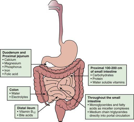
In addition to the length of the bowel, the anatomic location of the resection is also important. The jejunum has more circular folds and longer villi. The proximal 100-200 cm of jejunum is the main site for carbohydrate, protein, iron, and water-soluble vitamin absorption, whereas fat absorption occurs over a longer length of the small bowel. Depending on the region of the bowel resected, specific nutrient malabsorption can result. Vitamin B12 and bile salts are only absorbed in the distal ileum (Fig. 330-5). Jejunal resections are generally tolerated better than ileal resections because the ileum can adapt to absorb nutrients and fluids. Net sodium and water absorption is relatively much higher in the ileum. Ileal resection has a profound effect on fluid and electrolyte absorption due to malabsorption of sodium and water by the remaining ileum; ileal malabsorption of bile salts stimulates increased colonic secretion of fluid and electrolytes.

Treatment

After bowel resection, treatment of short bowel syndrome is initially focused on repletion of the massive fluid and electrolyte losses while the bowel initially accommodates to absorb these losses. Nutritional support is often provided via parenteral nutrition. A central venous catheter should be inserted to provide parenteral fluid and nutrition support. The ostomy or stool output should be measured and fluid and electrolyte losses adequately replaced. Measurement of urinary Na+ to assess body Na+ stores is useful to prevent Na+ depletion. Maintaining urinary Na+ higher than K+ ensures that Na+ intake is adequate. Use of oral glucose electrolyte solutions improves intestinal sodium absorption, particularly in patients without a colon.

After the initial few weeks following resection, fluid and electrolyte losses stabilize, and the focus of therapy shifts to bowel rehabilitation with the gradual reintroduction of enteral feeds. Continuous small-volume trophic enteral feeding should be initiated with a protein hydrolysate and medium-chain triglyceride–enriched formula to stimulate gut hormones and promote mucosal growth. Enteral feeding also increases pancreatobiliary flow and reduces parenteral nutrition-induced hepatotoxicity. As soon as possible, the infant should be given a small amount of water, and then formula by mouth to maintain an interest in oral feeding and minimize or avoid the development of oral aversion. As intestinal adaptation occurs, enteral feeding increases and parenteral supplementation decreases. The bowel mucosa proliferates and bowel lengthens with growth.

After achieving the maximal increase in bowel absorptive capacity, management of specific micronutrient and vitamin deficiencies and treatment of transient problems such as postinfectious mucosal malabsorption are required. GI infections such as rotavirus or small bowel bacterial overgrowth can cause setbacks in the progression to full enteral feeding in patients with marginal absorptive function. A marked increase in stool output or evidence of carbohydrate malabsorption (stool pH <5.5 and positive test for reducing substances) contraindicate further increases in enteral feeds. Slow advancement of continuous enteral feeding rates continues until all nutrients are provided enterally. Then the feeds can be altered to include increased oral or bolus feeding volumes.

In patients with large stool outputs, the addition of soluble fiber and antidiarrheal agents such as loperamide and anticholinergics can be beneficial, although these drugs can increase the risk of bacterial overgrowth. Cholestyramine can be beneficial for patients with distal ileal resection, but its potential depletion of the bile acid pool can increase steatorrhea. Bacterial overgrowth is common in infants with a short bowel and can delay progression of enteral feedings. Empirical treatment with metronidazole or other antibiotics is often useful. Diets high in fat and lower in carbohydrate may be helpful in reducing bacterial overgrowth as well as enhancing adaptation.

Complications

Long-term complications of short bowel syndrome include those of parenteral nutrition: central catheter infection, thrombosis, hepatic cholestasis and cirrhosis, and gallstones. Appropriate care of the central line to prevent infection and catheter-related thrombosis is extremely important. Some patients need long-term parenteral nutritional support, and lack of central line access is potentially life-threatening; inappropriate removal or changes of central lines in the neonatal period should be avoided. Other complications of terminal ileal resection include vitamin B12 deficiency, which might not appear until 1-2 yr after parenteral nutrition is withdrawn. Long-term monitoring for deficiencies of vitamin B12, folate, iron, fat-soluble vitamins, and trace minerals such as zinc and copper is important. Renal stones can occur as a result of hyperoxaluria secondary to steatorrhea (calcium binds to the excess fat and not to oxalate, so more oxalate is reabsorbed and excreted in the urine). Venous thrombosis and vitamin deficiency have been associated with hyperhomocystinemia in short bowel syndrome. Bloody diarrhea secondary to patchy, mild colitis can develop during the progression of enteral feedings. The pathogenesis of this “feeding colitis” is unknown, but it is usually benign and can improve with a hypoallergenic diet or treatment with sulfasalazine.

In patients who are unable to achieve full enteral feeding after several years of nutritional rehabilitation, surgical bowel lengthening procedures may be considered. In some children with complications of parenteral nutrition, especially impending liver failure, small intestinal and liver transplantation may be considered (Chapter 331).

330.8 Chronic Malnutrition

Primary malnutrition (i.e., undernutrition) is very common in developing countries and is directly related to increased disease burden and mortality (Chapter 43). In developed countries, chronic malnutrition occurs mainly as a result of decreased food intake, malabsorption syndromes, and increased nutritional needs in children with chronic diseases, and it affects 11-50% of hospitalized children. Child neglect and improper preparation of formula can result in severe malnutrition. Malnutrition can be identified by evaluating dietary intake, by medical history (anorexia, vomiting, dysphagia, mood and behavioral changes, abdominal pain, diarrhea), by anthropometric measurements (e.g., reduced weight per age and weight per height, BMI <5th percentile) by clinical signs of nutrient deficiencies (atrophic tongue in iron deficiency anemia or alopecia in zinc deficiency).

Malnourished children suffer from impaired immunity, poor wound healing, muscle weakness, and diminished psychologic drive. Malnutrition has short-term consequences (increased disability, morbidity, and mortalitiy) and long-term consequences (final adult size, lower IQ, economic productivity). Undernutrition in hospitalized children is related to increased infectious complications, delayed recovery, increased length of stay and costs, increased readmission rate, and increased mortality.

Nutritional rehabilitation in malnourished children is discussed in Chapter 43.

Chronic malnutrition complicated by diarrheal dehydration is a commonly observed phenomenon. Infectious diarrhea is common in tropical and subtropical countries, in the setting of poor hygiene practices, in immunocompromised hosts (HIV, congenital immunodeficiency), and when impairment of the immune response is due to chronic malnutrition itself. In children with chronic disorders, diarrhea may be related to the underlying disease and should be sought. Examples include noncompliance with a gluten-free diet in celiac disease, noncompliance with pancreatic enzyme treatment in cystic fibrosis, and disease relapse in inflammatory bowel disease (IBD). In the case of IBD, relapse should be diagnosed only after infectious diarrhea and C. difficile infection have been ruled out. Malnutrition per se can lead to exocrine pancreatic insufficiency, which, in turn, aggravates malabsorption and diarrhea.

In infants and children with severe malnutrition, many of the signs normally used to assess the state of hydration or shock are unreliable. Severe malnutrition might be accompanied by sepsis; thus, children with septic shock might not have diarrhea, thirst, or sunken eyes, but may be hypothermic, hypoglycemic, or febrile. The electrocardiogram (ECG) often shows tachycardia, low amplitude, and flat or inverted T waves. Cardiac reserve seems lowered, and heart failure is a common complication.

Despite clinical signs of dehydration, urinary osmolality may be low in the chronically malnourished child. Renal acidifying ability is also limited in patients with malnutrition.

Management of the diarrhea in chronically malnourished children is based on 3 principles: oral rehydration to correct dehydration, rapid resumption of regular feeds with avoidance of periods of nothing by mouth, and treating the etiology of the diarrhea.

When treating the dehydration, it must be remembered that in dehydrated and malnourished infants there appears to be overexpansion of the extracellular space accompanied by extracellular and presumably intracellular hypo-osmolality. Thus, reduced or hypotonic osmolarity oral rehydration solutions are indicated in this setting. When oral rehydration is not possible, the route of choice is nasogastric, and intravenous therapy should be avoided if possible.

Initial intravenous therapy in profound dehydration is designed to improve the circulation and expand extracellular volume. For patients with edema, the quality of fluid and the rate of administration might need to be readjusted from recommended levels to avoid overhydration and pulmonary edema. Blood should be given if the patient is in shock or severely anemic. Potassium salts can be given early if urine output is good. Clinical and ECG improvement may be more rapid with magnesium therapy.

Children with chronic malnutrition are at risk for the refeeding syndrome. Therefore, initial calorie provision should not exceed the previous daily intake and is usually begun at 50-75% of estimated resting energy expenditure, with rapid increase to caloric goals once there are no severe abnormalities in sodium, potassium, phosphorus, calcium, or magnesium. Correction of malnutrition and catch-up growth are not part of the primary treatment of these children, but a nutrition rehabilitation plan is necessary.

330.9 Enzyme Deficiencies

Michael J. Lentze and David Branski

Lactase Deficiency

Congenital lactase deficiency is rare and is associated with symptoms occurring on exposure to lactose in milk. Fewer than 50 cases have been reported worldwide. In patients with congenital lactase deficiency, 5 distinct mutations in the coding region of the LCT gene were found. In most patients (84%), homozygosity for a nonsense mutation, 4170T-A (Y1390X; OMIM 223000), designated Fin (major), was found.

Primary adult type-hypolactasia is caused by a physiologic decline in lactase actively that occurs following weaning in most mammals. The brush border lactase is expressed at low levels during fetal life; activity increases in late fetal life and peaks from term to 3 yr, after which levels gradually decrease with age. This decline in lactase levels varies between ethnic groups. Lactase deficiency occurs in ∼15% of white adults, 40% of Asian adults, and 85% of black adults in the United States. Lactase is encoded by a single gene (LCT) of ∼50 kb located on chromosome 2q21. C/T (−13910) polymorphisms of the MCM6 gene were found to be related to adult-type hypolactasia in most European populations. In 3 African populations—Tanzanians, Kenyans, and Sudanese—3 SNPs, G/C (−14010), T/G(−13915), and C/G(−13907) were identified with lactase persistence and have derived alleles that significantly enhance transcription from the lactase gene promoter in vitro.

Secondary lactose intolerance follows small bowel mucosal damage (celiac disease, rotavirus infection) and is usually transient, improving with mucosal healing.

Lactase deficiency can be diagnosed by H2-breath test or by measurement of lactase activity in mucosal tissue retrieved by small bowel biopsy. Diagnostic testing is not mandatory, and often simple dietary changes that reduce or eliminate lactose from the diet relieve symptoms.

Treatment of lactase deficiency consists of a milk-free diet. A lactose-free formula (based on either soy or cow’s milk) can be used in infants. In older children, low-lactose milk can be consumed. Addition of lactase to dairy products usually abbreviates the symptoms.

Live-culture yogurt contains bacteria that produce lactase enzymes and is therefore tolerated in most patients with lactase deficiency. Hard cheeses have a small amount of lactose and are generally well tolerated.

Sucrase-Isomaltase Deficiency

Sucrase-isomaltase deficiency is a rare autosomal recessive disorder with a complete absence of sucrase and reduced maltase digestive activity. The sucrase-isomaltase complex is composed of 1,927 amino acids encoded by a 3,364 bp mRNA. The gene locus on chromosome 3 has 30 exons spanning 106.6 kb. The majority of sucrase-isomaltase mutations result in a lack of enzyme protein synthesis (null mutation). Post-translational processing defects are also identified.

Approximately 2% of Europeans and Americans are mutant heterozygote. Sucrase deficiency is especially common in indigenous Greenlanders (estimated 5%) in whom it is often accompanied by lactase deficiency.

Symptoms of sucrase-isomaltase deficiency usually begin when the infant is exposed to sucrose or a glucose polymer diet. This can occur with ingestion of non–lactose based infant formula or on the introduction of pureed food, especially fruits and sweets. Diarrhea, abdominal pain, and poor growth are observed. Occasional patients present with symptoms in late childhood or even adult life, but careful history often indicates that symptoms appeared earlier. Diagnosis of sucrase-isomaltase malabsorption requires acid hydrolysis of stool for reducing substances because sucrase is a nonreducing sugar. Alternatively, diagnosis can be achieved with hydrogen breath test or direct enzyme assay of small bowel biopsy.

The mainstay of treatment is lifelong dietary restriction of sucrose-containing foods. Enzyme replacement with a purified yeast enzyme, sacrosidase (Sucraid), is a highly effective adjunct to dietary restriction.

Glucose-Galactose Malabsorption

More than 30 different mutations of the sodium/glucose co-transporter gene (SGLT1) are identified. These mutations cause a rare autosomal recessive disorder of intestinal glucose and galactose/Na+ co-transport system that leads to osmotic diarrhea. Because most dietary sugars are polysaccharides or disaccharides with glucose or galactose moieties, diarrhea follows the ingestion of glucose, breast milk, or conventional lactose-containing formulas. Dehydration and acidosis can be severe, resulting in death.

The stools are acidic and contain sugar. Patients with the defect have normal absorption of fructose, and their small bowel function and structure are normal in all other aspects. Intermittent or permanent glycosuria after fasting or after a glucose load is a common finding due to the transport defect also being present in the kidney. The presence of reducing substances in watery stools and slight glycosuria despite low blood sugar levels is highly suggestive of glucose-galactose malabsorption. Malabsorption of glucose and galactose is easily identified using the breath hydrogen test. It is safe to perform the 1st test with a dose of 0.5 g/kg of glucose; if necessary, a second test can be performed using 2 g/kg. Breath H2 will rise more than 20 ppm. The small intestinal biopsy is useful to document a normal villous architecture and normal disaccharidase activities. The identification of mutations of SGLT1 makes it possible to perform prenatal screening in families at risk for the disease.

Treatment consists of rigorous restriction of glucose and galactose. Fructose, the only carbohydrate that can be given safely, should be added to a carbohydrate-free formula at a concentration of 6-8%. Diarrhea immediately ceases when infants are given such a formula. Although the defect is permanent, later in life, limited amounts of glucose, such as starches or sucrose may be tolerated.

Exocrine Pancreatic Insufficiency

Disorders of exocrine pancreatic insufficiency are discussed in Chapter 341. Cystic fibrosis is the most common congenital disorder associated with exocrine pancreatic insufficiency. Although rare, the next most common cause of pancreatic insufficiency in children is Shwachman-Diamond syndrome. Other rare disorders causing exocrine pancreatic insufficiency are Blizzard-Johanson syndrome (severe steatorrhea, aplasia of alae nasi, deafness, hypothyroidism, scalp defects), Pearson bone marrow syndrome (sideroblastic anemia, variable degree of neutropenia, thrombocytopenia), and isolated pancreatic enzyme deficiency (lipase, colipase and lipase-colipase, trypsinogen, amylase). Deficiency of enterokinase—a key enzyme that is produced in the proximal small bowel and is responsible for the activation of trypsinogen to trypsin—manifests clinically as exocrine pancreatic insufficiency.

Autoimmune polyendocrinopathy syndrome type 1, a rare autosomal recessive disorder, is caused by mutation in the autoimmune regulator gene (AIRE). Chronic mucocutaneous candidiasis is associated with failure of parathyroid gland, adrenal cortex, pancreatic β-cells, gonads, gastric parietal cells, and thyroid gland. Pancreatic insufficiency and steatorrhea have been associated with this condition.

330.10 Liver and Biliary Disorders Causing Malabsorption

Anil Dhawan and David Branski

Absorption of fats and fat-soluble vitamins depends to a great extent on adequate bile flow providing bile acids to the small intestine. Most of the liver and biliary disorders lead to impairment of the bile flow, contributing to malabsorption of long-chain fatty acids and vitamins such as A, D, E, and K. In addition, severe portal hypertension can lead to portal hypertensive enteropathy, resulting in poor absorption of the nutrients. Decompensated liver disease leads to anorexia and increased energy expenditures, further widening the gap between calorie intake and net absorption, leading to severe malnutrition. Adequate management of nutrition is essential to improve the outcome with or without liver transplantation. This is usually achieved by using medium-chain triglyceride-rich milk formula, supplemental vitamins, and continuous or bolus enteral feed where oral intake is poor.

Vitamin D deficiency is commonly observed on biochemical tests, and children rarely present with pathologic fractures. Simultaneous administration of vitamin D with the water-soluble vitamin E preparation (d-α-tocopherol polyethylene glycol 1,000 succinate [TPGS]) enhances absorption of vitamin D. In young infants, oral vitamin D3 is given at a dose of 1,000 IU/kg/24 hr. After 1 mo, if the serum 25-hydroxyvitamin D level is low, the same dose of oral vitamin D is mixed with TPGS. 25-hydroxyvitamin D is then monitored every 3 mo, with adjustment of doses as necessary.

Vitamin E deficiency in patients with chronic cholestasis is not usually symptomatic, but it can manifest as a progressive neurologic syndrome, which includes peripheral neuropathy (manifesting as loss of deep tendon reflexes and ophthalmoplegia), cerebellar ataxia, and posterior column dysfunction. Early in the course, findings are partially reversible with treatment; late features might not be reversible. It may be difficult to identify vitamin E deficiency because the elevated blood lipid levels in cholestatic liver disease can falsely elevate the serum vitamin E level. Therefore, it is important to measure the ratio of serum vitamin E to total serum lipids; the normal level for patients <12 yr of age is >0.6, and for patients >12 yr it is >0.8. The neurologic disease can be prevented with the use of an oral water-soluble vitamin E preparation (TPGS, Liqui-E) at a dose of 25-50 IU/day in neonates and 1 IU/kg/day in children.

Vitamin K deficiency can occur as a result of cholestasis and poor fat absorption. In children with liver disease it is very important to differentiate between the coagulopathy related to vitamin K deficiency and one secondary to the synthetic failure of the liver. A single dose of vitamin K administered intravenously does not correct the prolonged prothrombin time in liver failure, but the deficiency state responds within a few hours. Easy bruising may be the 1st sign. In neonatal cholestasis, coagulopathy due to vitamin K deficiency can manifest with intracranial bleeds with devastating consequences, and prothrombin time should be routinely measured to monitor for deficiency in children with cholestasis. All children with cholestasis should receive vitamin K supplements.

Vitamin A deficiency is rare and is associated with night blindness, xerophthalmia, and increased mortality if patients contract measles. Serum vitamin A levels should be monitored and adequate supplementation considered.

In practice, children with cholestasis are prescribed twice the recommended daily allowance of the commonly available multivitamin preparations while awaiting blood levels.

330.11 Rare Inborn Defects Causing Malabsorption

Some congenital (primary) malabsorption disorders originate from a defect of integral membrane proteins, which fulfill a transport function as receptor or channel across the apical or basolateral membrane of enterocytes for nutritional components. Histologic examination of the small and large bowel is typically normal. Most of these disorders are inherited in an autosomal recessive pattern. Most are rare, and patients present with a broad phenotypic heterogeneity due to modifier genes and nutritional and other secondary factors.

Disorders of Amino Acid and Peptide Absorption

Owing to their ontogenic origins, enterocytes and renal tubules express amino acid transporter in common. Their highest intestinal transporter activity is found in the jejunum. The transporters causing Hartnup disease, cystinuria, iminoglycinuria, and dicarboxylic aminoaciduria are located in the apical membrane, and those causing lysinuric protein intolerance (LPI) and blue diaper syndrome are anchored in the basolateral membrane of the intestinal epithelium.

Dibasic amino acids, including cystine, ornithine, lysine, and arginine are taken up by the Na-independent SLC3A1/SLC7A9, which is defective in cystinuria. The overall prevalence of the disease is 1 in 7,000 newborns. This disorder is not associated with any GI or nutritional consequences because of compensation by alternative transporter. However, hypersecretion of cystine in the urine leads to recurrent cystine stones, which account for up to 1% of all urinary tract stones. Ample hydration, urine alkalinization, and cystine-binding thiol drugs can increase the solubility of cystine. Cystinuria type I is inherited as an autosomal recessive trait, and the transmission of type II is autosomal dominant with incomplete penetrance. Cystinuria type I has been described in association with 2p21 deletion syndrome and hypotonia-cystinuria syndrome.

Hartnup disease is characterized by malabsorption of neutral amino acids, including the essential amino acid tryptophan, with aminoaciduria, photosensitive pellagra-like rash, headaches, cerebellar ataxia, delayed intellectual development, and diarrhea. The clinical spectrum ranges from asymptomatic patients to severely affected patients with progressive neurodegeneration leading to death by adolescence. SLC6A19, which is the major luminal sodium-dependent neutral amino acid transporter of small intestine and renal tubules, has been identified as the defective protein. Its association with collectrin and angiotensin-converting enzyme (ACE) II is likely to be involved in the phenotypic heterogeneity of Hartnup disorder. Tryptophan is a precursor of NAD(P)H biosynthesis; therefore the disorder can be treated by nicotinamide in addition to a diet of 4 g protein/kg. The use of lipid-soluble esters of amino acids and tryptophan ethylester has also been reported.

In the blue diaper syndrome (indicanuria, Drummond syndrome) tryptophan is specifically malabsorbed and the defect is expressed only in the intestine and not in the kidney, in contrast to Hartnup disease. Intestinal bacteria convert the unabsorbed tryptophan to indican, which is responsible for the bluish discoloration of the urine after its hydrolysis and oxidation. Symptoms can include digestive disturbances such as vomiting, constipation, poor appetite, failure to thrive, hypercalcemia, nephrocalcinosis, fever, irritability, and ocular abnormalities. The molecular genetic defect of this disorder has not yet been characterized.

The underlying defect of iminoglycinuria is the malabsorption of proline, hydroxyproline, and glycine due to the proton amino acid transporter SLC36A2 defect, with a possible participation of modifier genes, one of which (SLC6A20), is present in the intestinal epithelium. This disorder is usually benign, but sporadic cases with encephalopathy, mental retardation, deafness, blindness, kidney stones, hypertension, and gyrate atrophy have been described.

The excitatory amino acid carrier SLC1A1 is affected in dicarboxylic aminoaciduria. This carrier is present in the small intestine, kidney, and brain, and transports the anionic acids L-glutamate, L– and D-aspartate, and L-cysteine. There are single case reports indicating that this disorder could be associated with hyperprolinemia and neurologic symptoms such as POLIP (polyneuropathy, ophthalmoplegia, leukoencephalopathy, intestinal pseudo-obstruction).

A histidine-specific transport system has also been proposed. A few patients have been reported with an intestinal and renal defect of this carrier. It has not been confirmed that patients with histidinuria, who have low plasma histidine levels, in contrast to histidinemia, develop neurologic symptoms (e.g., hearing loss, myoclonic seizures).

A methionine-preferring transporter in the small intestine was suggested to be affected in Smith-Strang disease (oasthouse urine disease), which is characterized by purple, red-brown-colored urine with a cabbage-like odor, containing 2-hydroxybutyric acid, valine, and leucine. The potential symptoms of methionine malabsorption include neurologic signs, white hair, and diarrhea. Large amounts of methionine and branched-chain amino acids are present in the feces but not in the urine. A low-methionine diet is recommended to alleviate the symptoms.

Among the diseases (see the earlier discussion of cystinuria) with a membrane transport defect of cationic amino acids (lysine, arginine, ornithine), lysinuric protein intolerance (LPI) is the 2nd most common, with a prevalence in Finland of 1 in 60,000. The y+LAT-1 (SLC7A7) carrier at the basolateral membrane of the intestinal and renal epithelium is affected, with failure to deliver cytosolic dibasic cationic amino acids into the paracellular space in exchange for Na+ and neutral amino. This defect is not compensated by the SLC3A1/SLC7A9 transporter (at the apical membrane), the latter being affected in cystinuria. The symptoms of LPI, which appear after weaning, include diarrhea, failure to thrive, hepatosplenomegaly, nephritis, respiratory insufficiency, alveolar proteinosis, pulmonary fibrosis, and osteoporosis. Abnormalities of bone marrow have also been described in a subgroup of LPI patients. The disorder is characterized by low plasma concentrations of dibasic amino acids (in contrast to high levels of citrulline, glutamine, and alanine) and massive excretion of lysine (as well as orotic acid, ornithine, and arginine in moderate excess) in the urine. Hyperammonemia and coma usually develop after episodic attacks of vomiting, after fasting, or following administration of large amounts of protein (or alanine load), possibly due to a deficiency of intramitochondrial ornithine. Some patients show moderate retardation. Cutaneous manifestations can include alopecia, perianal dermatitis, and sparse hair. Some patients avoid protein-containing food. Treatment includes orally administered citrulline (200 mg/kg/day), which is well absorbed from the intestine; dietary protein restriction (<1.5 g/kg/day); and carnitine supplementation. One patient with isolated lysinuria has been reported with growth failure, seizures, and mental retardation.

Disorders of Fat Transport

Abetalipoproteinemia, hypobetalipoproteinemia, and chylomicron retention disease are described in Chapter 80. The long-chain fatty acid (FATP4) and cholesterol transporters, the latter being called Niemann-Pick C1-like protein (NPC1L1), have been characterized at the intestinal brush border in knock-out mice models showing a hyperproliferative hyperkeratosis and an impaired fatty acid and cholesterol uptake. NPC1L1 is inhibited by ezetimibe, which is used to restrict the absorption of dietary cholesterol.

Tangier disease is characterized by the absence of high-density lipoprotein cholesterol (HDL-C), which is caused by mutations in the adenosine triphosphate (ATP)-binding cassette transporter A1 (ABCA1) gene. The failure of intracellular phospholipids and cholesterol efflux to lipid-poor apolipoprotein acceptors such as HDL predisposes to premature coronary heart disease and accumulation of cholesterol in liver, spleen, lymph nodes (tonsils), and small intestine.

Features of Tangier disease include orange tonsils, hepatosplenomegaly, relapsing neuropathy, orange-brown spots on the colon and ileum, diarrhea in association with decreased plasma cholesterol levels (apo A-I, apo A-II), and normal or elevated triglyceride levels. Specific therapy for Tangier disease has not yet been established.

In sitosterolemia defective efflux of sterol leads to increased absorption of dietary sterols; normally, <5% are retained by the GI tract. Patients carry mutations of the ABCG5 (sterolin-1) and ABCG8 (sterolin-2) transporters. The disorder is associated with tendon xanthomas, increased atherosclerosis, and hemolysis. Plasma levels of phytosterols (mainly sitosterol) are typically >10 mg/dL.

Disorders of Vitamin Absorption

Transporters and receptors of the intestinal epithelium have been described for water-soluble but not fat-soluble vitamins, the latter being absorbed primarily by enterocytes, by passive diffusion after emulsification of fats by bile salts. Transfer proteins (retinol-binding protein, RBP4 and α-tocopherol transfer protein, TTP1) have been involved in deficiency states of vitamins E (spinocerebellar ataxia) and A (ophthalmologic signs), respectively.

Vitamin B12 (cobalamin) is used exclusively by microorganisms and is acquired mostly from meat and milk. Its absorption starts with the removal of cobalamin from dietary protein by gastric acidity and its binding to haptocorrin. In the duodenum, pancreatic proteases hydrolyze the cobalamin-haptocorrin complex, allowing the binding of cobalamin to intrinsic factor (IF), which originates from parietal cells. The receptor of the cobalamin-IF complex is located at the apical membrane of the ileal enterocytes and represents a heterodimer consisting of cubulin and amnionless, with endocytic uptake of this ligand into endosomes, where it binds to megalin and forms a cobalamin–transcobalamin-2 complex (after cleavage of IF) for further transcytosis. As a cofactor for methionine synthase, cobalamin converts homocysteine to methionine. Cobalamin deficiency can be caused by inadequate intake of the vitamin (e.g., breast-feeding by mothers on a vegetarian diet), primary or secondary achlorhydria including autoimmune gastritis, exocrine pancreatic insufficiency, bacterial overgrowth (Chapter 330.4), ileal disease (Crohn disease, Chapter 328), ileal (or gastric) resection, infections (fish tapeworm), and Whipple disease (Chapter 333).

Clinical signs of congenital cobalamin malabsorption, which usually appear from a few months to 14 yr of age, are pancytopenia including megaloblastic anemia, fatigue, failure to thrive, and neurologic symptoms including developmental delay. Recurrent infections and bruising may be present. Laboratory evaluation indicates low serum cobalamin, hyperhomocysteinemia, methylmalonicacidemia, and mild proteinuria. The Schilling test is useful to differentiate between lack of IF and malabsorption of cobalamin. Three rare autosomal recessive disorders of congenital cobalamin deficiency affect absorption and transport of cobalamin (in addition to 7 other inherited defects of cobalamin metabolism). These include mutations of the gastric IF (GIF) gene with absence of IF (but normal acid secretion and lack of autoantibodies against IF or parietal cells), mutations of the amnionless (AMN) and cubilin (CUBN) genes (Imerslund-Grasbeck syndrome), and mutations in the transcobalamin 2 cDNA. These disorders require long-term parenteral cobalamin treatment: intramuscular injections of hydroxycobalamin 1 mg daily for 10 days and then once a month. High-dose substitution with oral cyanocobalamin (1 mg biweekly) does not seem to be sufficient for all patients with congenital cobalamin deficiency.

Folate is an essential vitamin required to synthesize methionine from homocysteine. It is found mainly in green leafy vegetables, legumes, and oranges. It is converted to 5-methyltetrahydrofolate (5MTHF) after its uptake by enterocytes. Secondary folate deficiency is caused by insufficient folate intake, villous atrophy (e.g., celiac disease, IBD), treatment with phenytoin, and trimethoprim among others (Chapter 448.1). Several inherited disorders of folate metabolism and transport have been described.

Hereditary folate malabsorption is characterized by a defect of the proton-coupled folate transporter (PCFT, formerly reported to be HCP1, a heme carrier) of the brush border, leading to impaired absorption of folate in the upper small intestine as well as impaired transport of folate into the central nervous system. Mutations of the reduced folate carrier (RFC1, SLC19A1) have not been found in this entity. Sulfasalazine and methotrexate are potent inhibitors of PCFT. Symptoms of congenital folate malabsorption are diarrhea, failure to thrive, megaloblastic anemia (in the 1st few months of life), glossitis, infections (Pneumocystis jirovecii) with hypoimmunoglobulinemia, and neurologic abnormalities (seizures, mental retardation, and basal ganglia calcifications). Macrocytosis, with or without neutropenia, multilobulated polymorphonuclear cells, increased LDH and bilirubin, increased saturation of transferrin, and decreased cholesterol can be found. Low levels of folate are present in serum and cerebrospinal fluid. Plasma homocysteine concentrations as well as urine excretion of formiminoglutamic acid and orotic acid are elevated. Long-lasting deficiency is best documented using red cell folate. Therapy involves large doses of oral (up to 100 mg/day) or systemic (intrathecal) folate.

The molecular basis of intestinal transport of other water-soluble vitamins such as vitamin C (Na+-dependent vitamin C transporter, SVCT1 and SVCT2), pyridoxine/vitamin B6, and biotin/vitamin B5 (Na+-dependent multivitamin transporter, SMVT) have been described; however congenital defects of these transporter systems have not yet been found in humans. A thiamine/vitamin B1-responsive megaloblastic anemia (TRMA) syndrome, which is associated with early-onset type 1 diabetes mellitus and sensorineural deafness, is caused by mutations of the thiamine transporter protein, THTR-1 (SLC19A2), present in the brush border.

Disorders of Electrolyte and Mineral Absorption

Congenital chloride diarrhea belongs to the more common causes of severe congenital diarrhea, with prevalence in Finland of 1: 20,000. It is caused by a defect of the SLC26A3 gene, which encodes a Na+-independent Cl/HCO3 exchanger within the apical membrane of ileal and colonic epithelium. Founder mutations have been described in Finnish, Polish, and Arab patients: V317del, I675-676ins, and G187X, respectively. The Cl/HCO3 exchanger absorbs chloride originating from gastric acid and the cystic fibrosis transmembrane conductance regulator (CFTR) and secretes bicarbonate into the lumen, neutralizing the acidity of gastric secretion.

Prenatal clinical signs of this disorder are a dilated small bowel that can mislead to a diagnosis of intestinal obstruction. Newborns with congenital chloride diarrhea present with severe life-threatening secretory diarrhea during the 1st weeks of life. Laboratory findings are metabolic alkalosis, hypochloremia, hypokalemia, and hyponatremia (with high plasma renin and aldosterone activities). Fecal chloride concentrations are >90 mmol/L and exceed the sum of fecal sodium and potassium. Early diagnosis and aggressive lifelong enteral substitution of KCl in combination with NaCl (chloride doses of 6-8 mmol/kg/day for infants and 3-4 mmol/kg/day for older patients) prevent mortality and long-term complications (such as urinary infections, hyperuricemia with renal calcifications, renal insufficiency, and hypertension) and allow normal growth and development. Orally administered proton pump inhibitors, cholestyramine, and butyrate can reduce the severity of diarrhea. The diarrheal symptoms usually tend to regress with age. However, febrile diseases are likely to exacerbate symptoms as a consequence of severe dehydration and electrolyte imbalances. (See Chapter 52 for fluid and electrolyte management.)

The classic form of congenital sodium diarrhea manifests with polyhydramnios, massive secretory diarrhea, severe metabolic acidosis, alkaline stools (fecal pH >7.5) and hyponatremia as a result of fecal losses of Na+ (fecal Na+ >70 mmol/L). Urinary secretion of sodium is low to normal. There is partial villous atrophy. The molecular genetic defect could not be located in the Na+-H+ exchangers (NHEs), which were thought to be impaired because they seem to be mainly responsible for Na+ absorption in the small intestine. In addition, a syndromic form of congenital sodium diarrhea with choanal or anal atresia, hypertelorism, and corneal erosions has been related to mutations of SPINT2, encoding a serine-protease inhibitor, whose pathophysiologic action on intestinal Na+ absorption is unclear. Some patients can be weaned from parenteral nutrition later in childhood but depend on oral sodium citrate supplementation.

The congenital form of acrodermatitis enteropathica manifests with severe deficiency of body zinc soon after birth in bottle-fed children or after weaning from breastfeeding. Clinical signs of this disorder are anorexia, diarrhea, failure to thrive, humoral and cell-mediated immunodeficiency (poor wound healing, recurrent infections), male hypogonadism, skin lesions (vesicobullous dermatitis on the extremities and perirectal, perigenital, and perioral regions, and alopecia), and neurologic abnormalities (tremor, apathy, depression, irritability, nystagmus, photophobia, night blindness, and hypogeusia). The genetic defect of acrodermatitis enteropathica is caused by a mutation in the Zrt-Irt-lik protein 4 (ZIP4, SLC39A4), normally expressed on the apical membrane, which enables the uptake of zinc into the cytosol of enterocytes. The zinc-dependent alkaline phosphatase and plasma zinc levels are low. Paneth cells in the crypt of the small intestinal mucosa show inclusion bodies. Acrodermatitis enteropathica requires long-term treatment with elemental zinc 1 mg/kg/day. Maternal zinc deficiency impairs embryonic, fetal, and postnatal development. Acquired forms of zinc deficiency are described in Chapter 51.

Menkes disease and occipital horn syndrome are both caused by mutations in the gene encoding Cu2+ transporting ATPase, alpha polypeptide (ATP7A), also called Menkes or MNK protein. ATP7A is mainly expressed by enterocytes, placental cells, and CNS and is localized in the trans-Golgi network for copper transfer to enzymes in the secretory pathway or to endosomes to facilitate copper efflux. Copper values in liver and brain are low in contrast to an increase in mucosal cells, including enterocytes and fibroblasts. Plasma copper and ceruloplasmin levels decline postnatally. Clinical features of Menkes disease are progressive cerebral degeneration (convulsions), feeding difficulties, failure to thrive, hypothermia, apnea, infections (urinary tract), peculiar facies, hair abnormalities (kinky hair), hypopigmentation, bone changes, and cutis laxa. Patients with the classic form of Menkes disease usually die before the age of 3 yr. A therapeutic trial with copper-histidinate should start before the age of 6 wk. In contrast to Menkes disease, occipital horn syndrome usually manifests during adolescence with borderline intelligence, craniofacial abnormalities, skeletal dysplasia (short clavicles, pectus excavatum, genu valgum), connective tissue abnormalities, chronic diarrhea, orthostatic hypotension, obstructive uropathy, and osteoporosis. It should be differentiated from Ehlers-Danlos syndrome type V.

Active calcium absorption is mediated by the transient receptor potential channel 6 (TRPV6) at the brush border membrane, calbindin, and the CaATPase, or the Na+-Ca++ exchanger for calcium efflux at the basolateral membrane within the proximal small bowel. A congenital defect of these transporters has not yet been described.

Intestinal absorption of dietary magnesium, which occurs via the transient receptor potential channel TRPM6 at the apical membrane, is impaired in familial hypomagnesemia with secondary hypocalcemia, which manifests with neonatal seizures and tetany.

Intestinal iron absorption consists of several complex regulated processes starting with the uptake of heme-containing iron by heme carrier protein 1 (HCP1) and Fe2+ (after luminal reduction of oxidized Fe3+) by the divalent metal transporter 1 (DMT1) at the apical membrane, followed by the efflux of Fe2+ by ferroportin 1 (also called iron-regulated transporter[ IREG1]) at the basolateral membrane of duodenal enterocytes. Mutations of the ferroportin 1 gene have been found in the autosomal dominant form of hemochromatosis type 4. Mutations of HFE (Cys282Tyr, His63Asn, Ser65Cys) of classic hemochromatosis reduce the endocytic uptake of diferric transferrin by the transferrin receptor-1 (TfR1) at the basolateral membrane of the intestinal epithelium. Hepcidin antimicrobial peptide (HAMP) encodes hepcidin, a hepatic peptide hormone, which inhibits the efflux of iron through ferroportin and can be induced by IL-6. It is the defective gene of juvenile hemochromatosis (type 2, subtype B).

Bibliography

Andrews GK. Regulation and function of Zip4, the acrodermatitis enteropathica gene. Biochem Soc Trans. 2008;36:1242-1246.

Baerlocher K, Solioz M. Disorders of copper, zinc and iron metabolism. In: Babu E, Duran M, Blaskovics ME, Gibson KM, editors. Physician’s guide to the laboratory diagnosis of metabolic diseases. ed 2. Heidelberg: Springer-Verlag; 2003:631-658.

Booth IW, Stange G, Murer H, et al. Defective jejunal brush-border Na+/H+ exchange: a cause of congenital secretory diarrhoea. Lancet. 1985;1:1066-1069.

Bor MV, Cetin M, Aytac S, et al. Long term biweekly 1 mg oral vitamin B12 ensures normal hematological parameters, but does not correct all other markers of vitamin B12 deficiency. A study in patients with inherited vitamin B12 deficiency. Haematologica. 2008;93:1755-1758.

Broer S. Amino acid transport across mammalian intestinal and renal epithelia. Physiol Rev. 2008;88:249-286.

Broer S, Bailey CG, Kowalczuk S, et al. Iminoglycinuria and hyperglycinuria are discrete human phenotypes resulting from complex mutations in proline and glycine transporters. J Clin Invest. 2008;118:3881-3892.

Brunham LR, Singaraja RR, Hayden MR. Variations on a gene: rare and common variants in ABCA1 and their impact on HDL cholesterol levels and atherosclerosis. Annu Rev Nutr. 2006;26:105-129.

Camargo SM, Singer D, Makrides V, et al. Tissue-specific amino acid transporter partners ACE2 and collectrin differentially interact with Hartnup mutations. Gastroenterology. 2009;136:872-882.

Drummond KN, Michael AF, Ulstrom RA, Good RA. The blue diaper syndrome: familial hypercalcemia with nephrocalcinosis and indicanuria; a new familial disease, with definition of the metabolic abnormality. Am J Med. 1964;37:928-948.

Grasbeck R. Imerslund-Grasbeck syndrome (selective vitamin B12 malabsorption with proteinuria). Orphanet J Rare Dis. 2006;1:17-23.

Heinz-Erian P, Muller T, Krabichler B, et al. Mutations in SPINT2 cause a syndromic form of congenital sodium diarrhea. Am J Hum Genet. 2009;84:188-196.

Hihnala S, Hoglund P, Lammi L, et al. Long-term clinical outcome in patients with congenital chloride diarrhea. J Pediatr Gastroenterol Nutr. 2006;42:369-375.

Hoglund P, Auranen M, Socha J, et al. Genetic background of congenital chloride diarrhea in high-incidence populations: Finland, Poland, and Saudi Arabia and Kuwait. Am J Hum Genet. 1998;63:760-768.

Kamoun P, Parvy P, Rabier D, et al. Dicarboxylic aminoaciduria. J Inherit Metab Dis. 1994;17:758.

Kellett GL, Brot-Laroche E, Mace OJ, Leturque A. Sugar absorption in the intestine: the role of GLUT2. Annu Rev Nutr. 2008;28:35-54.

Kleta R, Romeo E, Ristic Z, et al. Mutations in SLC6A19, encoding B0AT1, cause Hartnup disorder. Nat Genet. 2004;36:999-1002.

Lee PJ, Van’t Hoff WG, Leonard JV. Catch-up growth in Fanconi-Bickel syndrome with uncooked cornstarch. J Inherit Metab Dis. 1995;18:153-156.

Martens K, Jaeken J, Matthijs G, Creemers JW. Multi-system disorder syndromes associated with cystinuria type I. Curr Mol Med. 2008;8:544-550.

Nyhan WL, Ozand PT. Atlas of metabolic diseases. London: Chapman & Hall Medical; 1998.

Palacin M, Nunes V, Font-Llitjos M, et al. The genetics of heteromeric amino acid transporters. Physiology (Bethesda). 2005;20:112-124.

Patel SB, Zablocki CJ. Plant sterols and stanols: their role in health and disease. J Clin Lipidol. 2008;2:S11-S19.

Pietrangelo A. Hereditary hemochromatosis—a new look at an old disease. N Engl J Med. 2005;350:2383-2397.

Qiu A, Jansen M, Sakaris A, et al. Identification of an intestinal folate transporter and the molecular basis for hereditary folate malabsorption. Cell. 2006;127:917-928.

Santer R, Groth S, Kinner M, et al. The mutation spectrum of the facilitative glucose transporter gene SLC2A2 (GLUT2) in patients with Fanconi-Bickel syndrome. Hum Genet. 2002;110:21-29.

Santer R, Hillebrand G, Steinmann B, et al. Intestinal glucose transport: evidence for a membrane traffic-based pathway in humans. Gastroenterology. 2003;124:34-39.

Sperandeo MP, Andria G, Sebastio G. Lysinuric protein intolerance: update and extended mutation analysis of the SLC7A7 gene. Hum Mutat. 2008;29:14-21.

Veldhuis NA, Gaeth AP, Pearson RB, et al. The multi-layered regulation of copper translocating P-type ATPases. Biometals. 2009;22:177-190.

Whitehead VM. Acquired and inherited disorders of cobalamin and folate in children. Br J Haematol. 2006;134:125-136.

330.12 Malabsorption in Eosinophilic Gastroenteritis

The diagnosis of eosinophilic gastroenteritis is based on GI symptoms, GI eosinophilic infiltrates, and no demonstrable cause of the eosinophilia such as parasitic infection (most commonly Enterobius vermicularis in children) or a specific allergic response. Peripheral eosinophilia is variable and not uniformly considered a criterion for diagnosis. The majority (50-70%) of patients have a history of other allergic disorders, and others might have associated connective tissue diseases. Approximately 10% of patients with this disorder have an immediate family member with this disorder as well, suggesting that eosinophilic GI disorders stem from a genetic predisposition, common environmental factors, or, most likely, a combination. Hypersensitivity to specific food allergens has been postulated as an etiologic factor. Symptoms depend on the severity and location of eosinophilic inflammation. Any region or layer (mucosa, submucosa, and serosa) of the gut may be involved, alone or in combination.

Diagnosis requires panendoscopy and biopsies in combination with other imaging diagnostic procedures. Eosinophilic infiltrates dominate the histologic findings, and signs of other inflammatory diseases are absent; in particular, the crypt architecture remains normal, no parasites are identified, and no eggs or larvae are seen. An increase in mast cells and IgE-containing plasma cells may be observed. The mucosal layer might be involved focally, or it might not be involved at all, in which case mucosal biopsy will fail to establish the diagnosis. Small bowel capsule endoscopy is useful in that it characteristically reveals mucosal erythema with marked focal villous atrophy. The most common sites of involvement are the stomach and small intestine.

Diarrhea and subsequent malabsorption can occur if small bowel involvement with villous blunting is extensive. Eosinophilic gastroenteritis can cause abdominal pain and excessive gas, weight loss, and failure to thrive. Other than peripheral eosinophilia, hypoalbuminemia due to protein-losing enteropathy and iron deficiency anemia are the more common laboratory findings.

Nutritional exclusion (or elemental) diets and corticosteroids are the mainstay of treatment. Less well documented treatments such as mast cell stabilizers and leukotriene antagonists have been used in small, uncontrolled trials. Clinical trials using biological modalities such as monoclonal anti-IgE (omalizumab) and anti-IL-5 (SCH55700/reslizumab and mepolizumab) are anticipated for severe cases.

330.13 Malabsorption in Inflammatory Bowel Disease

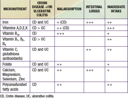
Crohn disease and ulcerative colitis represent the 2 forms of chronic, immune-mediated IBD that commonly affect pediatric patients (Chapter 328). Because the small bowel is involved in the majority of pediatric Crohn disease patients, malabsorption of nutrients is far more of a problem than in ulcerative colitis. At the time of diagnosis, significant weight loss is observed in up to 85% of pediatric patients with Crohn disease and in about 65% with ulcerative colitis, due to inadequate intake of energy and micronutrients as well as diarrhea and malabsorption. Consequently, growth failure due to chronic undernutrition is far more common in Crohn disease than in ulcerative colitis, affecting up to 40% of cases.

In addition to malabsorption, energy intake is lower in patients with Crohn disease compared to healthy controls, in part due to lesser appetite. Excessive levels of proinflammatory cytokines have been implicated in causing the anorexia as well as in mediating impaired growth. Affected children often have a lower desire to eat, because symptoms, including abdominal pain, nausea, vomiting, and diarrhea, can lead to reduced food intake. This can, in turn negatively affect nutritional status during a child’s critical period of growth and development. Patients with IBD are also at risk of developing nutritional deficiencies because of restrictive diets imposed by caregivers or by the patients themselves.

In children with active disease, inadequate intakes of energy and of a number of micronutrients have been observed. Reduced energy intake during active disease can contribute to poor weight gain and impaired growth. Patients with IBD, particularly Crohn disease, often have multiple nutritional deficiencies and are in negative nitrogen balance, due to decreased intake and malabsorption of macro- and micronutrients. Quantifying nutrient intake, determining micronutrient deficiencies, and ascertaining requirements for nutritional supplementation are essential components of successful management in pediatric IBD.

Optimizing nutritional status and growth are key priorities in the management of IBD in children and adolescents. Energy intake should meet the added costs of catch-up growth and are usually in the range of 40-70 kcal/kg ideal body weight per day. Protein requirements are higher in Crohn disease (1-1.5 g/kg/day). Bone mineral density deficit is common, even in pediatric patients who have not been exposed to systemic corticosteroid therapy. Osteoporosis or osteopenia is best assessed by bone densitometry, and levels of vitamin 25-hydroxyvitamin D should be monitored. Other micronutrient deficiencies that result from inadequate intake, malabsorption, and gut losses are shown in Table 330-10.

Enteral nutrition support is favored over parenteral for all but Crohn disease patients with extreme short gut. Patients requiring hospitalization for a severe relapse should receive nutrition support if they are already malnourished or their intake is likely to be severely curtailed for ≥1 wk. Preoperative nutrition support is essential to the prevention of morbidity and mortality. However, clinicians must be aware of the risk of the refeeding syndrome in patients with severe malnutrition. In ulcerative colitis, nutrition support is adjunctive therapy; there is no evidence that bowel rest or TPN influences the outcome of severe ulcerative colitis.