Published on 19/03/2015 by admin
Filed under Dermatology
Last modified 22/04/2025
This article have been viewed 2557 times
Bruce H. Thiers
Evidence Levels: A Double-blind study B Clinical trial ≥ 20 subjects C Clinical trial < 20 subjects D Series ≥ 5 subjects E Anecdotal case reports
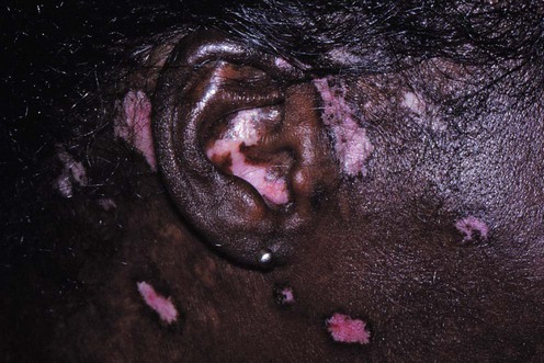
Discoid lupus erythematosus (DLE) is the most common form of chronic cutaneous lupus erythematosus (CCLE). Lesions predominate in sun-exposed areas, especially the face, scalp, upper chest, upper back, and extensor arms. Early lesions consist of sharply demarcated, erythematous, often hyperpigmented, hyperkeratotic papules and small plaques with adherent scale. The individual lesions spread peripherally, resulting in atrophy and central scarring which may be associated with alopecia, telangiectasia, and depigmentation.
The lesions of DLE are quite characteristic, especially in their later stages. When the diagnosis is in doubt, a skin biopsy should be performed. The histologic findings are usually diagnostic, although direct immunofluorescence examination can be obtained in questionable cases. A complete history and physical examination should be performed, looking for signs of systemic disease. Laboratory examinations to be obtained include a complete blood count with differential, erythrocyte sedimentation rate, serum chemistry profile, and urinalysis. Serum should be screened for antinuclear antibodies (ANA) and Ro(SSA)/La(SSB) antibodies. It should be emphasized that although, as mentioned above, DLE is the most common form of CCLE, most patients do not have systemic involvement. Risk factors for systemic disease include widespread skin lesions, anemia or leukopenia, and a positive ANA, especially when the titer is high. Despite the relative infrequency of internal involvement, aggressive treatment of DLE is warranted because the scarring from the disease can be devastating. The characteristic ‘carpet tack’ scale associated with lesions indicates follicular involvement, and the disease can result in permanent scarring alopecia. Moreover, the depigmentation in fully evolved lesions can be disfiguring, especially in dark-skinned individuals. The goal of therapy is to halt the inflammatory process quickly and effectively to prevent these changes. The predominance of lesions in exposed areas emphasizes the urgency for prompt effective therapy.
Patients should be counseled on the role of UV light in the provocation of skin lesions, and a program of sun avoidance and sunscreen use should be instituted. Corticosteroids, either topical or intralesional, are the cornerstone of initial therapy for patients with limited involvement. Hydroxychloroquine and other antimalarial drugs appear to afford a measure of photoprotection and are often quite effective, although their onset of action is relatively slow. Systemic retinoids are useful, especially for hyperkeratotic lesions. Dapsone, which is more commonly used for lesions of bullous lupus erythematosus, is occasionally effective for patients with DLE, as is another antibiotic, sulfasalazine. Cytotoxic agents are generally reserved for refractory cases. The role of thalidomide in the treatment of DLE is evolving. A possible place for biologic agents in the treatment of DLE has been suggested as increased knowledge is gained regarding the pathogenetic mechanisms responsible for the disease.
Autoantibody studies
Indicators of systemic disease
Walling HW, Sontheimer RD. Am J Clin Dermatol 2009; 10: 365–81.
This article reviews the three recognized subtypes of cutaneous lupus erythematosus, including the acute, subacute, and chronic forms, as well as the non-specific cutaneous manifestations of the disease. Diagnostic strategies which encompass histopathology, immunopathology, serology and other laboratory studies are discussed.
Kuhn A, Wozniacka A, Szepietowski JC, Glaser R, Lehmann P, Haust M, et al. J Invest Dermatol 2011; 131: 1622–30.
Kuhn A, Gensch K, Haust M, Meuth AM, Boyer F, Dupuy P, et al. J Am Acad Dermatol 2011; 64: 37–48.
Cutaneous lupus erythematosus can be precipitated and aggravated by exposure to UV light. Recommendation for use of a broad-spectrum sunscreen that includes protection against both UVB (sun protection factor 15 or higher) and UVA (e.g., containing oxybenzone, avobenzone, or ecamsule) is an essential disease management strategy.
Kuhn A, Gensch K, Haust M, Schneider SW, Bonsmann G, Gaebelein-Wissing N, et al. J Am Acad Dermatol 2011; 65: 54–64.
Avgerinou G, Papafragkaki DK, Nasiopoulou A, Arapaki A, Katsambas A, Stavropoulos PG. J Eur Acad Dermatol Venereol 2012; 26: 762–7.
The macrolactam immunosuppressive agents, tacrolimus and pimecrolimus, have been reported to be effective in the topical treatment of lesions of DLE, although most reports have consisted of uncontrolled case studies. This randomized controlled trial confirms that they may provide at least temporary benefit for patients with acute, edematous, non-hyperkeratotic lesions, particularly when cutaneous atrophy, either disease- or treatment-related, is a concern. Hypertrophic lesions may not respond well to calcineurin inhibitors or to other topical therapies, presumably because of limited penetration. Topical calcineurin inhibitors can also be used in combination with systemic therapy, e.g., hydroxychloroquine.
Wahie S, Daly AK, Cordell HJ, Goodfield MJ, Jones SK, Lovell CR, et al. J Invest Dermatol 2011; 131: 1981–6.
Piette EW, Foering KP, Chang AY, Okawa J, Ten Have TR, Feng R, et al. Arch Dermatol 2012; 148: 317–22.
Frances C, Cosnes A, Duhaut P, Zahr N, Soutou B, Ingen-Housz-Oro S, et al. Arch Dermatol 2012; 148: 479–84.
Antimalarial drugs are favored for long-term treatment of DLE and are effective in many patients for whom topical therapy alone is unsuccessful or impractical. In most patients, 6 weeks of treatment is needed before they begin to exert their effect. Hydroxychloroquine (200 mg once or twice daily) is most often used, chloroquine (250-500 mg daily) being reserved for unresponsive patients. Quinacrine was widely used in the past but has become increasingly difficult to obtain. Baseline lupus severity may predict response to hydroxychloroquine, whereas the deleterious role of cigarette smoking has likely been overstated. Treatment failure may be associated with subtherapeutic blood concentrations of the drug.
Al-Mutairi N, Rijhwani M, Nour-Eldin O. J Dermatol 2005; 32: 482–6.
Oral retinoids, either isotretinoin (1mg/kg/d in two divided doses) or acitretin (25-50 mg daily in one or two divided doses), are useful in the treatment of DLE, particularly the hypertrophic variety. Their teratogenic effects must be respected, especially because patients with DLE are often women of childbearing age. The long-term adverse effects of retinoids, including hypertriglyceridemia and possible bony abnormalities, must also be considered in constructing a treatment plan. As with other treatments for DLE, the disease occasionally flares, even with continued treatment.
Jessop S, Whitelaw DA, Delamere FM. Cochrane Database Syst Rev 2009; 9:CD002954.
Kuhn A, Ruland V, Bonsmann G. J Am Acad Dermatol 2011; 65: e179–93, e195–213.
Chang AY, Werth VP. Curr Rheumatol Rep 2011; 13: 300–7.
Kuhn A, Ochsendorf F, Bonsmann G. Lupus 2010; 19: 1125–36.
Knott HM, Martinez JD. Dermatol Clin 2010; 28: 489–99.
Evidence-based data to support the efficacy of drugs commonly used to treat DLE is lacking, with few randomized, controlled trials. Case reports have claimed efficacy for a variety of systemic drugs with diverse mechanisms of action, including antibiotics (e.g., sulfasalazine, dapsone) and biologic agents (e.g., etanercept, infliximab, rituximab) as well as older immune response modifiers (e.g., clofazimine). Immunosuppressive drugs (e.g., azathioprine, methotrexate, mycophenolate mofetil) have also been used in patients with disease refractory to conventional treatment.
Cortes-Hernandez J, Torres-Salido M, Castro-Marrero J, Vilardell-Tarres M, Ordi-Ros J. Br J Dermatol 2012; 166: 616–23.
Thalidomide (100-300 mg daily) has been used in the treatment of patients with cutaneous lupus erythematosus refractory to other modalities. The response is variable but may be quite favorable. It must be emphasized that the disease typically affects young women of childbearing age; thus, the teratogenic potential of the drug should not be ignored. Sensory neuropathy and thromboembolic events are other potential complications of thalidomide administration.
Treatment of Skin Disease Comprehensive Therapeutic Strategies 4e
WhatsApp us


 Sunscreens
 Topical or intralesional corticosteroids
 Topical immunosuppressive agents
 Antimalarial drugs
 Systemic retinoids
 Cytotoxic agents
 Immune response modifying agents
 Antibiotics