[level-membership-for-dermatology-category]
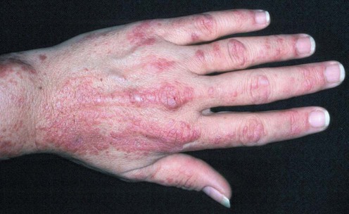
Dermatomyositis
Specific investigations


First-line therapies
 Sunscreens
 Topical corticosteroids
 Antimalarials – hydroxychloroquine or chloroquine
 Combination antimalarial therapy
 Systemic corticosteroids
 Immunosuppressive agents – methotrexate, azathioprine
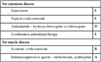
Second-line therapies
 Topical calcineurin inhibitors (tacrolimus or pimecrolimus)
 Methotrexate
 Mycophenolate mofetil
 Intravenous immunoglobulin
 Other immunosuppressive agents – cyclophosphamide, chlorambucil, mycophenolate mofetil, cyclosporine, tacrolimus
 Intravenous immunoglobulin
 Rituximab
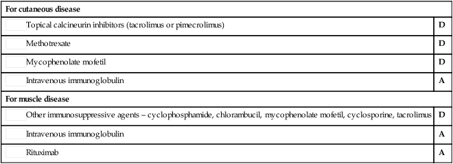
Third-line therapies
 Dapsone for cutaneous disease
 Thalidomide for cutaneous disease
 Anti-estrogen medication for cutaneous disease
 Diltiazem for calcinosis
 Etanercept
 Infliximab
 Total body irradiation
 Rituximab for cutaneous disease
 Leflunomide
 Sirolimus
 Stem cell transplantation
[/level-membership-for-dermatology-category][not-level-membership-for-dermatology-category]
Dermatomyositis
Specific investigations
Buy Membership for Dermatology Category to continue reading. Learn more here
[/not-level-membership-for-dermatology-category]
