[level-membership-for-dermatology-category]
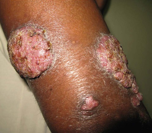
Chromoblastomycosis


First-line therapies
 Itraconazole
 Terbinafine
 Posaconazole
Second-line therapies
 Cryotherapy
 Surgical excision
 Thermotherapy
Third-line therapies
 Flucytosine
 Amphotericin B
 Voriconazole
[/level-membership-for-dermatology-category][not-level-membership-for-dermatology-category]
Chromoblastomycosis
[level-membership-for-dermatology-category]
[/level-membership-for-dermatology-category][not-level-membership-for-dermatology-category]
