Published on 18/03/2015 by admin
Filed under Dermatology
Last modified 22/04/2025
This article have been viewed 2319 times
Wanda Sonia Robles and Mahreen Ameen
Evidence Levels: A Double-blind study B Clinical trial ≥ 20 subjects C Clinical trial < 20 subjects D Series ≥ 5 subjects E Anecdotal case reports
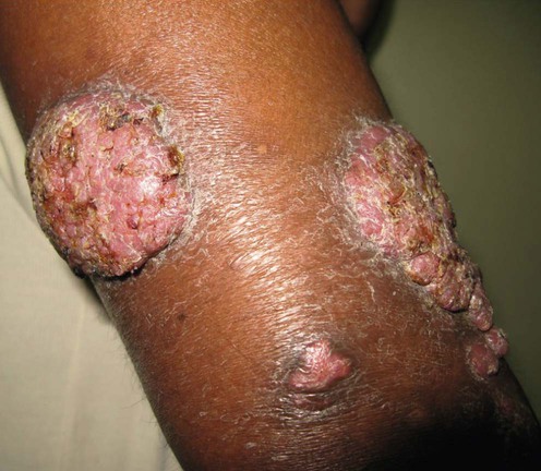
Chromoblastomycosis is a chronic fungal infection of cutaneous and subcutaneous tissues endemic in Central and South America, Africa, Australia, and Japan. It is caused by the traumatic implantation of pigmented (dematiaceous) fungi, which produce characteristic thick-walled sclerotic bodies (also known as muriform or Medlar bodies) in infected tissues. The commonest etiological agents are Fonsecaea pedrosoi, Phialophora verrucosa and Cladophialophora carrionii. Clinically, chromoblastomycosis presents as plaques, nodules or warty, exophytic lesions, which most commonly affect the feet and lower legs. Lesions are slow growing, and over years extend centripetally leaving central areas of scarring. Disease is usually localized but can spread through autoinoculation or lymphatic dissemination, producing metastatic lesions away from the primary site. Complications include ulceration, secondary bacterial infection, and lymphedema. Rarely, malignant transformation (squamous cell carcinoma) in chronic lesions and systemic involvement have been reported.
Chromoblastomycosis is a difficult-to-treat deep mycosis that is characterized by low cure rates and high rates of relapse, particularly in chronic and extensive disease. Studies report highly variable rates of clinical and mycological cure, ranging from 15% to 80%. The choice of treatment and outcome depends on the etiological agent, the extent of the lesions, clinical topography, and the presence of complications (dermal fibrosis and edema may reduce tissue antifungal drug levels). F. pedrosoi is the most common etiological agent, but has the lowest sensitivity to the major systemic antifungal agents. C. carrionii and P. verrucosa are much more sensitive and have been found to respond more favorably to treatment.
Surgical excision may be successful for small and localized lesions. It is performed with wide surgical margins and is usually accompanied by chemotherapy in order to reduce the risk of recurrence. Curettage and electrodesiccation is not recommended because it may promote lymphatic spread. Other physical modalities include cryosurgery using liquid nitrogen, and thermotherapy (applying local heat to produce controlled temperatures ranging from 42°C to 45°C, that inhibit fungal growth) using a variety of methods including benzene pocket warmers and pocket handkerchief type warmers. Cryosurgery and thermotherapy have the advantage that they are relatively inexpensive treatment options.
There are no comparative trials of antifungal chemotherapy for chromoblastomycosis. Itraconazole (100–400 mg daily) and terbinafine (250–500 mg daily) are considered first-line treatments, both drugs having shown high in vitro activity against the causative agents of chromoblastomycosis. They are typically given for long periods of time at high doses. Dual therapy with itraconazole and terbinafine is recommended if it is affordable and tolerated. It is not uncommon for more than one treatment modality to be used, such as oral antifungals combined with surgery, cryotherapy or thermotherapy. For example, itraconazole and/or terbinafine combined with cryosurgery is advocated for extensive disease. The antifungal is given first until there is a maximum reduction in lesion size, which usually requires 6 to 12 months of chemotherapy. The lesions then require several treatments with cryosurgery.
Of the other antifungal agents, ketoconazole is not recommended for treating chromoblastomycosis as it cannot be given at high doses for long treatment periods because of its toxicity profile. Despite a few cases in the early literature reporting successful treatment with fluconazole, it too is not recommended as in vitro studies have shown that it has little activity against black fungi. Flucytosine (converted into 5-fluorouracil in fungal cells) was an early treatment that demonstrated some degree of efficacy. It is associated with a high risk of developing resistance but this can be overcome if it is used in combination with another antifungal. It is also hepatotoxic and myelotoxic, requiring regular monitoring of serum levels. With the emergence of newer antifungals it is rarely used now except for resistant cases. Amphotericin B monotherapy is ineffective, and even in combination with other antifungals results are generally poor. However, amphotericin B and flucytosine dual therapy has demonstrated efficacy, in vitro studies having demonstrated synergistic activity between the two drugs. The new, second generation triazoles such as posaconazole and voriconazole are promising drugs in the management of deep cutaneous mycoses, but experience to date is limited by their prohibitively high costs in endemic settings.
Direct microscopy
Culture
Histopathology
A positive direct examination of scrapings in 10% potassium hydroxide will demonstrate the thick-walled, brown sclerotic cells that are pathognomonic of chromoblastomycosis, irrespective of the causative species. Specimens are more likely to yield a positive result if they include the ‘black dots’ visible on the surface of the lesion. Culture enables the identification of the causative agent. It is a slow-growing fungus and culture may be inconclusive due to poor morphological differentiation. Polymerase chain reaction (PCR) has been developed for the identification of Fonsecaea and C. carrionii. Serological tests such as ELISA can be useful in evaluating therapy response, but like PCR it is not widely available in most endemic settings. A biopsy demonstrates the typical sclerotic bodies in a granulomatous lesion with transepithelial elimination.
Bonifaz A, Paredes-Solis V, Saul A. Expert Opin Pharmacother 2004; 5: 247–54.
A review article highlighting the difficulties in treating this condition. The authors state that the best results for therapy have been obtained with combination and high-dose itraconazole and terbinafine for a minimum treatment period of 6 to 12 months.
López Martínez R, Méndez Tovar LJ. Clin Dermatol 2007; 25: 188–94.
A comprehensive review article. Itraconazole is considered the treatment of choice in combination with surgery in some cases.
Queiroz-Telles F, Esterre P, Perez-Blanco M, Vitale RG, Salgado CG, Bonifaz A. Med Mycol 2009; 47: 3–15.
The authors state that there is no treatment of choice but rather several treatment options: they suggest that physical therapies should augment chemotherapy and that systemic combination therapies may increase cure rate but may be associated with a higher risk of adverse effects.
Bonifaz A, Carrasco-Gerard E, Saúl A. Mycoses 2001; 44: 1–7.
This is a study of 51 cases diagnosed over a 17-year period in a tertiary referral center in Mexico. Ninety percent of the cases were caused by F. pedrosoi. The overall cure rate for all treatment modalities was 31%, and a further 57% showed clinical improvement. For large lesions, itraconazole proved to be the most effective treatment, and cryosurgery for small lesions. Itraconazole combined with cryosurgery was also effective.
Queiroz-Telles F, McGinnis MR, Salkin I, Graybill JR. Infect Dis Clin North Am 2003; 17: 59–85.
In this study from Brazil, 30 patients with chromoblastomycosis due mainly to F. pedrosoi were treated with itraconazole 200–400 mg daily depending on the clinical severity. Nineteen patients (63%) achieved clinical and mycological cure after 12 months of treatment (range 5–31 months). Treatment success depended on lesion size and extent.
This is the largest case series of itraconazole monotherapy for chromoblastomycosis.
Ungpakorn R, Reangchainam S. Clin Exp Dermatol 2006; 31: 245–7.
In this small study, six cases of F. pedrosoi infection in Thailand were treated with pulse itraconazole, 400 mg daily for 1 week each month. All achieved clinical and mycological cure. Four patients were cured after 12 pulses of treatment, and one after 15 pulses of treatment. The remaining patient required 20 pulses of itraconazole as well as cryosurgery. Disease severity and duration did not appear to be predictive of treatment response. This study demonstrated that the monthly pulse regimen is well tolerated and as effective as the conventional continuous 200–400 mg daily regimen for the treatment of F. pedrosoi. The authors recommend that treatment is continued until absence of organisms is proven by histology and tissue culture. Given that the total drug dosage is reduced, there is a marked reduction in cost of therapy by 50–75%. They also claim that pulse therapy is associated with higher compliance although therapy duration remains long.
The cost of long-term itraconazole therapy is expensive in endemic settings, making this an important and relevant study.
Mouchalouat Mde F, Gutierrez Galhardo MC, Zancopé-Oliveira RM, Monteiro Fialho PC, de Oliveira Coelho JM, Silva Tavares PM, et al. Int J Dermatol 2011; 50: 981–6.
The vast majority of these cases were caused by F. pedrosoi and disease duration varied from 4 months to 32 years. Systemic treatment included either itraconazole monotherapy 200–400 mg daily (n = 6) or itraconazole 200–400 mg daily combined with fluconazole 200 mg/day (n = 5) which were given for 12–60 months. There was an 80% cure rate and no relapse after 2 years of follow-up.
This study demonstrated good tolerability to combination azole therapy which was given for more severe disease forms which also responded well to drug therapy, suggesting that there may be a role for fluconazole in combination with itraconazole.
Esterre P, Inzan CK, Ramarcel ER, Andriantsimahavandy A, Ratsioharana M, Pecarrere JL, Roig P. Br J Dermatol 1996; 134: 33–6.
A multicenter study from Madagascar. Of 43 patients treated, 36 (F. pedrosoi, n = 29; C. carrionii, n = 7) could be completely evaluated. Approximately one-third of the patients had been resistant to previous treatment with thiabendazole. Oral terbinafine 500 mg daily was given for 6 to 12 months. Within 2 to 4 months of commencement of treatment, there was a marked clinical improvement with resolution of secondary bacterial infection, edema, and elephantiasis. There was mycological cure in 83% (24/29) of patients infected with F. pedrosoi at 12 months. Of those infected with C. carrionii, there was cure in one and clinical improvement in a further three. Total cure was observed even in imidazole-refractory patients or those with chronic disease present for over 10 years. There was a mild transient rise of hepatic enzymes in some patients, but no serious adverse effects were reported.
Bonifaz A, Saúl A, Paredes-Solis V, Araiza J, Fierro-Arias L. J Dermatolog Treat 2005; 16: 47–51.
Four cases from Mexico (F. pedrosoi, n = 3; P. verrucosa, n = 1) were treated with oral terbinafine 500 mg daily. Three cases achieved clinical and mycological cure after a mean treatment period of 7 months. Treatment was well tolerated with no abnormalities of liver enzymes.
Xibao Z, Changxing L, Quan L, Yuqing H. J Dermatolog Treat 2005; 16: 121–4.
Four cases from China (C. carrionii, n = 2; F. pedrosoi, n = 2) treated with terbinafine 500 mg daily for the first month followed by 250 mg daily were cured after 4 to 8 months of therapy without evidence of relapse at follow-up 6 months later. The total dosage of terbinafine ranged from 37.5 g to 60 g.
Gupta AK, Taborda PR, Sanzovo AD. Med Mycol 2002; 40: 529–34.
Four patients with long-standing disease refractory to standard therapies were treated with alternate week and combination therapy with itraconazole (200–400 mg daily) and terbinafine (250–1000 mg daily). Combination therapy proved to be more effective and was well tolerated.
The authors suggest that combination therapy with itraconazole and terbinafine may be synergistic, in vitro studies having already demonstrated this against other fungi. Larger studies are required to evaluate combination therapy with itraconazole and terbinafine. However, it must be noted that both drugs are expensive in endemic settings.
Negroni R, Tobon A, Bustamante B, Shikanai-Yasuda MA, Patino H, Restrepo A. Rev Inst Med Trop Sao Paulo 2005; 47: 339–46.
Cure was achieved in five of six patients with chromoblastomycosis refractory to standard antifungal therapies. They were treated with posaconazole 800 mg in divided doses. The maximum treatment period was for 34 months. Treatment was very well tolerated.
This new triazole demonstrates high efficacy and tolerability, but its use in most endemic settings is currently restricted due to its high cost.
Bonifaz A, Martinez-Soto E, Carrasco-Gerard E, Peniche J. Int J Dermatol 1997; 36: 542–7.
This study included 12 patients assigned to three different groups. Group 1, with small lesions, was treated with itraconazole 300 mg/day. Group 2, also with small lesions, was treated with one or more sessions of cryosurgery. Group 3, with large lesions, started treatment with itraconazole 300 mg/day until reduction of lesions was achieved, followed by one or more sessions of cryosurgery. The results showed complete clinical and mycological cure in two out of four patients in both groups 1 and 3. All four patients in group 2 achieved complete cure.
Although the case numbers were small, this study suggests that cryosurgery is a more suitable treatment option than antifungal therapy for small lesions.
Castro LG, Pimentel ER, Lacaz CS. Int J Dermatol 2003; 42: 408–12.
This retrospective study included 22 patients with Fonsecaea pedrosoi. Small lesions were frozen in a single session whereas larger lesions were frozen in small parts. The average number of cryosurgery treatments that each patient received was 6.7 (range 1–22 sessions). Nine patients (40.9%) were considered cured (clinically disease-free period of at least 3 years), and eight patients (36.4%) were under observation (clinically disease-free but less than 3 years of follow-up). The rest failed to clear with treatment.
This study suggests high efficacy for cryosurgery therapy. This study, as well as others, suggests that cryosurgery is a useful and inexpensive option for small lesions.
Tagami H, Ginoza M, Imaizumi S, Urano-Suehisa S. J Am Acad Dermatol 1984; 10: 615–19.
Four cases (F. pedrosoi isolated from three) were treated with topical application of tolerable heat from pocket warmers. Three patients responded after 2, 3, and 6 months of treatment. The fourth patient who received treatment in an irregular manner cleared only after a 12-month period.
Lopez CF, Alvarenga RJ, Cisalpino EO, Resende MA, Oliveira LG. Int J Dermatol 1978; 17: 414–18.
Twenty-three patients with chromoblastomycosis were treated with oral flucytosine for 2 to 67 months. Sixteen patients achieved clinical and mycological cure (59%). However, seven patients developed resistance, and they failed to respond with subsequent treatment with amphotericin B, calciferol or thiabendazole. Resistance appeared to occur particularly in those with long-standing lesions or widespread involvement.
Park SG, Oh SH, Suh SB, Lee KH, Chung KY. Br J Dermatol 2005; 152: 560–4.
This case from Korea was treated with liposomal amphotericin monotherapy for 3 months, which had some effect. The addition of 5-flucytosine 4 g daily resulted in marked clinical improvement after only 1 month. Amphotericin was then discontinued, and dual therapy with 5-flucytosine and itraconazole 200 mg daily was given for 12 months until mycological cure.
This case illustrates the low efficacy of amphotericin B monotherapy, and the synergistic activity in combination with 5-flucytosine. 5-Flucytosine is renally excreted and therefore careful monitoring is required when it is used with other nephrotoxic drugs. Despite the toxicity of both drugs this drug combination has been advocated by some mycologists as a very useful alternative to azoles.
Paniz-Mondolfi AE, Colella MT, Negrín DC, Aranzazu N, Oliver M, Reyes-Jaimes O, et al. Med Mycol 2008; 46: 179–84.
This report describes a case of extensive disease affecting the whole of the right lower limb of 22 years duration that had failed 15 years of previous therapy consisting of ketoconazole and fluconazole. The patient then achieved clinical and mycologoical cure with amphotericin (cumulative dose of 2150 mg) in combination with itraconazole 100 mg twice daily for approximately 3 months followed by a further 1 year of itraconazole 100 mg daily.
Criado PR, Careta MF, Valente NY, Martins JE, Rivitti EA, Spina R, et al. J Dermatolog Treat 2011; 22: 167–74.
Three patients with long-standing (10, 20, and 21 years) and extensive disease refractory to previous therapy with itraconazole and terbinafine were treated with voriconazole 200 mg twice daily for 12 months. Clinical response was evident after 30–50 days but at the end of treatment despite significant clinical improvement none of the patients achieved cure. All patients showed elevations of serum gamma-glutamyl transpeptidase during the treatment without clinical relevance and a single patient developed visual abnormalities and tremors requiring dose reduction.
This is the first report of the use of voriconazole for chromoblastomycosis. The partial response may be attributed to the severity of the disease. The report suggests that voriconazole represents an option for extensive disease refractory to conventional treatment.
Treatment of Skin Disease Comprehensive Therapeutic Strategies 4e
WhatsApp us


 Itraconazole
 Terbinafine
 Posaconazole
 Cryotherapy
 Surgical excision
 Thermotherapy
 Flucytosine
 Amphotericin B
 Voriconazole