Celiac plexus block

Published on 07/02/2015 by admin

Filed under Anesthesiology

Last modified 22/04/2025

Print this page

rate 1 star rate 2 star rate 3 star rate 4 star rate 5 star
Your rating: none, Average: 0 (0 votes)

This article have been viewed 3034 times

Celiac plexus block

David P. Martin, MD, PhD

Indications

The celiac plexus provides sensory innervation and sympathetic outflow to most of the upper abdominal viscera (see Chapter 40). Neurolytic blockade of the celiac plexus is most commonly used to control pain caused by pancreatic cancer, although it can be useful for managing pain related to malignancies of the gastrointestinal tract from the lower esophageal sphincter to the splenic flexure, as well as the liver, spleen, and kidneys. Although potentially long-lasting, neurolytic celiac plexus block is not “permanent” because the nerves in the plexus regenerate in 3 to 6 months. The block can be repeated in such circumstances, but many patients with pancreatic cancer do not outlive the effective duration of neurolytic celiac plexus block. The median survival after diagnosis with pancreatic cancer is 3 to 6 months. Most patients with pancreatic cancer still require some oral analgesics even after neurolytic celiac plexus block.

Temporary diagnostic blockade of the celiac plexus can be used to differentiate visceral pain from somatic pain. Visceral pain is poorly localized and can be referred to somatic areas. For example, pancreatic pain often presents as epigastric tenderness radiating to the back. Relief of pain after celiac plexus block suggests a visceral origin of the pain. If pain persists after celiac plexus block, it is more likely to be somatic in origin. In addition to its neurolytic and diagnostic uses, celiac plexus injection with a local anesthetic agent and a corticosteroid is sometimes used to treat the pain associated with chronic pancreatitis.

Anatomy

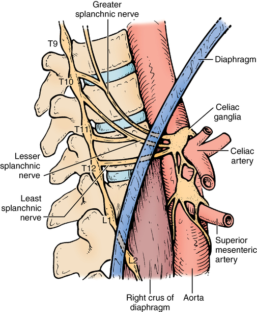
The celiac plexus is primarily a sympathetic nervous system structure that lies anterior to the aorta near the celiac arterial trunk (Figure 220-1). Preganglionic sympathetic fibers originate from the nerve roots of T5-T12 and combine to form the splanchnic nerves. The splanchnic nerves cross the crura of the diaphragm before joining the vagus nerve to form the celiac plexus anterior to the aorta. The location of the plexus varies from T12 to L2 vertebral levels; approaches to the block are directed at the T12-L1 level.

Effective visceral pain relief can be achieved by either blocking the splanchnic nerves before they pierce the diaphragm or blocking the nerves and ganglia anterior to the diaphragmatic crura. The splanchnic nerve block (retrocrural) is also termed the classic celiac plexus block, as opposed to true blockade of the plexus and ganglia (intercrural).

Procedure

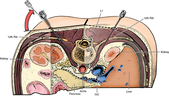
Several approaches to the celiac plexus have been described, including endoscopic, ventral, and dorsal. The endoscopic route is convenient when combined with endoscopic retrograde cholangiopancreatography (ERCP). The ventral approach can be advantageous if tumor blocks the dorsal route, but it has a higher risk of bowel injury and infection. The most common route used by anesthesia providers is via the dorsal approach and is performed with the patient in the prone position with a pillow under the hips. Landmarks are identified and marked on the skin surface, indicating the twelfth rib and the thoracolumbar spinous processes. Needles are inserted bilaterally at a site approximately 7.5 cm lateral to midline at a point 2 cm inferior to the twelfth rib. The initial pass is directed to contact the L1 vertebral body at an angle approximately 45 degrees from the sagittal plane (Figure 220-2). The path of the needle is approximately parallel to the inferior border of the twelfth rib, directed toward the middle of the L1 vertebral body. After noting the depth at which bone is contacted, the needle is withdrawn to skin level and redirected more steeply, so that it passes just lateral to the L1 body, and is then advanced an additional 1 to 2 cm. Ideal positioning is anterolateral to the body of the L1 vertebral body.

Once the needle is placed, careful aspiration is performed to exclude a vascular or intrathecal position. Proper drug distribution can be confirmed with the injection of radiocontrast dye under fluoroscopy. It is important to ensure that the injectant is not within the psoas muscle, which could result in blockade of the lumbar plexus. Bupivacaine, 0.25% to 0.5%, is a reasonable choice for the diagnostic nerve block. Typically, 10 to 15 mL is injected on each side.

For diagnostic blocks, the procedure ends at this point. At least 15 to 20 min must elapse until the effects of blockade can be assessed. In addition to pain relief, motor function should also be tested. If a neurolytic block is planned, the needles can be left in place during this assessment.

If pain is relieved and no motor deficits are observed after injecting a local anesthetic, it is reasonable to proceed with neurolysis. For neurolytic procedures, 50% to 100% alcohol is the most commonly used agent. Typically, 10 mL is injected on each side. A small volume of local anesthetic agent can be injected while withdrawing the needles to prevent alcohol from tracking to more superficial tissue.

Expected side effects

The procedure itself can cause local soreness and bruising. These symptoms are usually transient and can be treated with ice. Psoas spasm is not uncommon after neurolytic celiac plexus block and can be minimized by preventing the escape of neurolytic agent through the needle tract. Psoas spasm often responds well to intravenously or intramuscularly injected ketorolac.

Interruption of sympathetic innervation to the viscera can blunt normal postural hemodynamic reflexes, resulting in orthostatic hypotension. Patients should be cautioned that they may feel lightheaded upon standing. The sympathectomy can also cause increased gastrointestinal motility and possibly diarrhea. However, the effect of sympatholysis on intestinal motility can be beneficial in counteracting the constipation caused by orally administered opioids. Finally, celiac plexus block may mask early presenting symptoms of other intra abdominal diseases, such as cholecystitis and gastric ulceration.

Adverse effects

As with any injection, sterile technique should be observed with the performance of a celiac plexus block to minimize the risk of infection. Because of the close proximity of the celiac plexus to the aorta, vascular injury is possible; hematoma formation, aortic dissection, and distal (lower extremity) ischemia have been reported. Intravascular injection of a local anesthetic agent can cause mental status changes, seizures, and possible hemodynamic collapse.

Unintentional intrathecal or epidural spread can cause spinal nerve block. The spread of neurolytic agent to unintended nerve or vascular structures introduces the risk of permanent neurologic injury, including paralysis. Therefore, careful neurologic evaluation after injection of a local anesthetic is essential before injecting the neurolytic agent. The most common nerve injury after celiac plexus block is genitofemoral neuralgia. Despite these risks, celiac plexus block is relatively safe when performed by experienced physicians.