Bedside Assessment of the Patient

Published on 01/06/2015 by admin

Filed under Pulmolory and Respiratory

Last modified 22/04/2025

Print this page

rate 1 star rate 2 star rate 3 star rate 4 star rate 5 star
Your rating: none, Average: 5 (1 votes)

This article have been viewed 12366 times

Bedside Assessment of the Patient

Richard H. Kallet

Progress in the field of respiratory care has placed increasing demands on respiratory therapists (RTs) to develop competent bedside assessment skills. Decisions regarding when to initiate, change, or discontinue therapy depend on accurate clinical assessment. Although the physician has the ultimate responsibility for these decisions, RTs often participate in the clinical decision-making process. To fulfill this role effectively, the RT must assume responsibility for gathering and interpreting relevant bedside patient data.

Bedside assessment is the process of interviewing and examining a patient for signs and symptoms of disease and the effects of treatment. It is a cost-effective way of obtaining pertinent information about the patient’s health status. In many cases, bedside assessment provides the initial evidence that something is wrong and often helps establish the severity of the problem. In contrast to some diagnostic tests, bedside assessment techniques are of little risk to the patient.

Two key sources of patient data are the medical history and the physical examination. Data gathered initially by interview and physical examination help identify the need for subsequent diagnostic tests. After a tentative diagnosis is made, these assessment procedures also help the clinician to select the best approach to therapy. After a treatment regimen begins, these assessment procedures are repeated to monitor patient progress toward predefined goals.

The patient initially is assessed to identify the correct diagnosis. This initial assessment is most often performed by a physician. Exceptions may occur in emergency situations in which a physician is unavailable. In such cases, other health care personnel, such as nurses and RTs, may need to evaluate the patient rapidly to implement appropriate lifesaving treatment (e.g., cardiopulmonary resuscitation). After a tentative diagnosis is reached and the physician orders specific treatment, subsequent evaluations are made by health care personnel to monitor the patient during the hospital stay and to evaluate treatment results.

The skills of bedside assessment described here are not difficult to learn; however, mastery requires practice. Initially, students should practice the skills on healthy individuals. This practice helps improve technique and provides an understanding of normal variations. The ability to discriminate abnormal from the range of possible normal findings is an important skill that requires experience to master.

Interviewing the Patient and Taking a Medical History

Interviewing furnishes unique information because it provides the patient’s perspective. It serves the following three related purposes:

For these reasons, interviewing is a crucial aspect of general patient assessment.

Principles of Interviewing

Interviewing is a way of “connecting” with the patient. This connection is especially important for the patient who is under the stress of an illness because meaningful human contact lessens the patient’s sense of isolation. The factors that affect communication between the RT and the patient include the following:

Because of the above-listed factors, no two interviews are the same.

Although developing interviewing skills takes time and experience, beginners can get a head start by following a few basic guidelines and by becoming knowledgeable about the causes and characteristics of common cardiopulmonary symptoms. The following discussion provides some of the guidelines for interviewing and discusses common symptoms associated with diseases of the chest.

Structure and Technique for Interviewing

The ideal interview is one in which the patient feels secure and free to talk about important personal matters. Each interview should begin with the RT introducing himself or herself to the patient and stating the purpose of the visit. The introduction is done in the social space, approximately 4 to 12 feet from the patient. It begins the process of establishing a rapport with the patient and helps the patient feel more comfortable about answering personal questions. Pulling the curtain between the beds of a semiprivate room also may be helpful in making the patient feel more at ease with the interview (Box 15-1).

Box 15-1   Guidelines for Effective Patient Interviewing

Show Your Respect for the Patient’s Beliefs, Attitudes, and Rights

• Ensure the patient is appropriately covered.

• Position yourself so that eye contact is comfortable for the patient. (Ideally, patients should be sitting up, with their eye level at or slightly above yours.)

• Avoid standing at the foot of the bed or with your hand on the door because this may send the nonverbal message that you do not have time for the patient.

• Ask the patient’s permission before moving any personal items or making adjustments in the room.

• Remember that the patient’s dialog with you and his or her medical record are confidential. Share this information only with other health care providers who need to know about it, and do not share the information in a place where others can overhear the conversation.

• Be honest; never guess at an answer or information that you do not know; do not provide information beyond your scope of practice; providing new information to the patient is the privilege and responsibility of the attending physician.

• Make no moral judgments about the patient; set your values for patient care according to the patient’s values, beliefs, and priorities.

• Expect the patient to have an emotional response to illness and the health care environment.

• Listen, and then clarify and teach, but never argue.

• Adjust the time, length, and content of the interview to the patient’s needs.

Next, the RT moves into the personal space (2 to 4 feet from the patient) to begin the interview. In this space, the patient does not have to speak loudly in response to questions. The RT should assume a physical position at the same level with the patient (e.g., by sitting in a chair) before beginning the formal interview. Standing over the patient should be avoided because this position makes the patient feel inferior. Appropriate eye contact with the patient is essential for a quality interview. Eye contact gives the patient more confidence in the interviewer. Eye contact also allows the interviewer to see confusion, anger, frustration, and other emotions that may be expressed by the patient in response to questions.

Using neutral questions and avoiding leading questions during the interview is important. Asking the patient, “Is your breathing better now?” leads the patient toward a desired response and may elicit false information. Asking the patient, “How is your breathing now?” is a better way to get accurate information about the patient’s breathing (Box 15-2).

Box 15-2   Types of Questions Used in Patient Interviews

• Open-ended questions encourage patients to describe events and priorities as they see them, helping to bring out concerns and attitudes and to promote understanding. Questions such as “What brought you to the hospital?” or “What happened next?” encourage conversational flow and rapport, while giving patients enough direction to know where to start.

• Closed questions, such as “When did your cough start?” or “How long did the pain last?” focus on specific information and provide clarification.

• Direct questions can be open-ended or closed and always end in a question mark. Although they are used to obtain specific information, a series of direct questions or frequent use of the question “Why?” can be intimidating and cause the patient to minimize his or her responses to questions.

• Indirect questions are less threatening than direct questions because they sound like statements (e.g., “I gather your doctor told you to take the treatments every 4 hours”). Inquiries of this type also work well to confront discrepancies in the patient’s statements (e.g., “If I understood you correctly, it is harder for you to breathe now than it was before your treatment”).

• Neutral questions and statements are preferred for all interactions with the patient. “What happened next?” and “Can you tell me more about …?” are neutral, open-ended questions. A neutral, closed question may give the patient a choice of responses, while focusing on the type of information desired (e.g., “Would you say there was a teaspoon, a tablespoon, or a half cup?”). Leading questions, such as “You didn’t cough up blood, did you?” should be avoided because they imply an answer.

Common characteristics of symptoms can be identified by asking questions such as the following during the interview:

Identifying these characteristics of any new symptom can be helpful in recognizing the cause and potential therapy; this is primarily the role of the attending physician but sometimes falls to other clinicians in certain settings. Once the symptom or symptoms are established and therapy is started, other questions are used to evaluate the changes in the symptoms over the course of the hospital stay. For example, the clinician may ask, “Has the symptom changed in any way since admission?” or, “Does the therapy seem to make a difference?”

The best interview techniques are of no value if the interviewer is not knowledgeable about the pathophysiology and characteristics of common cardiopulmonary symptoms. The interview is a series of focused questions that pursue specific information related to a tentative diagnosis. The ability to ask the key questions at the right time comes from experience and familiarity with the signs and symptoms of lung disease.

Common Cardiopulmonary Symptoms

Dyspnea

Dyspnea is a general term describing the sensation of breathing discomfort. It is the most important symptom that the RT is called on to assess and treat. Dyspnea is a subjective experience and should not be inferred from observing the patient’s breathing pattern. Analogies often have been made between dyspnea and pain. Both sensations possess qualitatively distinct features and varying intensity. Similar to pain, dyspnea causes suffering. As breathing is the primordial sensation of life, dyspnea often is perceived as life-threatening and may provoke a profound sense of dread.

The term dyspnea also is used specifically to describe difficulty in the mechanical act of breathing. The simplest explanation is that the effort to breathe is proportionally greater than the tidal volume achieved. A person’s perception of breathing is a complex balance between the following three factors:

When the neuronal signals governing these sensations become unbalanced, breathing is perceived to be abnormal and unpleasant. The technical name for this imbalance is neuromechanical dissociation. Individuals normally experience this form of dyspnea only in unusual circumstances, such as when trying to breathe through a straw or when wearing a restrictive garment.

Breathlessness

Breathlessness is the specific sensation of an unpleasant urge to breathe. It is believed to be the conscious perception of intense neural discharge to the respiratory muscles. Breathlessness can be triggered by acute hypercapnia and acidosis and by hypoxemia. A normal experience of breathlessness is the unpleasant throbbing sensation that accompanies prolonged breath holding or feeling “winded” during strenuous physical exercise. However, it is not known how closely normal encounters with breathlessness resemble the sensation that arises during disease because dyspnea in patients with cardiopulmonary disease also is affected by other stimuli that contribute to both the quality and the intensity of the sensation. These include stimuli arising from hypoxemia, irritant receptors in the lungs and airways, and receptors in the blood vessels and heart.

Ultimately, dyspnea and breathlessness are magnified by the emotional distress that accompanies them. This distress is influenced by situation, knowledge, and control. In other words, a healthy person can quickly identify the source of breathlessness and arrest the symptom simply by stopping exercise or the breath hold. In contrast, a patient with cardiopulmonary disease may be unable to control the symptom, let alone be able to identify the source. These factors have a profound emotional impact that must be appreciated by the RT.

Positional Dyspnea

Dyspnea may be present only when the patient assumes the reclining position, in which case it is referred to as orthopnea. Orthopnea is common in patients with congestive heart failure (CHF); it apparently is caused by the sudden increase in venous return that occurs with reclining. The failing left ventricle is unable to accommodate the increased venous return, resulting in pulmonary vascular congestion and dyspnea. Orthopnea is also a symptom of bilateral diaphragmatic paralysis.

Dyspnea in the upright position is known as platypnea. This unusual symptom may accompany arteriovenous malformations in the lung, such as occur in chronic liver disease (hepatopulmonary syndrome), and some hereditary conditions. Platypnea may be accompanied by orthodeoxia, which is oxygen desaturation on assuming an upright position.

Language of Dyspnea

Dyspnea is a subjective experience, and patients possess a nuanced language to describe their sensations. RTs should ask specific questions about the quality and characteristics of the patient’s dyspnea. In this way, the RT might gain insight into the mechanism provoking dyspnea. As the patient describes the sensations, the RT should try to categorize each according to a particular aspect of breathing such as inspiration, expiration, respiratory drive, or lung volume. A remark such as, “I feel that my breath stops,” reflects a problem with inspiration, whereas the remark, “my breath does not go all the way out,” suggests a problem with expiration. Statements such as, “I can’t catch my breath,” suggest that respiratory drive is elevated (i.e., breathlessness).

Different types of lung diseases often evoke unique sensations that may provide clues about the underlying pathophysiology. Patients with asthma frequently complain of chest tightness. In contrast, patients with interstitial lung disease tend to focus on the sensations of increased work of breathing, shallow breathing, and gasping. Patients with CHF are seemingly unique in frequently feeling suffocated. Although the language used to describe dyspnea provides helpful clues, the RT should keep in mind that many lung diseases evoke common sensations.

Patients with cardiopulmonary disease frequently experience several unpleasant breathing sensations simultaneously. A particular sensation may be more prominent than others and may change over time. As mentioned previously, patients with asthma typically complain first about the sensation of chest tightness. However, as bronchoconstriction worsens and the lungs become more hyperinflated, patients often begin to focus more on the sensation of excessive work of breathing, air hunger, and the inability to take a deep breath.

Assessing Dyspnea in the Interview

The assessment of dyspnea is largely determined by the situation. When conducting an interview, the RT should pay particular attention to whether the patient can speak in full sentences. It may be very difficult for patients with severe dyspnea from any cause to speak more than a few words at a time. In this situation, the initial interview should be curtailed, and treatment should be initiated as soon as possible. Questions should be brief and limited to the quality and intensity of dyspnea and the circumstances of symptom onset. Also, the assessment of dyspnea should occur simultaneously with a gross examination of the patient’s breathing pattern (see later section of this chapter). Assessment of dyspnea that arises acutely in patients without a prior history of cardiopulmonary disease typically does not require the same detail as assessment in patients with long-standing cardiopulmonary or neurologic disease.

In patients with chronic cardiopulmonary disease, a detailed and systematic history should cover four major areas, as follows:

1. The RT should ask what activities of daily living tend to trigger episodes of dyspnea. For example, is dyspnea triggered by walking on flat surfaces, by climbing stairs, by bathing, by dressing?

2. The RT should ask how much exertion is required for the patient to stop to catch his or her breath with different activities. Does the patient need to stop after walking up one flight of stairs or one step? Dyspnea provoked by less strenuous activities indicates more advanced disease.

3. The RT should ask whether the quality or the sensation of breathing discomfort varies with different activities.

4. To gain a better understanding of the patient’s history, the RT should ask the patient to recall when dyspnea first began and how it has evolved over time. Has dyspnea progressed slowly or rapidly? How long has this progression taken place: over a period of months or years? Has there been a dramatic change in the intensity of dyspnea over recent months, weeks, days, or even within the past few hours?

Beyond the information gleaned, a detailed conversation about a patient’s struggle with dyspnea allows the patient to share his or her experience and decreases the patient’s sense of isolation.

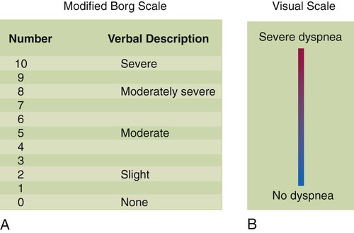
The intensity of dyspnea can be documented using a numeric intensity or visual analog scale (Figure 15-1). Such scales provide a way to evaluate the patient’s response to treatment over time. These scales are important because objective lung function measurements (e.g., pulmonary function tests, PaO2) seldom correlate with the degree of dyspnea in many patients.

Psychogenic Dyspnea: Panic Disorders and Hyperventilation

There are perplexing situations in which patients with normal cardiopulmonary function complain of intense dyspnea or suffocation. This condition is known as psychogenic hyperventilation syndrome and is associated with panic disorders. Hyperventilation may coincide with other symptoms such as chest pain, anxiety, palpitations and paresthesia (the sensation of tingling and numbness in the extremities that often accompanies respiratory alkalosis). This syndrome may be either sporadic or chronic and often is self-perpetuating.

Anxiety often is accompanied by breathlessness and hyperventilation. The resulting respiratory alkalosis amplifies the sensation of breathlessness and provokes more anxiety, increasing the intensity of hyperventilation. The classic homespun remedy of having the patient slowly rebreathe into a paper bag holds merit because this can arrest the respiratory alkalosis and help break the cycle. However, rebreathing techniques may require formal behavioral therapy and may not be appropriate in the hospital setting. This condition usually is treated clinically by administering judicious amounts of anxiolytic agents.

The RT always must approach any situation involving hyperventilation or dyspnea as if it had a pathophysiologic basis. The first priority is to measure the vital signs, including arterial oxygen saturation, and perhaps a 12-lead electrocardiogram and arterial blood gases. A psychogenic source should be considered only after a pathogenic source for hyperventilation or dyspnea has been ruled out. Intense pain or fear may provoke anxiety and hyperventilation. The RT must work in concert with nursing and physician colleagues to determine the root cause of any hyperventilation syndrome.

Cough

A cough is the most common, yet nonspecific symptom seen in patients with pulmonary disease. Coughing is a forceful expiratory maneuver that expels mucus and foreign material from the airways. It usually occurs when the cough receptors are stimulated by inflammation, mucus, foreign materials, or noxious gases. The cough receptors are located primarily in the larynx, trachea, and larger bronchi.

The effectiveness of a cough depends on the ability of the individual to take a deep breath, lung elastic recoil, expiratory muscle strength, and level of airway resistance. The ability to take a deep breath or exhale forcefully is often impaired in patients with neuromuscular disease. An effective cough also is impaired secondary to pain; this is typically seen in the early postoperative period in patients following upper abdominal surgery or thoracic surgery or after trauma. Often expiratory flow is limited by factors such as bronchospasm (e.g., asthma) and reduced lung elastic recoil (as in emphysema). Patients with an inadequate ability to cough because of impairment of these factors often have problems with retained secretions and are more prone to the development of pneumonia.

Important characteristics of the patient’s cough to identify include whether it is dry or loose, productive or nonproductive, and acute or chronic and whether it occurs more frequently at particular times (i.e., day or night). Knowledge of such details may help in determining the cause of the cough. A dry, nonproductive cough is typical for restrictive lung diseases such as CHF or pulmonary fibrosis. A loose, productive cough is more often associated with inflammatory obstructive diseases such as bronchitis and asthma. The most common cause of an acute, self-limited cough is a viral infection of the upper airway. Common causes of chronic coughing include asthma, postnasal drip, chronic bronchitis, and gastroesophageal reflux,1 although combinations of these often exist.2 Cough is also associated with the use of certain medications for hypertension (e.g., angiotensin-converting enzyme inhibitors).3

Sputum Production

Healthy airways produce mucus daily. Normally, the quantity of this mucus is minimal, and it is not enough to stimulate the cough receptors. Mucus is gradually moved to the hypopharynx by the mucociliary escalator, where it is either swallowed or expectorated. Disease of the airways may cause the mucous glands, which line the airways, to produce an abnormally increased amount of mucus, which usually stimulates the cough receptors and causes the patient to generate a loose, productive cough. This cough is seen in acute bronchitis or asthma attacks brought on by airway infection.

RTs need to be aware of the terminology associated with sputum. Technically, mucus from the tracheobronchial tree that has not been contaminated by oral secretions is called phlegm. Mucus that comes from the lung but passes through the mouth as it is expectorated is sputum. Because this is how most mucus samples from the lung are obtained, the term sputum is used in this chapter. Sputum that contains pus cells is said to be purulent, suggesting a bacterial infection. Purulent sputum appears thick, colored, and sticky. Sputum that is foul-smelling is said to be fetid. Sputum that is clear and thick is mucoid and is commonly seen in patients with airways disease (i.e., asthma). Changes in the color, viscosity, or quantity of sputum produced are often signs of infection and must be documented and reported to the physician.

Hemoptysis

Coughing up blood or blood-streaked sputum from the lungs is referred to as hemoptysis. Blood-streaked sputum is common in patients with pulmonary disease. Frank hemoptysis is the presence primarily of blood in the expectorant. Hemoptysis is characterized as massive when more than 300 ml of blood is expectorated over 24 hours, and this represents a medical emergency. Hemoptysis must be distinguished from hematemesis, which is vomiting blood from the gastrointestinal tract. Blood from the lung is often seen in patients with a history of pulmonary disease and may be mixed with sputum. Blood from the stomach may be mixed with food particles and occurs most often in patients with a history of gastrointestinal disease.

Nonmassive hemoptysis is caused most often by infection of the airways but also is seen in lung cancer, tuberculosis, blunt or penetrating chest trauma, and pulmonary embolism. Hemoptysis associated with infection usually is seen as blood-streaked, purulent sputum. Hemoptysis commonly is found in patients with bacterial pneumonia. Hemoptysis from bronchogenic carcinoma often is chronic and may be associated with a monophonic wheeze and cough. Common causes of massive hemoptysis include bronchiectasis, lung abscess, and acute or old tuberculosis.

Chest Pain

Most chest pain can be categorized as either pleuritic or nonpleuritic. Pleuritic chest pain usually is located laterally or posteriorly. It worsens when the patient takes a deep breath, and it is described as a sharp, stabbing type of pain. It is associated with diseases of the chest that cause the pleural lining of the lung to become inflamed, such as pneumonia or pulmonary embolism.

Nonpleuritic chest pain is located typically in the center of the anterior chest and may radiate to the shoulder or back. It is not affected by breathing, and it is described as a dull ache or pressure type of pain. A common cause of nonpleuritic chest pain is angina, which classically is a pressure sensation with exertion or stress and results from coronary artery occlusion. Other common causes of nonpleuritic chest pain include gastroesophageal reflux, esophageal spasm, chest wall pain (e.g., costochondritis), and gallbladder disease.

Mini Clini

Sudden Onset of Chest Pain

image Problem

The RT is called to the emergency department to see a 47-year-old man who came to the hospital with anxiety and chest pain. He is certain he is having a heart attack and demands immediate treatment. The attending physician is on the way to the hospital but has asked the nurse to call the RT in the interim. The RT places the patient on oxygen per protocol and asks him for details about the chest pain. The patient states that the pain is located laterally on the left side and increases with each inspiratory effort. The pain is sharp in nature. The patient’s vital signs are normal (including respiratory rate) except for a slight increase in heart rate. What is the most likely cause of this patient’s chest pain, given its characteristics? What should be done until the physician arrives?

Solution

Chest pain is a worrisome symptom because it can indicate a life-threatening problem or a less serious problem. Among the most serious problems are acute heart attack, pulmonary embolism, aortic dissection, and pneumothorax. In this case, the pain seems to be pleuritic. Angina, acute myocardial infarction, and aortic dissection are not likely causes. The pleuritic character of the pain is consistent with pulmonary embolism and pneumothorax, but the normal respiratory rate suggests that pulmonary embolism is not the likely cause of the chest pain. If pneumothorax is present, it must be small. The RT should continue oxygen therapy and monitor the patient until the attending physician arrives. The RT should ask the nurse to attach chest leads to monitor the patient’s heart rate and rhythm just in case the chest pain is related to heart disease. In addition, the RT should try to comfort the patient as much as possible.

Fever

Fever (an elevated body temperature secondary to disease) is a common complaint of patients with an infection of the airways or lungs. Fever may occur with a viral infection of the upper airway or bacterial pneumonia or tuberculosis. All patients with a fever need further assessment to determine the cause. When infection causes fever, the magnitude of temperature elevation may indicate the type and virulence of the infection. Low-grade fever typically accompanies common upper respiratory tract infections, whereas a high fever occurs with viral influenza infection.

Fever that occurs with a cough suggests a respiratory infection. An infection is even more likely to be the cause of the fever if the patient is producing purulent sputum. In this situation, a persistent fever of at least 38.9° C (102° F) for 2 days accompanied by chills is suggestive of pneumonia. However, the absence of coughing or sputum production does not rule out lung infection. Noninfectious causes of fever include head trauma (secondary to damage of the hypothalamus), cancer, immunologic disorders (e.g., sarcoidosis), an adverse reaction to certain medications (e.g., sulfa drugs), and thromboembolic disorders such as pulmonary embolism.

It was believed for many years that there was a link between fever and atelectasis in postoperative surgical patients. However, more recent evidence has shown no link between the formation of atelectasis and the development of fever (>101.3° F [>38.5° C]) during the first 72 hours after surgery.4

Patients with a significant fever have an increased metabolic rate and an increased oxygen (O2) consumption and carbon dioxide (CO2) production. The increased need for O2 intake and CO2 removal may cause tachypnea. The increased ventilatory demand caused by fever is particularly dangerous for patients with severe chronic cardiopulmonary disease because it may cause acute respiratory failure.

Pedal Edema

Swelling of the lower extremities is known as pedal edema. It most often occurs with heart failure, which causes an increase in the hydrostatic pressure of the blood vessels in the lower extremities. This increase in hydrostatic pressure causes fluid to leak into the interstitial spaces and leads to pedal edema, the degree of which depends on the level of heart failure. There are two subtypes of pedal edema. When pressure is applied with a finger on a swollen extremity, an indentation mark left on the skin is called pitting edema. Weeping edema is when a small fluid leak occurs at the point where pressure is applied.

Patients with chronic hypoxemic lung disease are especially prone to right-sided heart failure (cor pulmonale) because of the heavy demands placed on the right ventricle when hypoxemia causes severe pulmonary vasoconstriction. Eventually, the right side of the heart begins to fail and results in a backup of pressure into the venous blood vessels, especially in the dependent regions such as the lower extremities. This situation promotes high intravascular venous hydrostatic pressures and pedal edema. The patient often complains of “swollen ankles” in such cases.

Format for the Medical History

All health care practitioners must be familiar with the medical history of the patients they are treating, even if their reason for contact is simply to provide intermittent therapy. The medical history familiarizes clinicians with the signs and symptoms the patient exhibited on admission and the reason the therapy is being administered.

The RT should begin reviewing the patient’s chart by reading about the patient’s current medical problems. This information is found under the headings of chief complaint and history of present illness. This section of the medical history represents a detailed account of each of the patient’s major complaints. It is written by the physician after his or her interview with the patient at admission to the hospital.

The next step is to review the patient’s past medical history, which describes all past major illnesses, injuries, surgeries, hospitalizations, allergies, and health-related habits. This information provides a basic understanding of the patient’s previous experiences with illness and health care and may have an impact on decisions made during the current hospitalization. This section of the health history may be the place the interviewer records the patient’s history of cigarette and alcohol consumption.

An accurate determination of a patient’s smoking history is an extremely important aspect of assessing pulmonary health. The smoking history is often recorded in pack-years, which is determined by multiplying the number of packs smoked per day by the number of years smoked. Typically, a patient is asked how many cigarettes (on average) he or she smokes per day. Some patients express this in terms of packs of cigarettes, whereas others state the number of cigarettes. If a patient states that he or she has smoked a pack of cigarettes a day for 20 years, the patient has a 20 pack-year smoking history.

If patients describe their smoking in terms of the number of cigarettes, or fractions of a pack, the calculation is slightly more difficult. Two examples may help illustrate how to calculate pack-years of smoking. There are 20 cigarettes per pack. If a patient states he or she has smoked a pack and a half of cigarettes per day for 20 years, the smoking history is calculated as follows:

< ?xml:namespace prefix = "mml" />30 cigarettes/20 cigarettes per pack=1.5 packs×20 years=30 pack-years smoking history

image

If the patient states that he or she has smoked 15 cigarettes per day for 20 years:

15cigarettes/20cigarettes per pack=0.75 packs×20years=15pack-years smoking history

image

Next, the family and social/environmental history should be reviewed. This part of the medical history focuses on potential genetic or occupational links to disease and the patient’s current life situation. Pulmonary disorders such as asthma, lung cancer, cystic fibrosis, and chronic obstructive pulmonary disease (COPD) are believed to have a genetic link in many cases. A detailed occupational history is important in assessing pulmonary disorders that may result from inhaling dusts in the workplace, either organic (i.e., containing protein) or nonorganic (e.g., asbestos, silica). There is a strong link between asthma and poverty.

The review of systems is designed to uncover problem areas the patient forgot to mention or omitted. This information is usually obtained in a head-to-toe review of all body systems. For each body system, the interviewer obtains information about current, pertinent symptoms. During a review of the respiratory system, questioning would determine the presence or history of cough, hemoptysis, sputum production, chest pain, shortness of breath, and fever (Box 15-3).

Box 15-3   Outline of a Complete Health History

Demographic data (obtained from admission interview): Name, address, age, birth date, place of birth, race, nationality, marital status, religion, occupation, and source of referral

Date and source of history and estimate of the reliability of the historian

Brief description of the patient’s condition at the time the history or patient profile was taken

Chief complaint and reason for seeking treatment

History of present illness: Chronologic description of each symptom

Past medical history

Family history

Social and environmental history

Review of systems: Respiratory system

Patient’s printed name and signature

Finally, the medical record should be examined for information indicating any limits on the extent of care to be provided in the event of cardiac or respiratory arrest. This information is known as an advance directive, whereby the patient (or a legally authorized representative) has formalized his or her wishes for resuscitative efforts; this is typically referred to as the DNR status (“do not resuscitate”) or may be expressed as DNI (“do not intubate”). This information may be found either in the admission note or within the body of the physician progress notes. In addition to this descriptive note, there must be an order written by the physician clearly specifying how care should be limited in the event of a medical emergency.

The first priority of the RT reviewing the medical record is to ensure that all respiratory care procedures are supported by a physician order that is current, clearly written, and complete.

Physical Examination

A careful physical examination of the patient is essential for evaluating the patient’s problem and determining the effects of therapy. The physical examination consists of the following four general steps: (1) inspection (visually examining), (2) palpation (touching), (3) percussion (tapping), and (4) auscultation (listening with a stethoscope).

General Appearance

The first few seconds of an encounter with the patient usually helps reveal the severity of the current problem. For an experienced clinician, these initial impressions determine the course of subsequent assessment. If the patient’s general appearance indicates an acute problem, the rest of the examination may be abbreviated and focused until the patient’s condition is stabilized. If the initial impressions indicate that the patient is stable and not in immediate danger, a more complete assessment can be conducted (Box 15-4). Several indicators are important in assessing the patient’s overall appearance, including the patient’s level of consciousness (see later), facial expression, level of anxiety or distress, positioning, and personal hygiene.

The RT should look for specific characteristics when observing the body as a whole. Does the patient appear well nourished or emaciated? Weakness and emaciation (cachexia) are signs of general ill health and malnutrition. Is the patient sweating? Diaphoresis (sweating) can indicate fever, pain, severe stress, increased metabolism, or acute anxiety.

The general facial expression may help reveal pain or anxiety. Facial expression also can help in evaluating alertness, mood, general character, and mental capacity. More specific facial signs also can indicate respiratory distress. Simple observation of the patient’s anxiety level can indicate the severity of the current problem and whether cooperation can be expected. The patient’s position also may be useful in assessing the severity of the problem and the patient’s response to it. For example, a patient with severe pulmonary hyperinflation tends to sit upright while bracing his or her elbows on a table. This position helps the accessory muscles gain a mechanical advantage for breathing and is called tripodding. Finally, personal hygiene indicators can help determine both the duration and the impact of the illness on the patient’s daily activities.

Level of Consciousness

While observing the patient’s overall appearance, the RT should assess the patient’s level of consciousness (alertness). Evaluating the patient’s alertness is a simple but important task. If the patient appears conscious, the RT should assess the patient’s orientation to time, place, person, and situation. This assessment often is called evaluating the sensorium. An alert patient who can correctly tell the interviewer the current date, location, his or her name, and his or her situation (e.g., “I’m in the hospital because I fell and broke my hip”) is said to be “oriented × 4,” and the patient’s sensorium is considered normal. If the patient is not alert, the level of consciousness is assessed. The simple rating scale shown in Box 15-5 allows clinicians to describe the patient’s level of consciousness objectively, using common clinical terms.

Depressed consciousness may occur with poor cerebral blood flow (e.g., hypotension) or when poorly oxygenated blood perfuses the brain. As cerebral oxygenation acutely decreases, the patient initially becomes restless, confused, or disoriented. If hypoxia worsens, the patient may become comatose. However, patients with chronic hypoxia may adapt well and may have normal mental status despite significant hypoxemia. Abnormal consciousness also may occur in chronic degenerative brain disorders, as a side effect of certain medications, and in cases of drug overdose. Additional information on the evaluation of neurologic function is presented in Chapter 46 (see Glasgow Coma Scale score).

Vital Signs

Vital signs—the body temperature, pulse rate, respiratory rate, and blood pressure—are the most frequently used clinical measurements because they are easy to obtain and provide useful information about the patient’s clinical condition. Abnormal vital signs may reveal the first clue of adverse reactions to treatment. In addition, improvement in a patient’s vital signs is strong evidence that a treatment is having a positive effect. For example, a decrease in the patient’s breathing and heart rate toward normal after the application of O2 therapy suggests a beneficial effect.

Body Temperature

The average body temperature for adults is approximately 37° C (98.6° F), with daily variations of approximately 0.5° C (1° F). Body temperature normally varies over a 24-hour day and usually is lowest in the early morning and highest in the late afternoon. Metabolic functions occur optimally when the body temperature is normal.

Body temperature is kept normal by balancing heat production with heat loss. If the body were unable to discharge the heat generated by metabolism, the temperature would increase approximately 2° F (−16.7° C) per hour. The hypothalamus plays an important role in regulating heat loss and can initiate peripheral vasodilation and sweating (diaphoresis) to dissipate body heat. The respiratory system also helps remove excess heat through ventilation by warming the inspired air, which is subsequently exhaled.

An elevated body temperature (hyperthermia or hyperpyrexia) can result from disease or from normal activities such as exercise. Temperature elevation caused by disease is called fever, and the patient is said to be febrile. Fever increases the body’s metabolic rate, increasing both O2 consumption and CO2 production. This increase in metabolism must be matched by an increase in both circulation and ventilation to maintain homeostasis; this is why febrile patients often have increased heart and breathing rates. However, not all patients can easily accommodate the need for increased circulation and ventilation, and respiratory failure can result.

A body temperature below normal is called hypothermia. The most common cause of hypothermia is prolonged exposure to cold, to which the hypothalamus responds by initiating shivering (to generate heat) and vasoconstriction (to conserve heat). Other, less common causes of hypothermia include head injury or stroke, causing dysfunction of the hypothalamus; decreased thyroid activity; and overwhelming infection, such as sepsis.

Because hypothermia reduces O2 consumption and CO2 production, patients with hypothermia may exhibit slow, shallow breathing and reduced pulse rate. Mechanical ventilators in the control mode may need appropriate adjustments in the depth and rate of delivered tidal volumes as the body temperature of the patient varies above and below normal.

Body temperature is measured most often at one of the following four sites: mouth, axilla, ear (tympanic membrane), or rectum. The oral site is the most acceptable for an alert, adult patient, but it cannot be used with infants, comatose patients, or orally intubated patients. If a patient ingests hot or cold liquid or has been smoking, oral temperature measurement should be delayed for 10 to 15 minutes for accuracy. The axillary site is acceptable for infants or small children who do not tolerate rectal thermometers, but this site may underestimate core temperature by 33.8° F to 35.6° F (1° C to 2° C). The body temperature can also be assessed accurately with the use of a hand-held device to measure the temperature of the eardrum (tympanic membrane). Rectal temperatures are closest to actual core body temperature.

Pulse Rate

The peripheral pulse is evaluated for rate, rhythm, and strength (Box 15-6). The normal adult pulse rate is 60 to 100 beats/min, with a regular rhythm. A condition in which the pulse rate exceeds 100 beats/min is called tachycardia. Common causes of tachycardia are exercise, fear, anxiety, low blood pressure, anemia, fever, reduced arterial blood O2 levels, and certain medications. A condition in which the pulse rate is less than 60 beats/min is called bradycardia. Bradycardia is less common than tachycardia but can occur with hypothermia, as a side effect of medications, with certain cardiac arrhythmias, and with traumatic brain injury.

The amount of O2 delivered to the tissues depends on the ability of the heart to pump oxygenated blood. The amount of blood circulated per minute (cardiac output) is a function of heart rate and stroke volume. Pulmonary disease almost always causes a decrease in arterial O2 content and an increase in O2 consumption. In this situation, the heart tries to maintain adequate O2 delivery to the tissues by increasing cardiac output. Cardiac output is increased primarily by increasing the heart rate.

The radial artery is the most common site used to palpate the pulse. The second and third fingertip pads (but not the thumb) are used to palpate the radial pulse. Ideally, the pulse rate is counted for 1 minute, especially if the pulse is irregular. Essential pulse characteristics that should be noted and documented are described in Box 15-6.

Spontaneous ventilation can influence pulse strength, or amplitude. Normally, a slight decrease in pulse pressure is present with each inspiratory effort. This decrease is caused by negative intrathoracic pressure from respiratory muscle contraction during inspiration. The decrease in blood pressure is the result of decreased left ventricular filling from two mechanisms. First, negative intrathoracic pressure pools blood in the pulmonary circulation, which impedes left heart filling. Second, it simultaneously increases venous return (which increases right ventricular volume and pressure) and limits expansion of the left heart during diastole. This mechanism briefly reduces left ventricular stroke volume and decreases systolic blood pressure during inspiration. The slight decrease in pulse pressure (normally <10 mm Hg) with inspiration may not be noticeable with palpation. A significant decrease in pulse strength (>10 mm Hg) during spontaneous inhalation is called pulsus paradoxus, or paradoxical pulse. Pulsus paradoxus can be quantified with a blood pressure cuff (see later section) and is common in patients with acute obstructive pulmonary disease, especially patients experiencing an asthma attack. During respiratory distress, vigorous inspiratory efforts decrease stroke volume by impeding the strength of left ventricular contraction.5 Pulsus paradoxus also may signal a mechanical restriction of the pumping action of the heart, as can occur with constrictive pericarditis or cardiac tamponade.

Pulsus alternans is an alternating succession of strong and weak pulses. Pulsus alternans suggests left-sided heart failure and usually is not related to respiratory disease. The pulse also may be assessed by palpating the carotid, brachial, femoral, temporal, popliteal, posterior tibial, and dorsalis pedis pulses. The more centrally located pulses (e.g., the carotid and femoral) should be used when the blood pressure is abnormally low. If the carotid site is used, great care must be taken to avoid the carotid sinus area. Pressure on the carotid sinus area may cause strong parasympathetic stimulation resulting in bradycardia.

Respiratory Rate

The normal resting adult rate of breathing is 12 to 18 breaths/min. Tachypnea is defined as a respiratory rate greater than 20 breaths/min. Rapid respiratory rates are associated with exertion, fever, arterial hypoxemia, metabolic acidosis, anxiety, pulmonary edema, lung fibrosis, and pain. A respiratory rate less than 10 breaths/min is called bradypnea. Although uncommon, bradypnea may occur with traumatic brain injury or hypothermia, as a side effect of certain medications such as narcotics, with severe myocardial infarction, and in cases of drug overdose. In addition to respiratory rate, the pattern of breathing (see later section) is assessed.

The respiratory rate is counted by watching the abdomen or chest wall move in and out. With practice, even subtle breathing movements of a healthy individual at rest can be identified easily. In some cases, the RT may need to place a hand on the patient’s abdomen to confirm the breathing rate. Ideally, the patient should be unaware that the respiratory rate is being counted. One successful method for accomplishing this is for the RT to count the respiratory rate immediately after evaluating the patient’s pulse, while keeping the fingers on the patient’s wrist, giving the impression that the pulse rate is being counted.

Blood Pressure

The arterial blood pressure is the force exerted against the wall of the arteries as the blood moves through them. Arterial systolic pressure is the peak force exerted in the major arteries during contraction of the left ventricle. Arterial blood pressure typically increases with age. Generally, the normal range for systolic blood pressure in an adult is 90 to 140 mm Hg. Diastolic pressure is the force in the major arteries remaining after relaxation of the ventricles; it is normally 60 to 90 mm Hg. Pulse pressure is the difference between the systolic and diastolic pressures. A normal pulse pressure is 30 to 40 mm Hg. When the pulse pressure is less than 30 mm Hg, the peripheral pulse is difficult to detect.

Blood pressure is determined by the interaction of the force of left ventricular contraction, the systemic vascular resistance, and the blood volume (see Chapter 9). The blood pressure is recorded by listing systolic pressure over diastolic pressure (e.g., 120/80 mm Hg).

Hypertension is defined as arterial blood pressure persistently greater than 140/90 mm Hg. Hypertension is a common medical problem in adults, and in approximately 90% of cases the cause is unknown (primary hypertension). There are two subcategories of hypertension.6 Stage I hypertension occurs when the systolic blood pressure is 140 to 159 mm Hg or the diastolic blood pressure is 90 to 99 mm Hg. Stage II hypertension occurs when the systolic blood pressure is 160 mm Hg or greater or the diastolic blood pressure is 100 mm Hg or greater. In addition, there is a third category known as prehypertension, which is a systolic blood pressure between 120 mm Hg and 139 mm Hg or a diastolic blood pressure between 80 mm Hg and 89 mm Hg. This last category is not a disease state and does not require treatment but rather is used to assess the risk of eventually developing hypertension.

Mechanically, hypertension results from increased systemic vascular resistance or an increased force of ventricular contraction. Sustained hypertension can cause central nervous system abnormalities, such as headaches, blurred vision, and confusion. Other potential consequences of hypertension include uremia (renal insufficiency), CHF, and cerebral hemorrhage, leading to stroke. Acute, severe elevation of blood pressure can cause acute neurologic, cardiac, and renal failure and is called acute hypertensive crisis.

Hypotension is defined as a systolic arterial blood pressure less than 90 mm Hg or a mean arterial pressure less than 65 mm Hg.7 Hypotension also can be defined as a decrease of more than 40 mm Hg from baseline. This expanded definition acknowledges that patients with baseline hypertension may have inadequate tissue perfusion at a blood pressure that may be considered normal for most patients.

Shock is defined precisely as the inadequate delivery of O2 and nutrients to the vital organs relative to their metabolic demand.7 Hypotension is not synonymous with shock. In shock, vital body organs are in imminent danger of receiving inadequate blood flow (underperfusion) and impaired O2 delivery to the tissues (i.e., tissue hypoxia). For this reason, shock is usually treated aggressively with fluids, blood products, or vasoactive drugs, or a combination of these, depending on the cause and severity of shock.

There are two broad categories of hypotension and shock based on whether they are caused by a hypodynamic or hyperdynamic cardiovascular state.8 Hypodynamic states includes left ventricular failure (cardiogenic) and reduced blood volume (hypovolemia or hypovolemic) caused by either hemorrhage or severe fluid loss. Hyperdynamic states occur with profound systemic vasodilation (peripheral vascular failure) associated with overwhelming infection (septic shock), systemic allergic reaction (anaphylaxis), or severe liver failure.

When healthy individuals sit or stand up, there is little change in blood pressure. However, similar postural changes may produce an abrupt decrease in the blood pressure in hypovolemic patients. This condition is called postural hypotension and can be confirmed by measuring the blood pressure in both the supine and the sitting positions or on standing up. Postural hypotension is commonly caused by hypovolemia. A rapid decrease in arterial blood pressure caused by postural hypotension can reduce cerebral blood flow and lead to syncope (fainting). Postural hypotension generally is treated by administration of fluid.

A common technique for measuring arterial blood pressure requires a blood pressure cuff (sphygmomanometer) and a stethoscope (Figure 15-2). When the cuff is applied to the upper arm and pressurized to exceed systolic blood pressure, the brachial artery blood flow stops. As the cuff pressure is slowly released to a point just below the systolic pressure, blood flows intermittently past the obstruction. Partial obstruction of the blood flow creates turbulence and vibrations called Korotkoff sounds. Korotkoff sounds are heard with a stethoscope over the brachial artery distal to the cuff.

To measure the blood pressure, a deflated cuff is wrapped snugly around the patient’s upper arm, with the lower edge of the cuff 1 inch above the antecubital fossa. While palpating the brachial pulse, the clinician inflates the cuff to approximately 30 mm Hg above the point at which the pulse can no longer be felt. The clinician places the diaphragm of the stethoscope over the artery and deflates the cuff at a rate of 2 to 3 mm Hg/sec while observing the manometer.

The systolic pressure is recorded at the point at which the first Korotkoff sounds are heard. The point at which the sounds become muffled is the diastolic pressure. This muffling is the final change in the Korotkoff sounds just before they disappear. At this point, cuff pressure equals diastolic pressure, and turbulence ceases. When muffling begins and the sounds disappear at a wide interval, all three pressures are recorded (e.g., 120/80/60 mm Hg). The clinician must perform the procedure rapidly because the pressurized cuff impairs circulation to the forearm and hand.

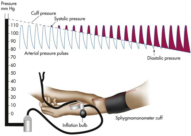
The systolic blood pressure usually decreases slightly with normal inhalation. However, a decrease in systolic pressure of more than 6 to 8 mm Hg during a resting inhalation is abnormal and is called paradoxical pulse, or pulsus paradoxus (see Chapter 9). Although simple palpation may be adequate to signal the presence of paradoxical pulse, it can be quantified only by auscultatory measurement. To obtain this measurement, the clinician inflates the cuff until the radial or brachial pulse can no longer be palpated. The clinician slowly deflates the cuff until sounds are heard on exhalation only (point 1). Next, the clinician reduces the cuff pressure until sounds are heard throughout respiration (point 2). The difference between points 1 and 2 indicates the degree of paradoxical pulse.

Most hospitals and clinics have adopted use of the digital blood pressure measuring devices. These devices do not require the health care provider to listen for the Korotkoff sounds and eliminate variances in recorded blood pressures based on human perception. They are considered to be very accurate and simply require the clinician to apply the blood pressure cuff correctly and press the start button. Subsequently, the device takes over and inflates and deflates the cuff automatically. The blood pressure and pulse rate are displayed on a digital screen.

Examination of the Head and Neck

Head

The patient’s face is inspected for abnormal signs that indicate respiratory problems. The most common facial signs are nasal flaring, cyanosis, and pursed-lip breathing. Nasal flaring occurs when the external nares flare outward during inhalation. This flaring is prevalent in neonates with respiratory distress and indicates an increased work of breathing.

When respiratory disease reduces arterial O2 content, cyanosis (a bluish discoloration of the tissues) may be detected, especially around the lips and in the oral mucosa of the mouth (central cyanosis). Cyanosis may be difficult to detect, especially in a poorly lit room or in people of color. Although central cyanosis suggests inadequate oxygenation (respiratory failure), further investigation is indicated. However, the absence of cyanosis does not ensure that oxygenation is adequate because a sufficient amount of desaturated hemoglobin (5 g) must exist before cyanosis can be identified.

Patients with COPD may use pursed-lip breathing during exhalation. Breathing through pursed lips during exhalation creates resistance to flow. The increased resistance creates a slight back pressure in the small airways during exhalation, which prevents their premature collapse and allows more complete emptying of the lung.

Neck

Inspection and palpation of the neck help determine the position of the trachea and the jugular venous pressure (JVP). Normally, when the patient faces forward, the trachea is located in the middle of the neck. The midline of the neck can be identified by palpating the suprasternal notch. The midline of the trachea should be directly below the center of the suprasternal notch.

The trachea can shift away from the midline in certain thoracic disorders. Generally, the trachea shifts toward an area of collapsed lung. Conversely, the trachea shifts away from areas with increased air or fluid (e.g., tension pneumothorax or large pleural effusion). Abnormalities in the lung bases generally do not shift the trachea.

JVP is estimated by determining how high the jugular vein extends above the level of the sternal angle. JVP reflects the volume and pressure of venous blood in the right side of the heart. Typically, the internal vein is assessed because it is more reliable. Individuals with obese necks may not have visible neck veins, even when the veins are distended.

When lying in a supine position, a healthy individual has neck veins that are full. When the head of the bed is elevated gradually to a 45-degree angle, the level of the blood column descends to a point no more than a few centimeters above the clavicle. With elevated venous pressure, the neck veins may be distended as high as the angle of the jaw, even when the patient is sitting upright.

JVP may vary with breathing. Under normal circumstances, the blood column descends toward the thorax during inhalation and ascends with exhalation. For this reason, JVP should always be estimated at the end of exhalation. Under abnormal conditions (e.g., cardiac tamponade), the JVP may increase during inhalation. This is called Kussmaul sign and is rare.

Jugular venous distention (JVD) is present when the jugular vein is enlarged and it can be seen more than 3 to 4 cm above the sternal angle. The most common cause of JVD is right heart failure (cor pulmonale). Right heart failure frequently occurs in patients with advanced COPD because of chronic hypoxemia. Hypoxemia causes chronic pulmonary vasoconstriction and hypertension, which leads to right heart failure from the excessive workload. Other conditions associated with JVD include left heart failure, cardiac tamponade, tension pneumothoraces, and mediastinal tumors.

The neck is a common place for the physician to palpate for enlarged lymph nodes, which is known as lymphadenopathy. Lymphadenopathy occurs with various medical disorders, including infection, malignancy, and sarcoidosis. Tender lymph nodes in the neck suggest a nearby infection. The lymph nodes are not tender when malignancy is the cause.

Examination of the Thorax and Lungs

Inspection

The chest should be inspected visually to assess the thoracic configuration and the pattern and effort of breathing. For adequate inspection, the room must be well lit, and the patient should be sitting upright. When the patient is too ill to sit up, the clinician should carefully roll the patient to one side to examine the posterior chest. Inspection, palpation, percussion, and auscultation of the patient’s chest require that the patient be disrobed. Consequently, the clinician should make every effort to respect the patient’s modesty (especially for female patients) and drape the chest when possible.

Thoracic Configuration

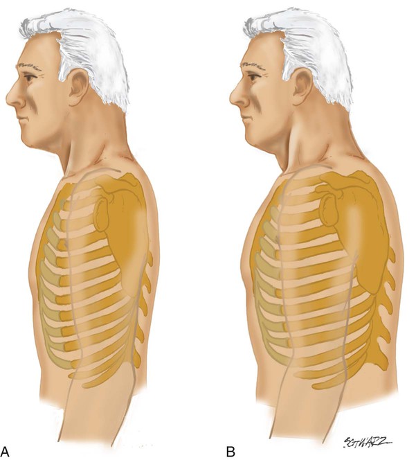
The anteroposterior (AP) diameter of the average adult thorax is less than the transverse diameter. Normally, the AP diameter increases gradually with age but may prematurely increase in patients with COPD. This abnormal increase in AP diameter is called barrel chest and is associated with emphysema. When the AP diameter increases, the normal 45-degree angle of articulation between the ribs and spine is increased, becoming more horizontal (Figure 15-3). Other abnormalities of the thoracic configuration are listed in Table 15-1.

TABLE 15-1

Abnormalities of Thoracic Configuration

Name Condition
Pectus carinatum Abnormal protrusion of sternum
Pectus excavatum Depression of part or entire sternum, which can produce a restrictive lung defect
Kyphosis Spinal deformity in which the spine has an abnormal AP curvature
Scoliosis Spinal deformity in which the spine has a lateral curvature
Kyphoscoliosis Combination of kyphosis and scoliosis, which may produce a severe restrictive lung defect as a result of poor lung expansion

Breathing Pattern and Effort

At rest, a healthy adult has a consistent rate and rhythm of breathing. Breathing effort is minimal on inhalation and passive on exhalation. Abnormal breathing patterns can be broken down into two broad categories. First are breathing patterns directly associated with thoracic or pulmonary diseases that increase work of breathing. Second are patterns primarily associated with neurologic disease (see Chapter 14). Table 15-2 describes common abnormal patterns of breathing.

TABLE 15-2

Abnormal Breathing Patterns

Breathing Pattern Characteristics Causes
Apnea No breathing Cardiac arrest, narcotic overdose, severe brain trauma
Apneustic breathing Deep, gasping inspiration with brief, partial expiration Damage to upper medulla or pons caused by stroke or trauma; sometimes observed with hypoglycemic coma or profound hypoxemia
Ataxic breathing Completely irregular breathing pattern with variable periods of apnea Damage to medulla
Asthmatic breathing Prolonged exhalation with recruitment of abdominal muscles Obstruction to airflow out of the lungs
Biot’s respiration Clustering of rapid, shallow breaths coupled with regular or irregular periods of apnea Damage to medulla or pons caused by stroke or trauma; severe intracranial hypertension
Cheyne-Stokes respiration Irregular type of breathing; breaths increase and decrease in depth and rate with periods of apnea; variant of “periodic breathing” Most often caused by severe damage to bilateral cerebral hemispheres and basal ganglia (usually infarction); also seen in patients with CHF owing to increased circulation time and in various forms of encephalopathy
Kussmaul breathing Deep and fast respirations Metabolic acidosis
Paradoxical breathing Abdominal paradox: Abdominal wall moves inward on inspiration and outward on expiration Abdominal paradox: Diaphragmatic fatigue or paralysis
  Chest paradox: Part or all of the chest wall moves in with inhalation and out with exhalation Chest paradox: Typically observed in chest trauma with multiple rib or sternal fractures
    Also found in patients with high spinal cord injury with paralysis of intercostal muscles
Periodic breathing Breathing oscillates between periods of rapid, deep breathing and slow, shallow breathing without periods of apnea Same causes as Cheyne-Stokes respiration

Cardiopulmonary or thoracic diseases that increase work of breathing typically cause recruitment of the accessory muscles of ventilation. Common causes of an increase in the work of breathing include narrowed airways (e.g., COPD, asthma), “stiff lungs” (e.g., severe pneumonia, pulmonary edema), or a stiff chest wall (e.g., ascites, anasarca, pleural effusions). Increased work of breathing also can result in retractions. Retractions are an intermittent sinking inward of the skin overlying the chest wall during inspiration. They occur when the ventilatory muscles contract forcefully enough to cause a large decrease in the intrathoracic pressure. Retractions may be seen between the ribs, above the clavicles, or below the rib cage. These are called intercostal, supraclavicular, or subcostal retractions. Retractions are difficult to see in obese patients.

Another form of retraction is tracheal tugging, which is caused by extreme negative pressure that pulls the trachea downward during inspiration. This phenomenon is noted by observing the downward movement of the thyroid cartilage toward the chest during inspiration. Typically, this movement occurs in concert with recruitment of the accessory muscles of inspiration, primarily the sternocleidomastoid muscles of the neck.

Generally, two archetypal abnormal breathing patterns exist that provide clues about the underlying pulmonary problem. These patterns fall into two categories: (1) patterns characterized by rapid, shallow breathing and (2) patterns marked by a relatively brief inspiratory phase coupled with an abnormally prolonged exhalation. Rapid, shallow breathing typically occurs in patients with increased lung stiffness, such as patients with pulmonary edema or severe pneumonia. Obstruction of the intrathoracic airways slows lung emptying and results in a prolonged expiratory phase as patients attempt to minimize gas trapping inside the lungs. This prolonged expiratory phase alters the normal ratio of inspiratory to expiratory time from 1 : 2 to 1 : 4 or greater; this always occurs with activation of the expiratory muscles. Obstruction of the extrathoracic upper airway (as with epiglottitis or croup) usually results in a prolonged inspiratory time because airways outside the thorax tend to narrow more on inhalation. Patients with diabetic ketoacidosis often breathe with a deep and rapid pattern that is called Kussmaul breathing.

The diaphragm may be nonfunctional in patients with spinal injuries or neuromuscular disease and may be severely limited in patients with COPD. When the diaphragm is nonfunctional or limited, the accessory muscles of ventilation become active to maintain adequate gas exchange. Heavy use of accessory muscles is reliable evidence of significant cardiopulmonary disease.

In patients with emphysema, the lungs lose their elastic recoil and become hyperinflated. Over time, the hyperinflation forces the diaphragm into a low, flat position. Contraction of a flat diaphragm tends to draw in the lateral costal margins instead of expanding them (Hoover sign) and does little to help move air into the thorax. Ventilation eventually must be achieved by other means and involves heavy use of the accessory muscles. The accessory muscles must assist ventilation by raising the anterior chest in an effort to increase thoracic volume. The severity of lung disease in this situation is often reflected by the magnitude of accessory muscle activity.

Diaphragmatic fatigue is found in many types of chronic and acute pulmonary diseases. When it occurs acutely, diaphragmatic fatigue often manifests with distinctive breathing patterns.9 The first sign of acute diaphragmatic fatigue is tachypnea. Sometimes tachypnea is followed by a breathing pattern in which the diaphragm and rib cage muscles alternately power breathing in an attempt to give each muscle group some rest (respiratory alternans). This pattern is noted by the upward motion of the diaphragm during inspiration on a series of breaths, followed by diaphragmatic contractions and inward movement of the abdominal wall on the following series of breaths. When the diaphragm is relaxed, contraction of the rib cage muscles sucks the diaphragm upward and the abdomen inward during inspiration. The opposite phenomenon occurs on breaths when the diaphragm is active. When the rib cage muscles are relaxed, the chest wall may appear to sink in as the abdomen protrudes during diaphragmatic contraction; this often gives the impression that the chest has a rocking motion. Finally, abdominal paradox occurs with complete diaphragmatic fatigue, as the diaphragm is drawn upward into the thoracic cavity with each inspiratory effort of the rib cage muscles. An abdominal paradox also occurs when the diaphragm is paralyzed.

These patterns are not always associated with impending muscle fatigue. Rather, they may be adaptations to high workloads when the respiratory muscle strength is normal.10 Also, patients with respiratory distress often have tachypnea, along with recruitment of the expiratory muscles. This situation can make it difficult to discern accurately the presence and type of abnormal breathing pattern. The RT must be careful about offering definitive therapeutic suggestions (e.g., absolute need for mechanical ventilation) when he or she perceives the presence of these abnormal breathing patterns.

Palpation

Palpation is the art of touching the chest wall to evaluate underlying structure and function. It is used in selected patients to confirm or rule out suspected problems suggested by the history and initial examination findings. Palpation is performed to evaluate vocal fremitus, estimate thoracic expansion, and assess the skin and subcutaneous tissues of the chest.

Vocal and Tactile Fremitus

The term vocal fremitus refers to the vibrations created by the vocal cords during speech. These vibrations are transmitted down the tracheobronchial tree and through the lung to the chest wall. When these vibrations are felt on the chest wall, it is called tactile fremitus. Assessing vocal fremitus requires a conscious, cooperative patient. Both vocal and tactile fremitus increase in intensity when the lung becomes consolidated (e.g., filled with inflammatory exudate) as in pneumonia. However, if the consolidated area is not in communication with an open airway, speech cannot be transmitted, and fremitus is absent or decreased. In addition, fremitus is reduced in patients who are obese or overly muscular.

Vocal and tactile fremitus decrease in intensity when either fluid or air collects in the pleural space (e.g., pleural effusion or pneumothoraces). Similarly, in patients with emphysema, the lungs become hyperinflated, which reduces the density of lung tissue. Because the density is low, speech vibrations transmit poorly through the lung, resulting in a bilateral reduction in fremitus.

To assess for tactile fremitus, the RT asks the patient to repeat the word “ninety-nine” while the RT systematically palpates the thorax. The palmar aspect of the fingers or the ulnar aspect of the hand can be used for palpation. If one hand is used, it should be moved from one side of the chest to the corresponding area on the other side. The anterior, lateral, and posterior portions of the chest wall are evaluated.

Thoracic Expansion

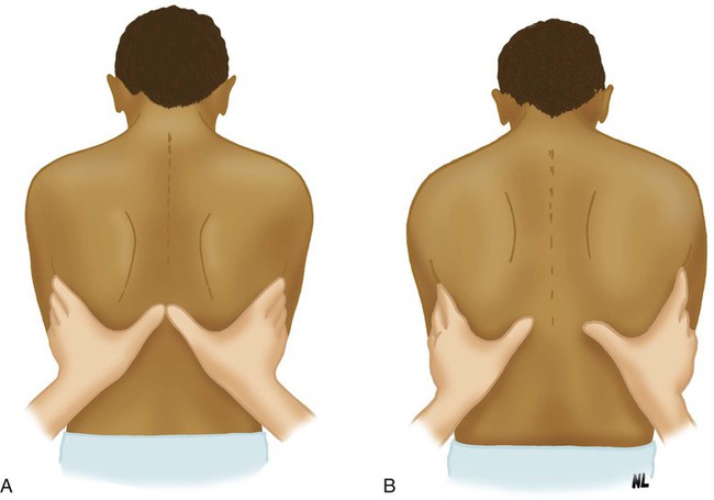
The normal chest wall expands symmetrically during deep inhalation. This expansion can be evaluated on the anterior and posterior chest. To evaluate expansion anteriorly, the RT places his or her hands over the anterolateral chest, with the thumbs extended along the costal margin toward the xiphoid process. To evaluate posteriorly, the RT positions the hands over the posterolateral chest with the thumbs meeting at the T8 vertebra (Figure 15-4). The patient is instructed to exhale slowly and completely. When the patient has exhaled maximally, the RT gently secures his or her fingertips against the sides of the patient’s chest and extends the thumbs toward the midline until the tip of each thumb meets at the midline. The RT next instructs the patient to take a full, deep breath and notes the distance the tip of each of the thumbs moves from midline. Normally, each thumb moves an equal distance of approximately 3 to 5 cm.

Diseases that affect the expansion of both lungs cause a bilateral reduction in chest expansion. Reduced expansion commonly is seen in neuromuscular disorders and COPD. Unilateral reduction in chest expansion occurs with respiratory diseases that reduce the expansion of one lung or a major part of one lung. This condition can occur with lobar consolidation, atelectasis, pleural effusion, or pneumothorax.

Percussion of the Chest

Percussion is the art of tapping on a surface to evaluate the underlying structure. Percussion of the chest wall produces a sound and a palpable vibration useful in evaluating underlying lung tissue. The vibration created by percussion penetrates the lung to a depth of 5 to 7 cm below the chest wall. This assessment technique is not performed routinely on all patients but is reserved for patients with suspected conditions for which percussion could be helpful (e.g., pneumothorax).

The technique most often used in percussing the chest wall is called mediate, or indirect, percussion. A right-handed RT places the middle finger of the left hand firmly against the patient’s chest wall, parallel to the ribs, with the palm and other fingers held off the chest. The RT uses the tip of the middle finger of the right hand or the lateral aspect of the right thumb to strike the finger against the chest near the base of the terminal phalanx with a quick, sharp blow. Movement of the hand striking the chest is generated at the wrist, not at the elbow or shoulder.

The percussion note is clearest if the RT remembers to keep the finger on the patient’s chest firmly against the chest wall and to strike this finger and then immediately withdraw. The two fingers should be in contact for only an instant. As one gains experience in percussion, the feel of the vibration becomes as important as the sound in evaluating lung structures.

Percussion Over Lung Fields

Percussion of the lung fields is performed systematically, consecutively testing comparable areas on both sides of the chest. Percussion over the bony structures and over the breasts of female patients has no diagnostic value and should not be performed. Asking patients to raise their arms above their shoulders helps move the scapulae laterally and minimize their interference with percussion on the posterior chest wall.

The sounds generated during percussion of the chest are evaluated for intensity (loudness). Percussion over normal lung fields produces a moderately low-pitched sound that can be heard easily. This sound is described as normal resonance or tympanic. When the percussion note is louder and lower than normal, the sound is said to be increased resonance or hypertympanic. Percussion may produce a sound with characteristics just the opposite of resonance, referred to as decreased resonance, dampened, or dull.

Clinical Implications

By itself, percussion of the chest is of little value in making a diagnosis. However, when considered along with other findings, percussion can provide essential information. In modern practice, chest percussion enables rapid bedside assessment of abnormalities inside the chest and may aid in the decision to obtain chest radiographic studies.

Any abnormality that increases lung tissue density, such as pneumonia, tumor, or atelectasis, results in a loss of resonance and decreased resonance to percussion over the affected area. Pleural spaces filled with fluid, such as blood or water, also produce decreased resonance to percussion. Increased resonance can be detected either when the lungs are hyperinflated (e.g., asthma or emphysema) or when the pleural space contains large amounts of air (pneumothorax).

Unilateral problems are easier to detect than bilateral problems because the normal side provides a normal standard for immediate comparison. The unilateral decrease in resonance heard when percussing an area of consolidation is easier to detect than the subtle bilateral increase in resonance heard with bilateral hyperinflation.

Percussion of the chest has clinically important limitations. Abnormalities that are small or deep below the surface are not likely to be detected during percussion of the chest. Many clinicians do not routinely use chest percussion to evaluate lung resonance.

Auscultation of the Lungs

Auscultation is the process of listening for bodily sounds. Auscultation over the thorax is performed to identify normal and abnormal lung sounds and to evaluate the effects of therapy. Because auscultation can be performed quickly and is noninvasive, it is a particularly useful tool in many clinical situations. Auscultation is performed with a stethoscope to enhance sound transmission from the patient’s lungs to the examiner’s ears. The clinician always must ensure that the room is as quiet as possible whenever performing auscultation.

Stethoscope

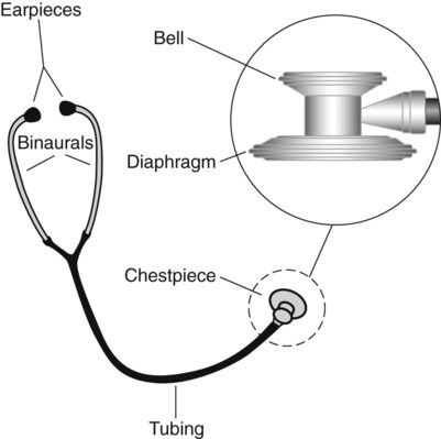
A stethoscope has the following four basic parts: (1) a bell, (2) a diaphragm, (3) tubing, and (4) earpieces (Figure 15-5). The bell detects a broad spectrum of sounds and is very useful for listening to low-pitched sounds (e.g., heart sounds). Proper technique for listening to heart sounds is to place the bell lightly against the chest; this avoids stretching the skin, which makes auscultating heart sounds more difficult by inadvertently filtering out low-frequency sounds. Using the bell to auscultate the lungs is also helpful when emaciation causes rib protrusion that restricts placement of the diaphragm flat against the chest.

image
FIGURE 15-5 Acoustic stethoscope.

The diaphragm is preferred for auscultation of the lungs because most lung sounds are high frequency. The ideal tubing should be thick enough to exclude external noises and approximately 25 to 35 cm (11 to 16 inches) in length. Longer tubing may impair sound transmission.

The clinician should examine his or her stethoscope regularly for cracks in the diaphragm, wax or dirt in the earpieces, and other defects that may interfere with the transmission of sound. The stethoscope should always be cleaned with hospital-approved disinfectant after every patient contact to minimize contamination with microorganisms.11,12 Patients who are placed in contact isolation and patients who are in protective isolation because of immunosuppression should have a dedicated stethoscope in the room to prevent cross infection.

Technique

When possible, the patient should be sitting upright in a relaxed position. The patient should be instructed to breathe a little more deeply than normal through an open mouth. Exhalation should be passive. The bell or diaphragm is placed directly against the chest wall when possible because clothing may produce distortion. The tubing must not be allowed to rub against any objects because this may produce extraneous sounds, which could be mistaken for adventitious lung sounds (discussed later).

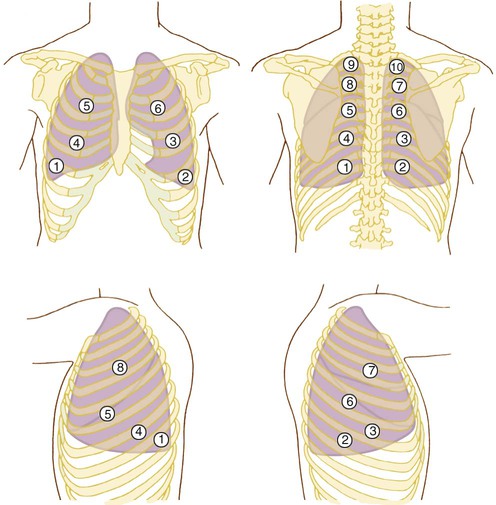
Auscultation of the lungs should be systematic and include all lobes on the anterior, lateral, and posterior chest. Auscultation should begin at the lung bases with comparison of breath sounds side to side, working upward toward the lung apexes (Figure 15-6). It is important to begin at the bases because certain abnormal sounds that occur only in the lower lobes may be altered by several deep breaths. At least one full ventilatory cycle should be evaluated at each stethoscope position. If abnormal sounds are present, the clinician should listen to several breaths to clarify the characteristics.

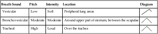
The clinician should listen for and distinguish among the key features of breath sounds. The clinician should identify the pitch (vibration frequency), the intensity (loudness), and the duration of the inspiratory and expiration components of the sound. The acoustic characteristics of breath sounds can be illustrated in breath sound diagrams (Figure 15-7). The features of normal breath sounds are described in Table 15-3. One must be familiar with normal breath sounds before one can expect to identify the subtle changes that may signify respiratory disease.

TABLE 15-3

Characteristics of Normal Breath Sounds

Breath Sound Pitch Intensity Location Diagram
Vesicular Low Soft Peripheral lung areas image
Bronchovesicular Moderate Moderate Around upper part of sternum, between the scapulae image
Tracheal High Loud Over the trachea image

image

Terminology

In healthy individuals, the sounds heard over the trachea have a loud, tubular quality. These are referred to as tracheal breath sounds. Tracheal breath sounds are loud sounds with an expiratory component equal to or slightly longer than the inspiratory component.

A variation of the tracheal breath sounds can be heard around the upper half of the sternum on the anterior chest and between the scapulae on the posterior chest. These sounds are not as loud as tracheal breath sounds, are slightly lower in pitch, and have equal inspiratory and expiratory components. They are referred to as bronchovesicular breath sounds.

When auscultating over the lung parenchyma of a healthy individual, soft, muffled sounds are heard. These normal breath sounds, referred to as vesicular breath sounds, are lower in pitch and intensity than bronchovesicular breath sounds. Vesicular sounds are heard primarily during inhalation, with an exhalation component approximately one-third the duration of inhalation (see Table 15-3).

Respiratory disease may alter the intensity of normal breath sounds heard over the lung fields. Breath sounds are described as diminished when the intensity decreases and as absent in extreme cases. They are described as harsh when the intensity increases. When the expiratory component of harsh breath sounds equals the inspiratory component, they are described as bronchial breath sounds.

Adventitious lung sounds are added sounds or vibrations produced by the movement of air through abnormal airways. Adventitious lung sounds are classified as either discontinuous or continuous. Discontinuous adventitious lung sounds are intermittent, crackling, or bubbling sounds of short duration. Discontinuous adventitious lung sounds are referred to as crackles, whereas continuous adventitious lung sounds are described with the term wheezes; a wheeze is a quasimusical sound. The term rhonchi is encountered frequently. Rhonchi is derived from the Latin word meaning “wheezing.” This term has had a confusing history in clinical practice, and its use is not recommended.

Another continuous type of adventitious lung sound, heard primarily over the larynx and trachea during inhalation, is stridor. Stridor is a loud, high-pitched sound and sometimes can be heard without a stethoscope. Most common in infants and small children, stridor is a sign of obstruction in the trachea or larynx. Stridor is most often heard during inspiration.

When abnormal lung sounds are heard, their location and specific features should be documented. Abnormal lung sounds may be high-pitched or low-pitched, loud or faint, scant or profuse, and inspiratory or expiratory (or both). Faint or low-intensity crackles are often referred to as fine crackles; more pronounced or more intense crackles are referred to as coarse crackles.

Mechanisms and Significance of Lung Sounds

The exact mechanisms that produce normal and abnormal lung sounds are not fully known. However, there is sufficient agreement among investigators to allow a general description.

Normal Breath Sounds

Lung sounds heard over the chest of a healthy individual are generated primarily by turbulent airflow in the larger airways. Turbulent airflow creates audible vibrations in the airways, producing sounds that are transmitted through the lungs and chest wall. As this sound travels to the lung periphery and the chest wall, it is altered by the filtering properties of normal lung tissue. Normal lung tissue acts as a low-pass filter, which means it preferentially passes low-frequency sounds. If you place the diaphragm portion of your stethoscope over the chest wall of a friend and listen while he or she speaks, this filtering (attenuation) effect will be evident. The voice sounds are muffled and difficult to understand because of attenuation. This filtering effect explains the characteristic differences between tracheal breath sounds, heard directly over the trachea, and vesicular sounds, heard over the lung periphery. Normal vesicular lung sounds essentially are attenuated tracheal breath sounds.

Wheezes and Stridor

Wheezes and stridor represent vibrations of airway wall that are caused when air flows at high velocity through a narrowed airway. Airway diameter can be reduced by bronchospasm, mucosal edema, inflammation, tumors, foreign bodies, and pulmonary edema. This narrowing initially causes an increase in the velocity of airflow, which causes the lateral wall pressure to decrease. This decrease in pressure causes the lateral walls of the narrowed airway to pull closer together, and airflow stops. When airflow stops, the lateral wall pressure increases, and the airway opens back to the previous position. This cycle repeats many times per second and causes the airway walls to vibrate and make a musical type of adventitious lung sound similar to a reed instrument.

It is useful to monitor the pitch and duration of wheezing. Improved expiratory flow is associated with a decrease in the pitch and length of the wheezing. If high-pitched wheezing is present during the entire expiratory time before treatment but becomes lower pitched and occurs only late in exhalation after therapy, the pitch and duration of the wheeze have diminished. This change suggests that the degree of airway obstruction has decreased.

Wheezing may be monophonic (single note) or polyphonic (multiple notes). A monophonic wheeze indicates that a single airway is partially obstructed. Monophonic wheezing may be heard during inhalation and exhalation or during exhalation only. Polyphonic wheezing suggests that many airways are obstructed, such as with asthma, and is heard only during exhalation. Bronchitis and CHF with pulmonary edema can also cause polyphonic wheezing.

Stridor is a serious adventitious lung sound that indicates that the upper airway is compromised. It may occur in patients of any age but most often is heard from the neck of children. In children, laryngomalacia is the most common cause of chronic stridor, whereas croup is the most common cause of acute stridor. Generally, inspiratory stridor is consistent with narrowing above the glottis, whereas expiratory stridor indicates narrowing of the lower trachea.

Crackles

Crackles occur when airflow moves secretions or fluid in the airways. In this situation, coarse crackles are usually heard during inspiration and expiration. These crackles often clear when the patient coughs or when the upper airway is suctioned. Crackles also may be heard in patients without excess secretions. These crackles occur when collapsed airways pop open during inspiration. Airway collapse or closure can occur in peripheral bronchioles or in larger, more proximal bronchi.

Larger, more proximal bronchi may close during expiration when there is an abnormal increase in bronchial compliance or when the retractile pressures around the bronchi are low. In this situation, crackles usually occur early in the inspiratory phase and are referred to as early inspiratory crackles (Figure 15-8). Early inspiratory crackles are usually scant but may be loud or faint. They often are transmitted to the mouth and are not silenced by a cough or a change in position. They frequently occur in patients with COPD (chronic bronchitis, emphysema, or asthma) and usually indicate severe airway obstruction.

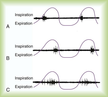
Peripheral airways may close during exhalation when the surrounding intrathoracic pressure increases and when surfactant levels are diminished. Fine, late inspiratory crackles are produced by the sudden opening of peripheral airways, usually late in the inspiratory phase. They are more common in the dependent lung regions, where the peripheral airways are most prone to collapse during exhalation. They may clear with changes in posture or if the patient performs several deep inspirations. Late inspiratory crackles are most common in patients with respiratory disorders that reduce gas volume of the lung, such as atelectasis, pneumonia, pulmonary edema, and pulmonary fibrosis (Table 15-4).

TABLE 15-4

Application of Adventitious Lung Sounds
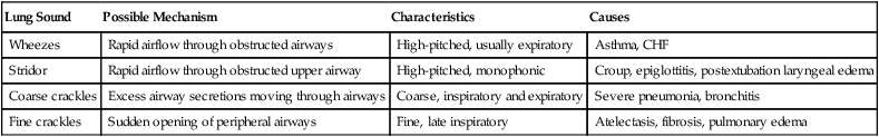
Lung Sound Possible Mechanism Characteristics Causes
Wheezes Rapid airflow through obstructed airways High-pitched, usually expiratory Asthma, CHF
Stridor Rapid airflow through obstructed upper airway High-pitched, monophonic Croup, epiglottitis, postextubation laryngeal edema
Coarse crackles Excess airway secretions moving through airways Coarse, inspiratory and expiratory Severe pneumonia, bronchitis
Fine crackles Sudden opening of peripheral airways Fine, late inspiratory Atelectasis, fibrosis, pulmonary edema

image

Voice Sounds

If chest inspection, palpation, percussion, or auscultation suggests a lung abnormality, evaluation of vocal resonance may be useful in further assessment. Vocal resonance is produced by the same mechanism as vocal fremitus. Vibrations created by the vocal cords during speech travel down the airways and through the peripheral lung units to the chest wall. The patient is instructed to repeat the words “one,” “two,” “three,” or “ninety-nine” while the clinician listens over the chest wall with a stethoscope, comparing one side with the other side. Normal, air-filled lung tissue filters the voice sounds, producing a significant reduction in intensity and clarity. Pathologic abnormalities in lung tissue alter the transmission of voice sounds, causing either increased or decreased vocal resonance. Increased vocal resonance occurs with lung consolidation, whereas decreased vocal resonance occurs with hyperinflated lung or with pneumothorax.

Cardiac Examination

Because of the close relationship between the heart and lungs, chronic lung diseases often cause cardiac problems. The techniques for physical examination of the chest wall overlying the heart (precordium) include inspection, palpation, and auscultation. Most clinicians examine the precordium at the same time they assess the lungs.

Inspection and Palpation

Inspection and palpation of the precordium help identify normal or abnormal pulsations. Pulsations on the precordium are created by ventricular contraction. Detection of pulsations depends on the force of ventricular contraction, the thickness of the chest wall, and the quality of the tissue through which the vibrations travel.

Normally, left ventricular contraction is the most forceful and generates a visible, palpable pulsation during systole. This pulsation is called the point of maximal impulse (PMI). In healthy individuals who are not obese (or overly muscular), the PMI can be felt and visualized near the left midclavicular line in the fifth intercostal space. The PMI shifts laterally with left ventricular hypertrophy.

Right ventricular hypertrophy (a common finding in COPD) often produces a systolic thrust called a heave that is felt (and possibly visualized) near the lower left sternal border. To identify the PMI, the clinician places the palmar aspect of the right hand over the lower left sternal border. Right ventricular hypertrophy may be the result of chronic hypoxemia, pulmonary valve disease, or primary pulmonary hypertension.

In patients with chronic pulmonary hyperinflation (emphysema), the PMI may be difficult to locate. Because of the increase in AP diameter and the changes in lung tissue, systolic vibrations are not well transmitted to the chest wall.

The PMI may shift either left or right, following deviations in the position of the lower mediastinum, which may be caused by pneumothorax or lobar collapse. Typically, the PMI shifts toward lobar collapse but away from a tension pneumothorax. The PMI in patients with emphysema and low flat diaphragms may be shifted centrally to the epigastric area.

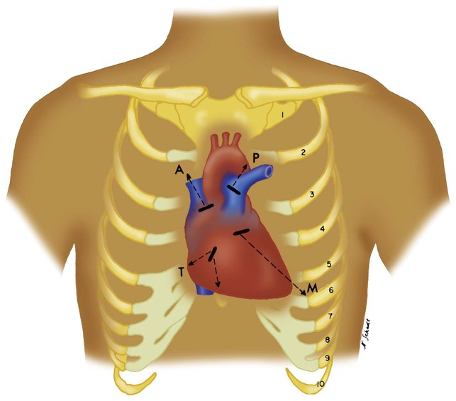
The second left intercostal space near the sternal border is referred to as the pulmonic area and is palpated to identify accentuated pulmonary valve closure. Strong vibrations may be felt in this area with the presence of pulmonary hypertension or valvular abnormalities (Figure 15-9). Rapid blood flow through a narrowed valve or backflow through an incompetent valve may produce palpable vibrations known as thrills. Thrills are usually accompanied by a murmur (see discussion later).

Auscultation of Heart Sounds

Normal heart sounds are created by closure of the heart valves (see Chapter 9). The first heart sound (S1) is produced by closure of the mitral and tricuspid (atrioventricular [AV]) valves during contraction of the ventricles. When systole ends, the ventricles relax, and the pulmonic and aortic (semilunar) valves close, creating the second heart sound (S2). Because pressures in the left side of the heart are higher, mitral valve closure is louder and contributes more to S1. For the same reason, closure of the aortic valve usually is more significant in producing S2. If either the AV valves or the semilunar valves do not close together, a split heart sound is heard. A slight splitting of S2 is normal; it occurs because of increased venous return during spontaneous breathing.

A third heart sound (S3) can be heard during diastole and is produced by rapid ventricular filling immediately after systole. S3 is a low-intensity, low-pitched sound best heard over the apex of the heart. In young, healthy children, S3 is considered normal and is called physiologic S3. Otherwise, S3 is abnormal. In older patients with heart disease, S3 may signify CHF.

A fourth heart sound (S4) is produced by mechanisms similar to the mechanisms that produce S3. S4 may occur in healthy individuals or may be considered a sign of heart disease. S4 differs from S3 only in its timing during the cardiac cycle. S4 occurs later, just before S1, whereas S3 occurs just after S2. A patient with heart disease who has S3 and S4 is said to have a gallop rhythm.

Abnormal Heart Sounds

Reduced intensity of heart sounds may result from cardiac or extracardiac abnormalities. Extracardiac factors include alteration in the tissue between the heart and the surface of the chest. Pulmonary hyperinflation, pleural effusion, pneumothorax, and obesity make identification of both S1 and S2 difficult. The intensity of S1 and S2 also decreases when the force of ventricular contraction is poor, as in heart failure, or when valvular abnormalities exist.

Pulmonary hypertension may cause two abnormalities in heart sounds. First, it increases the intensity of S2 from a more forceful closure of the pulmonic valve (this is also referred to as a loud P2). Second, S2 splitting may be absent. An increased P2 is identified best over the pulmonic area of the chest (see Figure 15-9).

Cardiac murmurs occur whenever the heart valves are incompetent or stenotic. Murmurs are classified as either systolic or diastolic. Systolic murmurs are produced by an incompetent AV valve or a stenotic semilunar valve. An incompetent AV valve typically produces a high-pitched “whooshing” noise simultaneously with S1. This noise is caused by a backflow of blood through the AV valve into the atrium. In contrast, a stenotic semilunar valve produces a crescendo-decrescendo sound created by an obstruction of blood flow out of the ventricle during systole.

Diastolic murmurs are created by either an incompetent semilunar valve or a stenotic AV valve. An incompetent semilunar valve allows a backflow of blood into the ventricle simultaneously with, or immediately after, S2. A stenotic AV valve obstructs blood flow from the atrium into the ventricles during diastole and creates a turbulent murmur.

A murmur also may be created by rapid blood flow across normal valves, such as occurs with heavy exertion. Murmurs are created by the following: (1) a backflow of blood through an incompetent valve, (2) a forward flow of blood through a stenotic valve, and (3) a rapid flow of blood through a normal valve.

Heart sounds can be auscultated by using the bell or diaphragm pieces of the stethoscope. The heart sounds may be easier to identify when the patient leans forward or lies on the left side. This positioning moves the heart closer to the chest wall. When peripheral pulses are difficult to identify, auscultation over the precordium may provide important additional information. Normally, the rate heard over the precordium (the apical rate) should be the same as the palpated peripheral pulse. In patients with atrial fibrillation, the apical rate often is higher than the peripheral pulse (pulse deficit). During atrial fibrillation, the irregular rhythm causes frequent weak ventricular contractions that cannot be detected at peripheral locations.

Abdominal Examination

The abdomen should be inspected and palpated for evidence of distention and tenderness. Abdominal distention and pain impair diaphragmatic movement and may contribute to or cause respiratory insufficiency. Abdominal dysfunction may inhibit deep breathing and coughing and promote atelectasis. Of particular concern is the presence of intraabdominal hypertension, which is defined as intraabdominal pressure greater than 12 mm Hg and is found in 18% of critically ill patients.13 Abdominal compartment syndrome occurs when intraabdominal pressures are greater than 20 mm Hg and often requires emergency decompressive surgery. This syndrome causes profound atelectasis and hypoxemia, hypotension, and renal failure.

Intraabdominal hypertension is a common finding in patients with blunt or penetrating abdominal trauma, ruptured aortic aneurysm, bowel infarction, and end-stage liver failure. It is suspected when gross examination of the abdomen reveals very pronounced abdominal distention. Intraabdominal pressure is measured by connecting an intraarterial pressure catheter to the culture port of a Foley urine catheter.

The presence of an enlarged liver (hepatomegaly) is a frequent cause of right lower lobe atelectasis and pleural effusion. Hepatomegaly is a common finding in patients with liver disease and patients with cor pulmonale.

Examination of the Extremities
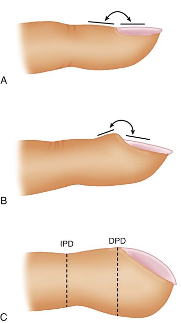
Respiratory disease may cause several abnormalities of the extremities, including digital clubbing, cyanosis, and pedal edema.

Clubbing

Clubbing of the digits is a significant manifestation of cardiopulmonary disease. Clubbing is a painless enlargement of the terminal phalanges of the fingers and toes that develops over time. As the process advances, the angle of the fingernail to the nail base increases, and the base of the nail feels “spongy.” The profile view of the digits allows easier recognition of clubbing (Figure 15-10), but sponginess of the nail bed is the most important sign. Causes of clubbing include infiltrative or interstitial lung disease, bronchiectasis, various cancers (particularly lung cancer),14 congenital heart problems that cause cyanosis, chronic liver disease, and inflammatory bowel disease. COPD alone, even when hypoxemia is present, does not lead to clubbing. Clubbing of the digits in a patient with COPD indicates that something other than obstructive lung disease is occurring.

Cyanosis

Examination of the digits for cyanosis is part of the initial assessment and is done whenever hypoxemia is suspected. Cyanosis can be detected easily because of the transparency of the fingernails and skin.

Cyanosis becomes visible when the amount of unsaturated hemoglobin in the capillary blood exceeds 5 to 6 g/dl; this may be caused by a reduction in arterial or venous O2 content, or both. Cyanosis of the digits is referred to as peripheral cyanosis or acrocyanosis and may involve extensive portions of limbs. This condition is mainly the result of poor perfusion, especially in the extremities. When capillary blood flow is poor, the tissues extract more O2, reducing the venous O2 content and increasing the amount of reduced hemoglobin. The extremities are usually cool to the touch when peripheral cyanosis is a sign of poor perfusion.

Peripheral Skin Temperature

When systemic perfusion is poor (as in heart failure or shock), compensatory vasoconstriction in the extremities helps shunt blood to the vital organs. This reduction in peripheral perfusion causes the extremities to become cool to the touch. The extent to which the coolness to touch extends back toward the torso is an indication of the degree of circulatory failure. In contrast, patients with high cardiac output and peripheral vascular failure (as occurs in septic shock) may have warm, dry skin.

Summary Checklist

• The interview is used to obtain important diagnostic information and to build a rapport between the health care provider and the patient.

• Dyspnea is the sensation that occurs when breathing effort is excessive relative to the tidal volume achieved. The work of breathing increases with reduced lung compliance and narrowed airways. Breathlessness is the unpleasant urge to breathe and is the sensation associated with a heightened drive to breathe.

• Cough is one of the most common symptoms of lung disease and occurs when the cough receptors that line the larger airways are stimulated by foreign material, mucus, noxious gases, or inflammation.

• The most common cause of hemoptysis (spitting up blood from the lung) is infection.

• Vital signs provide reliable assessment information about the general condition of the patient and the patient’s response to therapy.

• Rapid, shallow breathing indicates pathologic changes in the lung consistent with a reduction in the gas volume of the lungs.

• A prolonged expiratory phase suggests that the intrathoracic airways are narrowed.

• Normal breath sounds are generated by turbulent airflow in the larger airways.

• Crackles are generated by the sudden opening of closed airways or by the movement of excessive airway secretions with breathing.

• Wheezes are produced by the rapid vibration of narrow airways as gas passes through at high velocity.

• Cor pulmonale causes jugular venous distention, hepatomegaly, a loud P2, and pedal edema.

• Central cyanosis is a sign of hypoxemia caused by respiratory failure, whereas peripheral cyanosis indicates circulatory failure.