[level-membership-for-dermatology-category]
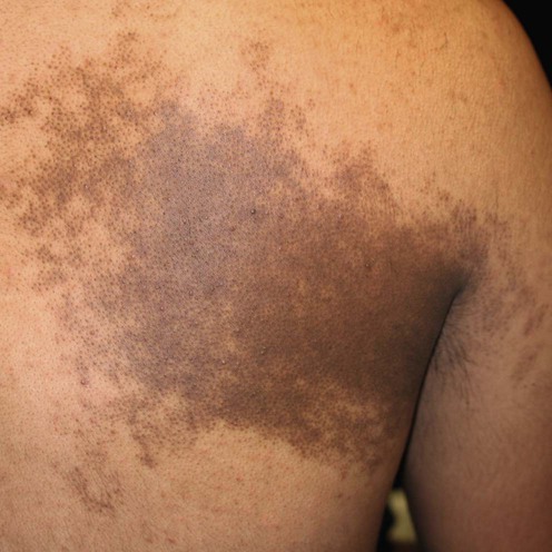
Becker’s nevus
Michael P. Loosemore, Adisbeth Morales-Burgos, Elnaz F. Firoz, Bahar F. Firoz and Leonard H. Goldberg


First-line therapies


Reduction of hyperpigmentation
 Erbium:YAG laser
 Long-pulse alexandrite laser
 Erbium-doped fiber laser (Fraxel)
 Q-switched ruby laser (QSRL)
 Normal mode ruby laser
 Frequency-doubled QSNd:YAG
 Electrolysis
Third-line therapies
 Spironolactone
 Corrective camouflage
[/level-membership-for-dermatology-category][not-level-membership-for-dermatology-category]
Becker’s nevus
Michael P. Loosemore, Adisbeth Morales-Burgos, Elnaz F. Firoz, Bahar F. Firoz and Leonard H. Goldberg
