Basic Principles of Cranial Surgery for Brain Tumors

Published on 26/03/2015 by admin

Filed under Neurosurgery

Last modified 22/04/2025

Print this page

rate 1 star rate 2 star rate 3 star rate 4 star rate 5 star
Your rating: none, Average: 0 (0 votes)

This article have been viewed 1691 times

CHAPTER 115 Basic Principles of Cranial Surgery for Brain Tumors

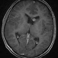
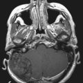
Preoperative Imaging Interpretation

Interpretation of the preoperative imaging studies is important in terms of planning the surgery, setting surgical goals, and educating the patient. Most patients have already had gadolinium-enhanced magnetic resonance imaging (MRI) scans when they are seen by the neurosurgeon. When evaluating the MRI scan, answers to the following questions are invaluable:

Answering these six questions allows the surgeon to identify the most likely pathology, assess the urgency of the problem, plan the surgical approach, and schedule any additional preoperative imaging studies that are needed.

While studying the MRI findings and answering the six questions, surgeons should begin to organize their thoughts about the biology of the tumor. For example, is this a slow-growing tumor, such as an intrinsic low-grade astrocytoma, or a more aggressive intrinsic brain tumor? The presence of brain edema, mass effect, and contrast enhancement argues against a slow-growing process and favors a high-grade, fast-growing tumor. Surgeons must be aware, however, that there are always exceptions to such rules. The surgical issues and approach will differ, depending on the probable diagnosis, and surgeons must constantly test their diagnostic hypothesis by looking for clues that suggest a different diagnosis. For example, meningiomas are enhancing lesions that can reach a large size and have a significant mass effect, but the surgical goals differ from those in patients with high-grade gliomas. Although the extradural location leads the surgeon to the diagnosis of meningioma, the pace and severity of symptoms provide important clues about the biology of the tumor. Slow-growing tumors can reach very a large size and cause impressive shifts in normal brain structures without causing many symptoms. Growth of the tumor over a long period allows the brain to accommodate and adjust and maintain its function at a normal or nearly normal level. Symptoms that arise over a short period and increase in severity suggest a more ominous pathology and a fast-growing lesion. Correlating the pace and severity of symptoms with the radiologic findings is critical to formulating a surgical treatment plan and its timing.

Timing of Surgery

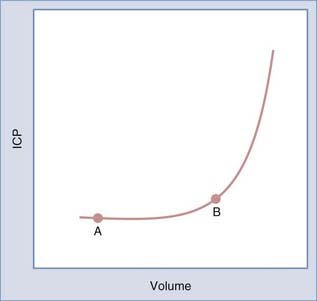
The timing of surgery is dictated by the type of symptoms and their pace. Progressive symptoms over a short period indicate that the tumor and edema are expanding faster than the brain is able to compensate. Recognition of this situation is critical because such patients are at risk for rapid deterioration. The Monro-Kellie principle, introduced to neurosurgery by Cushing, states that changes in brain volume cause reciprocal changes in the blood and cerebrospinal fluid (CSF) compartments within the rigid cranial sphere to maintain intracranial pressure (ICP) within a normal range.1 As brain volume increases secondary to a mass such as a tumor, a compensatory decrease in CSF or blood volume occurs to maintain ICP within the normal range. As the tumor expands, ICP increases slowly until there is no further displaceable volume (i.e., CSF or blood). At this point, further increases in brain volume result in an accelerated increase in ICP. The relationship between volume and pressure is depicted by the volume-pressure curve in Figure 115-1. As a tumor grows and expands, a patient moves along the curve from point A to point B. During this time, compensatory changes occur, including displacement of CSF and blood, as well as compression of normal brain tissue, to maintain ICP in the normal range. At point B, the ability of the brain to further compensate is limited. Thus, a small additional increase in volume results in a larger increase in pressure. It is important to recognize patients who are approaching point B on the volume-pressure curve because they are at risk for a rapid increase in ICP with small additional increases in volume. This rapid increase in ICP can lead to rapid neurological decline.

It is unusual for a patient with a brain tumor to need emergency surgery. The two exceptions are patients with markedly increased ICP secondary to the tumor mass and brain edema and those with acute or subacute obstructive hydrocephalus. In both circumstances, ICP is elevated. Patients with increased ICP can have marked alterations in mental status. Patients who prefer to keep their eyes closed or sleep are nearing a critical point on the volume-pressure curve. Surgeons should be wary of a patient who can perform a neurological examination adequately when stimulated but who falls back to sleep when unstimulated. A patient in this condition may need emergency surgery, especially if the source of the increased ICP is the tumor mass itself rather than brain edema. Patients with obstructive hydrocephalus need either a temporary ventriculostomy before a planned resection or a ventriculoperitoneal shunt. A third option is to proceed directly to tumor resection, with the goal of relieving the hydrocephalus. Patients with marked brain edema require high doses of steroids, which usually results in neurological improvement.

Surgical Planning: Imaging Studies

After deciding that the patient needs surgery, the surgeon must determine whether additional imaging studies are needed to better define the relationship between the tumor and the patient’s normal anatomy. The studies that might be obtained are (1) angiography or magnetic resonance angiography or venography to assess the relationship of the tumor to the blood vessels, (2) MRI for use with a frameless stereotactic system, (3) functional MRI to assess the tumor’s relationship to areas controlling eloquent brain functions, and (4) diffusion tensor imaging to assess the relationship of critical cortical pathways to the tumor.image

The use of angiography for the diagnosis and evaluation of brain tumors has diminished significantly with the advent of MRI. The location of large vessels within the subarachnoid space is well seen on T2-weighted and fluid-attenuated inversion recovery (FLAIR) MRI scans. On both sequences the vessels are black. The displacement and orientation of these vessels with respect to the tumor can usually be well seen on MRI. An important use for angiography or magnetic resonance angiography or venography is evaluation of the venous sinus system in patients with extra-axial dural tumors, particularly meningiomas. These tumors can often partially or completely occlude the large dural sinuses. This information can be important in planning the surgical exposure and resection. Frequently, magnetic resonance venography provides the information, thus making angiography unnecessary. If the dural sinus has been occluded by the tumor, magnetic resonance venography will show the cortical draining veins entering the sinus in front of or behind the tumor. Those entering anterior to the tumor drain anteriorly, and those posterior drain posteriorly. If the image on magnetic resonance venography is unclear and the surgical plan includes removal of a segment of a dural sinus, angiography should be performed. Finally, preoperative angiographic embolization can be beneficial with certain dural tumors. If the tumor has a significant dural blood supply, embolization can decrease the vascularity of the tumor and make surgery safer. Preoperative embolization represents another indication for preoperative angiography.

Frameless stereotaxy has become an important part of intracranial tumor surgery. The intraoperative correlation of anatomy with the preoperative MRI is helpful when planning the skin incision, bone flap, cortical incision, and approach to the tumor, as well as to maximize resection of the tumor. Typically, routine enhanced MRI is the imaging modality used interactively in the operating room. However, additional imaging modalities can now be placed onto the navigation system and used to increase the likelihood of a successful operation (i.e., maximal tumor resection with no neurological sequelae). Such modalities include functional MRI, diffusion tensor imaging with fiber tract images, positron emission tomography, and ultrasonography. It is critical to understand the limitation of this technology in terms of the brain shifts that occur during surgery, as well as the accuracy and reliability of using the different imaging modalities. Although most useful with intra-axial tumors, we have found it valuable for extra-axial tumors also, particularly meningiomas. image

For intra-axial tumors, frameless stereotaxy provides feedback to the surgeon about the degree of tumor resection. The extent of tumor resection as judged by postoperative imaging studies is greater when frameless stereotaxy is used. An invaluable use of frameless stereotaxy is for resection of low-grade astrocytomas. These tumors typically do not enhance and can be very difficult to grossly distinguish from normal brain. Furthermore, the extent of the tumor is often well defined by the high signal seen on T2-weighted MRI. These T2-weighted images can be loaded onto the frameless stereotactic system to allow the surgeon to map out the extent of the tumor on the cortex. The depth of the tumor can be defined by using the navigation probe as a biopsy needle and passing it through the middle of the tumor until the tip is at the deepest edge of the tumor. By leaving the tip at this point, the surgeon resects tissue around the probe until the tip is reached. With this technique, the deep edge is reached before any significant brain shift, and the subsequent resection is carried to that depth. By defining the extent of resection before removing any tissue, the probability of gross total resection of the tumor, as defined by the change in signal on T2-weighted images, is much greater. As tumor and CSF are removed during the resection, the brain drops away from the skull, and this must be taken into account when interpreting the data provided by the stereotactic system. The surgeon must use other sources of information in these circumstances, including (1) the appearance of the cavity wall in comparison to the tumor’s gross appearance, (2) the relationship of the cavity margins to surrounding normal sulci, and (3) how far the cortical surface has dropped away from the inner table.

The natural evolution from frameless stereotaxy is intraoperative MRI. Intraoperative MRI allows the surgeon to use an updated MRI scan obtained during surgery to determine whether additional resection is necessary. The usefulness of this tool is limited by the quality of the images, which is dependent on the strength of the magnet. The added benefit in terms of improved tumor resection and better outcomes remains to be determined.

Finally, an emerging imaging technology is localization of function on brain MRI scans, known as functional MRI. Functional MRI is becoming easier to obtain and thus more commonly used. Its value resides in its ability to identify the relationship of eloquent brain functions, speech and motor, to the tumor. This information is used to assess the patient’s risk with surgery and to assist the surgeon in approaches to resection. There are limitations in functional MRI that involve the specificity of the data. The study is based on changes in blood flow that occur when carrying out a task, so the accuracy of the result depends on the assumption that the measured increased blood flow identifies the area of the brain responsible for the activity being measured. This assumption may not be correct in the setting of certain tumors in which there is increased blood flow and perfusion such that there is an uncoupling of the relationship between blood flow and function around the tumor. It is imperative that the surgeon discuss the functional MRI results with an experienced radiologist.

Surgical Preparation

We prefer to pretreat patients with steroids for several days before surgery when there is symptomatic brain edema or mass effect. The typical dose is 16 mg/day or 4 mg four times a day. In patients with high ICP, higher doses of steroids are used, up to 120 mg/day (20 mg every 4 hours). The upper limit of the dosage of dexamethasone (Decadron), at least in the short term, has not been established. In patients with significant brain edema, there appears to be a dose threshold that must be surpassed before symptomatic benefit is derived. Therefore, dexamethasone should be started at a high dose in patients with large amounts of brain edema and then reduced after neurological improvement has occurred.

In general, patients respond within 24 hours of beginning steroid treatment. This clinical improvement parallels the increased compliance measured in patients 24 hours after beginning steroids. Investigative studies in patients and animals indicate that dexamethasone decreases tumor capillary permeability and tumor blood volume.24 These actions alter the configuration of the volume-pressure curve such that further increases in tumor volume result in a smaller increase in ICP.

In addition to steroids, an anticonvulsant is started, usually phenytoin (Dilantin) or levetiracetam (Keppra). Patients with high-grade lesions are operated on within 7 to 10 days of initial evaluation. Dexamethasone is generally started after the first imaging study and maintained until surgery. In patients undergoing more elective surgery, such as those with schwannomas and meningiomas, surgery may not be performed for several weeks or longer. These patients may be given steroids beginning 3 days before surgery. However, if there is only minimal edema, steroids are typically first given at time of surgery. In addition to clearing any cardiac or pulmonary issues before surgery, the surgeon should review the patient’s medications to check for any that might be contraindicated for surgery, particularly aspirin and some of the antipsychotics.

Once the patient is under general anesthesia, an arterial line and Foley catheter are placed. For many but not all craniotomies, central venous access is indicated. In particular, central venous access is important when the patient’s head is going to be significantly higher than the heart. There is a real risk of venous air embolism when the head is positioned significantly higher than the heart, and the central venous line is helpful in treating this problem. In addition to anesthetic needs, neuromonitoring electrodes for evoked potentials, electromyography, and electroencephalography are placed on the patient. Somatosensory evoked potentials (SEPs) are the most common neurophysiologic monitoring tool used in craniotomies for brain tumors. One derivation of SEPs is to localize the motor strip with a strip electrode. This is valuable when planning where to make the cortical incision for an intrinsic tumor located near the central sulcus. If frameless stereotaxy is to be used, registration of the machine to the patient is carried out after the patient’s head has been positioned in the head-holding device.

Positioning

Positioning is an important but often overlooked part of the surgical procedure. The four body positions are supine, lateral, prone, and sitting. Sitting is rarely used today. In the past, the sitting position was used primarily for pineal tumors, but it has now been replaced by the prone Concorde position (see Chapter 126 for pineal tumors).

Several general principles apply to all craniotomies for tumor removal. First, the position of the patient should not put any extremity, muscle, or body part at risk for injury. For example, it is better to position the patient laterally for a posterior frontal-parietal lesion than to turn the head in the supine position such that the neck and shoulder are under stress and stretch. Included in this principle is the use of padding and protection of the elbows, hands, and eyes.

The second general principle is that the position should promote venous drainage from the brain back to the heart. This includes positioning the head so that the neck is supple and elevating the head so that it is above the level of the heart. In the prone and lateral positions, where the neck is flexed and sometimes bent laterally, the neck veins can be compromised. Poor venous drainage results in increased blood volume within the intracranial cavity. If the surgeon is dealing with a large tumor with vasogenic edema, the extra venous blood will result in a full and swollen brain and make the surgery more difficult. The corollary of this principle can be used to achieve brain relaxation when the brain is full and pushing out through the dural opening. Elevation of the patient’s head and improvement in venous return can lead to brain relaxation.

The third principle is that whenever possible, the approach through the brain to the tumor should be perpendicular to the floor. Positioning the patient in this way results in a minimal amount of brain retraction during resection. In addition, this is often the most comfortable position for the surgeon. Positioning the patient in this way is most useful for intrinsic supratentorial tumors. For extra-axial tumors, patient position is again aimed at allowing the surgeon to remove the tumor with minimal brain retraction. However, for extra-axial lesions, this is usually a position in which gravity, as well as removal of CSF, will assist in brain relaxation.

Incision

Once the patient is positioned, the incision is planned. The incision needs to provide adequate bone exposure and thus brain exposure to safely remove the tumor. In addition, the blood supply to the scalp must be left intact. Frequently, frameless stereotaxy is used at this stage of the procedure to make a smaller incision and a smaller craniotomy, which have advantages in terms of patient morbidity and satisfaction. In fact, a properly placed linear incision directly over an intrinsic lesion provides excellent bone exposure and is time efficient. For superficial lesions, a linear incision is an excellent approach. For deeper intrinsic tumors such as gliomas, we favor a larger craniotomy. These tumors often recur into adjacent brain, and an initial small incision and craniotomy may be inadequate to expose the area needed for a second surgery; an initial larger craniotomy minimizes this possibility. Along the same lines, in patients who are going to receive radiation therapy and chemotherapy and who may undergo more than one craniotomy, the viability of the skin at the suture line is greater when the incision is not directly under the bone cut. When the incision is directly under the bone cut, the healed suture line becomes thin and can result in wound-healing problems at repeated craniotomy.

When preparing the head, the hair around the incision is shaved. Options include shaving the entire area or just a strip along the planned incision site. The shaved area is then prepared with a prescrub, followed by an iodine-based final preparation. At the time of the incision or just before, the patient is given dexamethasone (10 to 20 mg), mannitol (0.5 g/kg body weight), and an antibiotic (cefazolin [Ancef] or clindamycin). During the procedure, the patient is hyperventilated to an end-tidal PCO2 of 25 to 30 mm Hg. If additional brain relaxation is necessary, a second dose of mannitol can be given, or a dose of furosemide (Lasix) can be administered.

Craniotomies

Frontotemporal

imageA frontotemporal craniotomy is a standard approach to intrinsic tumors located in the frontal or temporal lobes. It is also used for tumors in the suprasellar area and the anterior skull base. A modification of this approach is the orbitozygomatic or modified orbitozygomatic approach, which removes the orbital rim to facilitate access to the skull base and minimize brain retraction. The patient is positioned according to the principles stated earlier: a relaxed neck to promote venous drainage, elevation of the head above the level of the heart to promote venous drainage, and turning of the head so that the approach to an intrinsic tumor is perpendicular to the floor. For tumors in the suprasellar area, the head is positioned so that the neck is extended about 30 degrees from the axis of the cervical spine, which results in the malar eminence being the highest point in the field. This position allows gravity to pull the frontal lobes away from the skull base and thus reduces the amount of retraction necessary to expose the suprasellar area.

The standard incision begins at the root of the zygoma and extends perpendicular to the superior temporal line, at which point it arcs forward to the midline at the hairline. This allows the skin flap to be retracted out of the way for bone removal. The skin incision can be modified to meet the exposure needs for the particular tumor location. To expose tumors in the posterior frontal lobe, the arc of the incision can be extended more posteriorly. To expose more temporal lobe, the incision can be curved from the root of the zygoma over the ear and then curved anteriorly to end at the hairline just above the superior temporal line. When mobilizing the skin flap, the pericranium and temporalis muscle are flapped back with the skin. If the tumor requires only frontal lobe exposure, the temporalis muscle is left attached to the bone, and the skin and subcutaneous tissue are dissected anteriorly off it.

The bone is removed by placing bur holes at the root of the zygoma and at the frontozygomatic process and two holes posteriorly. After stripping the dura off the inner table with a Penfield No. 3 dissector, the holes are connected with a high-speed drill (craniotome). Because of the sphenoid wing, it is often impossible to use a craniotome to connect the bur hole at the root of the zygoma and the one at the frontozygomatic process. A cutting bur is used to thin out the bone between these two holes, followed by a Kerrison punch to complete the bone cut. As long as the outer table of the sphenoid wing is drilled away, the wing will crack as the bone flap is elevated. If exposure of the anterior temporal lobe is necessary, a subtemporal craniectomy is carried out with bone-removing hand instruments. This bone removal includes the extension of the sphenoid wing that projects between the frontal and temporal lobes. For approaches to the suprasellar region, additional bone over the orbit, including the orbital roof, is removed with a high-speed bur. The roof of the orbit is often very uneven and can obstruct the surgeon’s view through the microscope. Thus, removing this bone permits less brain retraction when exposing the suprasellar area. If the periorbita is inadvertently exposed while drilling the orbital roof, a small piece of Surgicel is tucked into the bone defect to hold the periorbita within the orbit.

Before opening the dura, it is tacked up to the bone margin with nylon suture passed through holes made in the bone. These tack-up sutures limit epidural bleeding and thus minimize the chance of postoperative epidural hematoma. The dura is then opened with the flap based anteriorly on the sphenoid wing.

Parietal-Occipital

imageParietal-occipital craniotomy allows exposure of the entire parietal lobe, the posterior temporal lobe, and the occipital lobe. Tumors located in the parietal-occipital area are best approached with this craniotomy. In addition, intraventricular tumors located in the posterior horn of the ventricle, as well as thalamic tumors, are approached with this craniotomy. The patient is positioned in the lateral position on a beanbag with an axillary roll. The head is positioned so that the approach to the tumor is perpendicular to the floor. For lateral parietal tumors, the head should be neutral with the long axis of the body. For occipital tumors, the head should be turned toward the floor. For approaches to ventricular and periventricular tumors, the head is bent toward the ipsilateral shoulder to bring the superior parietal lobule to the highest point in the field.

A miter-shaped incision beginning above the ear and extending superiorly to the midline and then posteriorly along the midline to the inion is made. Five to six bur holes are placed when elevating the bone flap. Three holes are placed just off the midline and the others laterally. Once the bone flap has been elevated and the dura tacked up to the bone edge, the dura is opened in cruciate fashion based on the sagittal and transverse sinuses.

For tumors in the occipital pole, a smaller craniotomy provides adequate exposure. Patients are positioned in the lateral position, and the head is turned toward the floor to bring the occipital area directly into the surgeon’s view. An alternative position is straight prone on chest rolls. A horseshoe-shaped incision is made and flapped inferiorly. Bur holes are placed adjacent to the sagittal and transverse sinuses. The dura is opened in cruciate fashion, with the base of the dural leaves toward the sinus.

Parasagittal

imageIntra-axial and extra-axial tumors located in the parasagittal region are approached with this craniotomy. This is also the craniotomy for intraventricular tumors in the anterior horn and body of the lateral ventricle, as well as for tumors located in the region of the foramen of Monro and third ventricle.

The patient is positioned supine with a roll that extends under both shoulders. The head is flexed, and the bed is flexed to elevate the vertex. The head is normally maintained in a neutral position with no turning.

The skin incision depends on the tumor’s location along the sagittal plane, as well as the patient’s hairline. The two options are a trap-door–shaped incision or a bicoronal incision. In patients with receding hairlines and intraventricular tumors or tumors anterior to the coronal suture, a bicoronal incision is necessary. For tumors posterior to the coronal suture, a trap-door incision is used. The bone flap is elevated in similar fashion regardless of where along the sagittal plane the tumor is located.

For intrinsic tumors, the bone flap does not cross the midline. The medial bur holes are placed adjacent to the sagittal sinus. For meningiomas in the parasagittal area, it is advisable to expose across the midline to obtain control of the sagittal sinus in the event that the tumor involves the sinus. When planning to extend the bone flap across the midline over the sagittal sinus, bur holes are placed on both sides of the sinus or directly over it. On the contralateral side of the sinus, a third bur hole is placed between the two where the cut across the sinus will be made. This hole is used to free the sinus from the inner table when elevating the bone flap. The bone cuts over the sinus are made last. Once the bone flap is elevated, the exposed sinus is covered with Surgicel, followed by thrombin-soaked Gelfoam and a cottonoid strip.

For approaches to the ventricle using a transcallosal approach, the bone flap should be 6 to 7 cm long and located two thirds in front of and one third behind the coronal suture. The bone flap is extended across the midline to allow retraction of the falx, as well as the ipsilateral hemisphere.

The dura is opened in a trap-door fashion based on the sagittal sinus. When opening the dura near the midline, the surgeon must be wary of the cortical veins that drain into the sagittal sinus. These veins often attach and run in the dura before reaching the sagittal sinus. One of these veins may be injured inadvertently when opening the dura. Injury to veins posterior to the coronal suture can lead to venous infarction and hemiparesis. The dural opening should be started laterally and then directed medially. The surgeon should look beneath the dura to locate the cortical veins as the midline is approached. When a vein is encountered, the dural opening can be directed around the vein to leave the vein intact and still provide adequate exposure for tumor resection.

Midline Cerebellar

imageThis approach is used for tumors located in the fourth ventricle, in the vermis, or in the pineal region. The exposed cerebellum includes a portion of both cerebellar hemispheres, so a tumor located medially in the cerebellar hemisphere could also be approached with this craniotomy.

The patient is positioned prone on chest rolls with the head flexed. The degree of flexion should be such that two fingers can be placed between the mandible and the sternum. Flexing the head causes the endotracheal tube to be pushed farther in. Therefore, the breath sounds should be rechecked after the final positioning has been completed to make sure that the endotracheal tube has not been pushed into the right mainstem bronchus. The incision is a midline incision from the inion to the spinous process of C2. Access to the occipital area should be included in the prepared field in the event of acute hydrocephalus during surgery. If this does occur, a bur hole placed in the occipital area, known as a Dandy bur hole, is used to pass a ventriculostomy into the occipital horn of the lateral ventricle. It is always best to mark the location of the bur hole on the skin before placing the drapes because it can be difficult to find the proper site with all the drapes in place.

The bone removal involves several centimeters of the suboccipital bone on both sides of the midline. The craniectomy should include removal of the foramen magnum. The superior extent of the bone removal should extend up to the transverse sinus. Usually, the lamina of C1 does not need to be removed. However, to get adequate retraction of the soft tissue, the lamina of C1 must be exposed. The suboccipital bone is thinned with a high-speed bur and then removed with bone-biting instruments (Leksell rongeur, Kerrison punch). The craniectomized bone defect can be reconstructed with titanium mesh at the end of the procedure, although this is not necessary. In young patients and children, a craniotomy can easily be carried out. To do this, a high-speed drill is used to thin out the bone below the transverse sinus in the midline. This allows the midline keel to be removed with a Kerrison punch. The high-speed drill is also used to thin out the bone on each side of the foramen magnum. The craniotome is used to connect the superior trough with the holes above the foramen magnum. The bone flap is then elevated. The dura is usually very adherent to the ring of the foramen magnum and needs to be dissected off with a curet to complete elevation of the bone flap.

This midline approach is modified when planning an approach to the pineal region. In this case, the incision is extended higher to expose the skull above the transverse sinuses. The bone removal involves cutting across the sagittal sinus and both transverse sinuses. The safest technique is to place bur holes on both sides of the sinuses, then thin out the bone over the sinuses, and finally use a Kerrison punch to complete the bone removal over the sinus. A Penfield No. 3 dissector can be used to free the sinuses and the torcular from the inner table. A craniotome is then used to connect the bur holes above and below the sinuses.

The dura is opened in a V or Y shape and tacked up superiorly. Retraction works best with a Budde halo-type system, which allows retraction in opposite directions. Retraction in opposite directions is necessary both for vermian or fourth ventricular tumors in which the cerebellar hemispheres are retracted laterally and for pineal region tumors in which the vermis is retracted caudally and the tentorium cephalad. Primary closure of the dura is often difficult, and a dural graft is used to repair it. To avoid postoperative CSF leakage, the watertight dural closure is reinforced with fibrin glue.

Lateral Cerebellar

Tumors located in the lateral cerebellar hemisphere and tumors in the cerebellopontine angle are exposed with this approach.image

The patient is placed in a lateral or park bench position. A beanbag is used to support the patient’s back. An axillary roll is placed just under the axilla to reduce pressure on the brachial plexus. The head is flexed, turned slightly down toward the floor, and then bent laterally toward the floor. The degree to which the head is turned depends on where the tumor is located. The head is turned more if the tumor is located in the cerebellar hemisphere than if it is located in the cerebellopontine angle.

There are several options for skin incisions. A linear paramedian incision works well. The other options include a hockey stick incision or a sigmoid incision. A craniectomy is carried out to expose the edge of the transverse and sigmoid sinuses superiorly and laterally. Inferiorly, bone is removed up to but not including the foramen magnum. Medially, bone is removed to the midline keel. The transverse-sigmoid junction is located by identifying the asterion, which is the junction of the temporoparietal, parieto-occipital, and occipitomastoid sutures.

The dura is opened in cruciate fashion based on the two venous sinuses. The cerebellum is often pushed up against the dura, with a small amount of space left in which to work. After opening the dura, a brain retractor is used to elevate the inferior cerebellum to open the cisterna magna. Removal of CSF from the cisterna magna results in excellent relaxation of the cerebellum. Once the lesion has been removed, the dura is closed primarily in watertight fashion. Titanium mesh can be used to repair the craniectomy defect.

Tumor Removal

Removal of the tumor is the culmination of the operative process. All the presurgical planning, positioning of the patient, and size of the craniotomy are aimed at putting the surgeon in the best position to safely remove the tumor. Before beginning the surgery, the surgeon should have established the goal of surgery and mentally reviewed the intraoperative steps necessary to remove the tumor. The surgeon should also review the important regional anatomy and its relationship to the tumor, as well as the location of important blood vessels and the eloquent cortical areas. Many surgeons have commented on the value of mentally visualizing the surgery beforehand to prepare themselves.

The surgical goals differ according to the pathology. This is where interpretation of the MRI scan and correlation with the tumor biology are so important. For an extra-axial meningioma, the goal of surgery is complete removal of the tumor and its dural origin. For a brain metastasis, the goal is complete removal of the tumor. For an intrinsic glioma, the goal is resection of the gross tumor, which for a high-grade glioma correlates with the enhancing portion of the tumor. For a low-grade intrinsic glioma, the goal may be different—resection of the area of abnormal T2-weighted signal change. However, the principle of “first do no harm” must be kept in mind. The goal is maximal tumor resection without causing significant long-term neurological morbidity. In other words, surgeons must use their judgment in deciding when to leave residual tumor if the risk for neurological morbidity is high.

Intraoperative adjuvants that facilitate maximal safe surgical resection include intraoperative navigation with frameless stereotactic systems, intraoperative ultrasonography, and intraoperative MRI. These tools provide imaging feedback that the surgeon can use to evaluate the extent of resection. These tools should be used as adjuvants to complement the visual feedback that the surgeon is receiving from the gross appearance of the tumor. 5-Aminolevulinic acid (5-ALA) is a fluorescent marker that accumulates in malignant glioma tissue and can be used to evaluate the extent of resection. Intraoperative use of this marker guides the tumor resection such that studies have demonstrated more complete glioma resection, based on postoperative MRI, when 5-ALA was used than when it was not used.5

For dural meningiomas, the goal of surgery is total tumor removal, including the dural enhancement. The ability to resect the dural origin depends on the location of the tumor. It may not be possible if the dural involvement includes a wall of a patent dural sinus or if the tumor is arising from the skull base. When resection of the dural origin is not possible, cauterization with bipolar cautery or laser is used. Because meningiomas displace the brain, the tumor can be removed without any brain retraction. The general approach is to cauterize the exposed capsule of the tumor and then internally debulk the tumor. An ultrasonic aspirator is the preferred tool for tumor removal. The laser may work best for tumors that are very vascular. Some meningiomas are calcified and fibrotic to the degree that a knife is needed to cut out the internal portion of the tumor. As the internal debulking is carried out, the remaining outer shell is folded toward the center of the tumor to allow the brain to be dissected off the capsule. As the brain is dissected away, cottonoids are placed between the tumor and brain. A cottonoid can be an effective tool to push the brain off the tumor capsule. As additional capsule is exposed, it is cauterized, which devascularizes the tumor and shrinks the capsule. The dissection should proceed circumferentially around the tumor. Usually, additional internal debulking is required to completely dissect the capsule away from the brain. At this point, the remaining tumor is the thin capsule. The point of dural attachment is attacked with the bipolar cautery, and the tumor is separated from the dura. The remaining shell of tumor and capsule is then lifted out. The dural attachment is removed or cauterized, depending on its location.

For brain metastases, the goal is gross total resection. These tumors are typically subcortical in location and often have a well-defined capsule. Because they grow as noninfiltrating masses, metastases located in eloquent brain tissue can be removed safely without causing neurological worsening and often with improvement. When located in eloquent brain, placement of the cortical incision is critical for removing the lesion safely. Frameless stereotaxy or ultrasonography is essential to localize the lesion. When possible, sulcal dissection is used to avoid a cortical incision. The tumor can usually be removed in one piece. Once the lesion is identified, suction and bipolar cautery are used to work around the lesion at the tumor–white matter interface. Cottonoids or cotton balls are used to maintain the dissection plane once it is established. As with meningiomas, the surgeon wants to work circumferentially around the lesion. As with all tumor surgery, minimal brain retraction is the goal. For larger metastases, especially those located in eloquent areas, internal debulking with an ultrasonic aspirator or suction is advisable to minimize retraction and manipulation of normal brain tissue. At the end of the resection, all walls of the tumor cavity should be inspected to ensure complete removal.

For high-grade gliomas, the goal of surgery is to remove the enhancing portion of the tumor, which has been shown to be all tumor cells with no functional brain tissue admixed. Thus, this portion of the tumor can be safely removed with minimal neurological morbidity. One key to minimizing new neurological deficits is to select an approach through a silent area of the brain. Although these tumors are subcortical in location, they often come very close to the cortical surface. Frequently, the safest approach is the area where the tumor is closest to the surface. When the brain is exposed, involved gyri may appear swollen, enlarged, blanched, or highly vascular. In addition to these visual clues about the tumor’s location, intraoperative frameless stereotaxy or ultrasonography can be useful in planning entry into the tumor.

Once the tumor is entered, the general approach is to work from within the abnormal-appearing tissue out to where the tissue begins to appear white. Tumors located at the lobar poles can be removed via a lobectomy approach in which the cortical incision is beyond the enhancing tumor margin. With high-grade gliomas, it is advisable to avoid working through narrow passages into the tumor. When possible, a large cortical opening into the tumor allows better visualization for tumor resection and for achieving hemostasis at the end of resection.

At the end of any tumor resection, hemostasis must be achieved. Obvious bleeding vessels are treated with bipolar cautery. The tumor cavity is packed with thrombin-soaked cotton balls and left for 5 to 10 minutes. During this time, the surgeon can be attaching the plates to the bone flap. The cotton balls are then removed one at a time. After removing each cotton ball, the area is irrigated with room-temperature solution to look for any bleeding. If bleeding is seen, an attempt is made to control it with bipolar cautery. As the last cotton ball is removed, the irrigation fluid from the tumor cavity should be clear. If there is persistent bleeding, several minutes of irrigation with room-temperature solution frequently results in complete hemostasis. If bleeding persists, the cavity can be repacked with thrombin-soaked cotton balls and the process repeated. After removing the cotton balls, Avitene often mixed with thrombin is packed into the cavity, and again 5 to 10 minutes is allowed to pass. The Avitene is washed out, and once again the cavity is inspected for any signs of bleeding. The dura should be closed only when immaculate hemostasis has been achieved. Persistent bleeding from a wall of a high-grade glioma cavity usually indicates the presence of additional tumor. In this case, resection of the residual tumor may be necessary to achieve hemostasis. Surgicel can be used as a hemostatic agent when achieving hemostasis is difficult.

Postoperative Care

After surgery, most patients spend 1 day in the neurological critical care unit before returning to the regular hospital floor. During this time, the patient is monitored for signs and symptoms of elevated ICP.

Routine postoperative orders for patients with brain tumors include high-dose steroids, H2 blockers, antibiotics for 24 hours, anticonvulsants, and intravenous fluids at two-thirds maintenance requirements. Pain after a craniotomy is usually mild and can be alleviated with acetaminophen, although stronger pain medicine can be used safely in an intensive care unit setting with frequent nurse monitoring.

Important parameters that should be monitored are vital signs; sodium, potassium, glucose, and anticonvulsant levels; and the neurological examination. Anticonvulsant levels should be checked when the patient is admitted from the operating room because levels are often subtherapeutic as a result of the induced diuresis during intracranial surgery. As mentioned earlier, the most valuable parameter to follow is the neurological examination, particularly the level of consciousness, pupillary responses, and the motor examination. Any changes in the neurological examination should be investigated to determine the cause, and the neurosurgeon should be notified.

Headache as a postoperative symptom is treated with acetaminophen initially. Narcotics can be used carefully in these patients in a closely monitored setting. High doses of narcotics should be avoided and can be dangerous because these drugs can alter the patient’s mental status and thus make it difficult to perform the neurological examination. They can also depress respirations and lead to elevation of PCO2, increased cerebral blood flow, and increased ICP. Postoperative headaches may also be due to caffeine withdrawal. These headaches are best prevented with a small dose of caffeine.

Two important clinical situations can arise in the early postoperative period: the patient may fail to awaken after surgery, or a delayed neurological deficit or a decrease in level of consciousness may develop. When a patient fails to awake properly, the first cause to rule out is residual anesthesia. Bilateral miotic pupils, although possibly caused by brainstem compression, generally support residual narcotics as the cause of the slow arousal. At this point the anesthetist should be consulted. If the anesthetist is in agreement, naloxone should be administered. Rapid narcotic reversal is not without risk because it has been associated with cardiac arrhythmias, hypertension, and pulmonary edema. However, reversal is safe when done in a controlled setting with cardiac monitoring. With reversal of residual anesthesia, the patient should awaken long enough to be examined. The examination should be detailed enough to rule out an acute hematoma by looking for major, focal hemispheric deficits. As the reversal agents are metabolized, the patient may fall back to sleep and fail to protect the airway or fail to respirate adequately. Thus, patients who respond to narcotic reversal should be monitored closely. While determining whether residual anesthesia is present, blood for electrolyte, glucose, and arterial pH testing should be sent to the laboratory because abnormalities in any of these chemistries can also depress consciousness. If the patient fails to make steady neurological improvement, an emergency computed tomographic scan should be obtained. Because early treatment of a symptomatic postoperative hematoma is beneficial, any question about the cause of slow arousal after surgery should result in an emergency computed tomographic scan of the head to rule out a hematoma in the surgical bed and assess the degree of cerebral edema. Significant postoperative hematomas occur in 1% to 2% of intracranial procedures. Hematomas that produce a significant mass effect can cause a depressed level of consciousness and should be surgically evacuated. However, a hematoma that fills the resection cavity but is not exerting significant pressure on the surrounding brain may not need evacuation.

Another cause of depressed level of consciousness is an increase in cerebral edema, which can be detected with computed tomography. Usually, the maximal postoperative swelling occurs between days 1 and 5 after surgery. Nevertheless, immediate progression of the cerebral edema, along with an increase in mass effect and ICP, develops in some patients with high-grade brain tumors. Patients at risk for the development of significant postoperative edema are those with deep intrinsic tumors in which only minimal resection was possible, those with infiltrating tumors involving a large amount of white matter, and those with extensive edema before surgery. Progressive cerebral edema can also be treatment related. Significant cerebral edema can develop in patients who have chemotherapy-impregnated polymers or radioactive seeds implanted. Regardless of the cause of the cerebral edema, management is similar. These patients should receive maximal medical treatment of the edema and elevated ICP. Steroids should be given at maximal doses (20 mg every 4 hours). Furosemide should be administered on a regular schedule to maintain urine output at greater than 100 mL/hr. During active diuresis, serum sodium and osmolarity should be monitored to avoid problems with excessive dehydration (electrolyte disturbances and renal failure). The goal of these treatments is to control ICP during maximal postoperative swelling.

The second important clinical situation to recognize is the development of a new major neurological deficit or deterioration in the level of consciousness. In this situation, an emergency computed tomographic scan of the head should be obtained. Similar to the situation described earlier, the scan should be assessed for increased mass effect secondary to a hematoma or progressive cerebral edema. Depending on the findings, the treatment is surgical evacuation of a hematoma or maximal medical therapy to control the cerebral edema.

A third possible cause of delayed deterioration in postoperative patients is a venous infarct. These occur most commonly after compromise of the vein of Labbé or the vein of Trolard but can also occur with compromise of any large cortical vein draining into the sagittal sinus. Typically, the patient has no signs or symptoms immediately after surgery. Over the first 72 hours, venous congestion develops within the drainage field of the compromised vein and results in increased pressure and infarction. As an example, compromise of the vein of Labbé leads to a swollen temporal lobe that can shift medially and compress the brainstem. These patients need aggressive medical management of ICP. In patients who continue to deteriorate despite maximal medical therapy, anterior temporal lobectomy should be considered.