Autonomic dysreflexia

Published on 07/02/2015 by admin

Filed under Anesthesiology

Last modified 22/04/2025

Print this page

rate 1 star rate 2 star rate 3 star rate 4 star rate 5 star
Your rating: none, Average: 0 (0 votes)

This article have been viewed 3944 times

Autonomic dysreflexia

Michael E. Johnson, MD, PhD

Autonomic dysreflexia (AD, also referred to as autonomic hyperreflexia) is a potentially life-threatening emergency. It occurs in at least two thirds of patients with a spinal cord injury at T6 or above and is characterized by acute hypertension, usually accompanied by bradycardia, in response to a noxious stimulus below the level of the spinal cord lesion. Distention of the bladder or bowel is a frequent cause of AD.

Pathophysiology

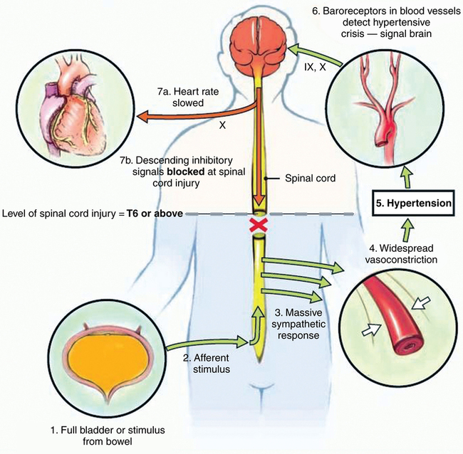
AD results from unopposed sympathetic efferent outflow in response to noxious afferent input below the level of the spinal cord injury, with reflex activation of parasympathetic outflow above the T6 dermatomal level. The pathways involved are summarized in Figure 178-1.

Afferent stimuli from an insult below the level of the spinal cord injury can ascend via spinothalamic and posterior columns to activate sympathetic neurons up to the level of the cord injury, discharging these neurons as an independent reflex at the level of the cord. Ordinarily this would elicit compensatory bulbospinal sympathetic inhibition via descending spinal pathways, but these pathways are now blocked by the spinal cord injury, resulting in unopposed vasoconstriction below the injury. With a spinal cord injury at T6 or higher, noxious stimuli result in intense constriction of splanchnic vascular beds and the vasculature in the lower extremities, leading to an exaggerated hypertensive response. Cord injury below T10 does not cause AD, whereas patients with injuries at the T6 to T10 levels may have a mild blood pressure elevation without full-blown AD. AD can occur in patients with incomplete spinal cord injuries, but the AD is more severe in those with a complete injury.

Baroreceptors in the aortic arch and carotid sinus respond to hypertension by activating brainstem vasomotor reflexes, resulting in increased parasympathetic activation via the intact cranial nerve X effector pathway, which is unaffected by the spinal cord injury, usually resulting in bradycardia. Tachycardia is also possible but occurs less frequently, presumably depending on the balance between catecholamines that diffuse into the bloodstream after sympathetic neuron activation below the spinal cord lesion and vagal outflow. Parasympathetic activation also causes vasodilation above the level of the cord injury.

Although AD has been reported to occur in the acute phase of a spinal cord injury, it generally becomes evident 1 to 6 months after the initial injury. This delayed occurrence is attributed to injury-induced changes in the structure and electrophysiology of both primary afferents and spinal neurons, as well as increased sensitivity of the peripheral vasculature to α-adrenergic stimulation, which heightens the exaggerated sympathetic response to noxious stimuli.

Clinical features

Acute hypertension is the sign of AD that is of greatest concern, causing the major morbid conditions associated with AD (e.g., myocardial ischemia, arrhythmia, congestive heart failure, cerebral ischemia, cerebral hemorrhage, and hypertensive encephalopathy). Initial blood pressure elevation may be mild and disguised by the fact that resting blood pressure in patients with high spinal cord injuries is usually low. A blood pressure of 120/80 mm Hg in a patient whose normal pressure is 90/60 mm Hg should raise concern. Blood pressures as high as 250 to 300/100 to 130 mm Hg have been reported during AD. Bradycardia and other arrhythmias often accompany the hypertension.

Other signs and symptoms can vary among patients and even among episodes of AD in the same patient and may be masked by sedative or anesthetic drugs. In an awake patient, AD often presents with the triad of severe headache, profuse sweating, and cutaneous flushing above the level of the spinal cord injury. The skin below the level of the injury may be pale and cool with piloerection. Nasal congestion, anxiety, malaise, nausea, and visual disturbances may also occur. These reactions are generally consistent with marked sympathetic activation below the cord injury, with cephalad reflex parasympathetic activity. However, the hyperhydrosis of AD is most common on the face and neck, above the level of cord injury, rather than below, where sympathetic outflow is maximal. The mechanism is not well understood, although it could involve a direct effect of catecholamines spilling over into the bloodstream, a central effect of excess catecholamines passing the blood-brain barrier, or a direct effect of intense parasympathetic stimulation on the forehead and upper lip, the only area in humans where there is parasympathetic as well as sympathetic innervation of sweat glands.

Prevention and treatment

AD is a potential concern during any surgical procedure in which innervation of the surgical field is below the level of the spinal cord injury. Anything that would elicit pain in a patient who does not have a spinal cord injury can cause AD in an at-risk patient. A patient’s history of AD should alert the anesthesia provider to the risk of the patient subsequently developing AD and to its potential magnitude in a specific patient, but any patient with a spinal cord injury at T6 or above should be considered at risk, even in the absence of previous episodes of AD. Pelvic visceral pain is a particularly potent stimulus of AD, and thus, urologic and bowel operations and childbirth are the most frequent causes of significant AD that most anesthesia providers will encounter. The magnitude of AD increases with the magnitude of the sensory stimulus and with increasing distance between the level of the cord lesion and the level of the dorsal root entry zone of the stimulus.

Prevention of AD is the ideal and can be accomplished with a dense regional block or a deep inhalation anesthetic. (This may require education of the patient and surgeon to accept the need for an anesthetic for a procedure that would not elicit any conscious sensation of pain in the patient.) Sevoflurane has been shown to prevent AD in at-risk patients undergoing transurethral litholapaxy with a half-maximal effective concentration (EC50) of 3.1% and an EC95 of 3.8% in 50% N2O. Although both epidurally and spinally administered local anesthetics have been used successfully to prevent AD, epidural anesthesia may not block the larger sacral nerve roots as effectively as does spinal anesthesia. It is difficult to assess the level of neuraxial block in a patient with a high cord injury, so a spinal anesthetic confirmed by cerebrospinal fluid return during placement may offer more assurance of an adequate block than would an epidural anesthetic. Anesthesia providers may encounter technical challenges in accessing the subarachnoid space in patients with spinal cord injuries and who have low resting blood pressures, but, in practice, accessing the subarachnoid space has not proved to be problematic in most patients. Topical local anesthetic agents administered alone prior to performing superficial rectal and urinary procedures has not been uniformly effective in preventing AD. Neither parenterally nor epidurally administered opioids, nor N2O, is consistently effective in preventing AD, except for epidural meperidine, which also has some local anesthetic properties. Other intravenously administered anesthetic agents have not been extensively tested in patients with AD.

When AD does occur, it is a medical emergency and must be treated rapidly. Removal of the triggering stimulus by a temporary halt in the surgical procedure may reverse AD and allow institution of more potent prophylaxis and treatment. In mild cases, nonpharmacologic measures such as elevating the head and torso, loosening tight clothing, and relieving inadvertent bladder or bowel distention may suffice. The diagnosis of AD in the setting of an operation below the spinal cord injury in a susceptible patient is usually straightforward, but other potential causes of acute hypertension should also be considered. In a laboring parturient, preeclampsia can also cause severe hypertension, but in patients with AD, the blood pressure elevation is usually much more marked during uterine contraction, with decline during relaxation. It should also be kept in mind that obstruction of a urinary catheter and bowel impaction are frequent causes of AD in nonanesthetized patients and can occur in any susceptible patient during any surgical procedure, including those perfomed on sites above the level of the spinal cord injury.

Multiple pharmacologic agents have been used to treat AD, but for many of them, their use is supported by only anecdotal case reports. The use of sublingually administered nifedipine was widely recommended in the past but has fallen into disfavor because of reports of severe adverse reactions when nifedipine is given for acute blood pressure control in patients without AD. Case reports have shown that nitroglycerin, nitroprusside, and other nitrates have been used effectively, although the use of sildenafil or other phosphodiesterase inhibitors in the previous 24 h needs to be ruled out first. Sildenafil alone is not effective in treating AD. The oral α-adrenergic receptor blocking agents terazosin and prazosin are effective in the long-term prevention of AD outside the operating room, and intravenously administered phentolamine is acutely effective, but the effect of phenoxybenzamine is inconsistent. Intravenously administered prostaglandin E1 and hydralazine are effective for acute treatment of AD, although hydralazine appears to be more likely to cause excessive hypotension. Labetolol and metoprolol have been used successfully in individual cases.

AD can continue into the postoperative period; therefore, patients who develop AD require careful monitoring and continued treatment. AD can also present de novo in the postanesthesia care unit, so susceptible patients should be appropriately monitored.