Asthma

Published on 12/06/2015 by admin

Filed under Pulmolory and Respiratory

Last modified 22/04/2025

Print this page

rate 1 star rate 2 star rate 3 star rate 4 star rate 5 star
Your rating: none, Average: 0 (0 votes)

This article have been viewed 2720 times

5

Asthma

Chapter 4 discussed the normal structure of airways and considered several aspects of airway function. The most common disorders disrupting the normal structure and function of the airways—asthma and chronic obstructive pulmonary disease—are discussed here and in Chapter 6, respectively. Several other miscellaneous diseases affecting airways are covered in Chapter 7.

Asthma is a condition characterized by episodes of reversible airway narrowing associated with contraction of smooth muscle within the airway wall. It is a common disorder that affects approximately 7% to 10% of the population. Although asthma can occur in any age group, it is particularly common in children and young adults and probably is the most common chronic disease in these age groups.

The primary feature patients with asthma appear to have in common is hyperresponsiveness of the airways, that is, an exaggerated response of airway smooth muscle to a wide variety of stimuli. The hyperresponsiveness is likely due in part to underlying airway inflammation with a variety of types of inflammatory cells, especially eosinophils. The particular constellation of stimuli triggering attacks often varies among patients, but the net effect (bronchoconstriction) is qualitatively similar. Because asthma is by definition a disease with at least some reversibility, the patient experiences exacerbations (attacks) interspersed between intervals of diminished symptoms or symptom-free periods. During an attack, the diagnosis usually is straightforward. During a symptom-free period, the diagnosis may be more difficult to make and may require provocation or challenge tests to induce airway constriction.

Etiology and Pathogenesis

Despite the high prevalence of asthma in the general population and the many advances that have been made in treating the manifestations of the disease, a great deal about its etiology and pathogenesis remains speculative. This section focuses on two major questions: (1) What causes certain people to have airways that hyperreact to various stimuli? (2) What is the sequence of events from the time of exposure to the stimulus until the time of clinical response?

Predisposition to Asthma

Potential factors that may predispose an individual to developing asthma are either inherited or acquired. There has been significant interest in and investigation of genetic and environmental factors that may contribute to the development of asthma, but the roles of these factors and their possible interactions have not been fully elucidated.

Genetics

A substantial proportion of patients with asthma have an underlying history of allergies (allergic rhinitis and eczema) along with accompanying markers for allergic disease, such as positive skin tests and elevated immunoglobulin E (IgE) levels. In these patients, the asthma frequently is exacerbated by exposure to various allergens to which the patients have been previously sensitized. Patients with an allergic component to their asthma often have a strong family history of asthma or other allergies, suggesting that genetic factors may play a role in the development of asthma as well as the underlying allergic diathesis (often called atopy). However, no simple pattern of mendelian inheritance suggesting a single gene as responsible for either atopy or asthma has been identified.

Epidemiologic studies have confirmed an increased frequency of asthma and atopy in first-degree relatives of asthmatic subjects compared with control subjects, and studies in twins indicate a much higher concordance for asthma in monozygotic than in dizygotic twins. Attempts to identify chromosomal regions carrying genes associated with asthma have found a number of such regions, particularly on the long arm of chromosomes 5, 11, and 12 (5q, 11q, and 12q, respectively) and on the short arm of chromosome 6 (6p). Examples of candidate genes proposed to be involved in the predisposition to asthma include the β-subunit gene of the high-affinity receptor for IgE (on chromosome 11q), a gene cluster for production of various cytokines (on chromosome 5q), and the gene encoding the β2-adrenergic receptor (on chromosome 5q). More recently, a disintegrin and matrix metalloproteinase gene called ADAM33 on the short arm of chromosome 20 (20p) has attracted interest as a major candidate gene for asthma and bronchial hyperresponsiveness. Despite these intriguing associations, there is general agreement that the genetic influences in asthma are complex, varying according to the population being studied, and that multiple genes, gene products, and environmental exposures likely interact in the pathogenesis of the disease.

Acquired (Environmental) Factors

A variety of environmental factors that might predispose an individual to develop asthma, most likely interacting with one or more genetic factors, have been proposed. Exposure to allergens, possibly at a critical time during childhood, may be an important environmental factor. Some of these exposures are to common environmental allergens, such as those derived from house dust mites, domestic animals, and cockroaches. These allergens are found indoors, often concentrated in bedding and carpets, and are present throughout the year. Another potential environmental factor is maternal cigarette smoking. An increased risk for early-onset asthma is found in children whose mothers smoke, possibly related to increasing the immune responsiveness of the child.

Viral respiratory tract infections precipitate airway inflammation and trigger acute exacerbations of asthma, but their potential role as an inducer or cause of asthma in the absence of other factors is controversial. One theory suggests that early childhood viral infections are causally associated with later development of asthma. A contrary view (the so-called hygiene hypothesis) suggests that exposure to microbes and microbial byproducts (e.g., endotoxin) during childhood protects against development of asthma by shifting the immunologic profile of helper T (TH) cells toward a TH1 response (responsible for cellular defense) and away from a TH2 response (which mediates allergic inflammation). It is also possible some infections increase the chance of developing asthma, whereas others decrease the risk.

Finally, a recent line of inquiry to explain the increasing prevalence of asthma throughout industrialized parts of the world has turned to a possible role for vitamin D deficiency among pregnant women. Vitamin D is believed to have an immunoregulatory role, and it has been hypothesized that deficiency of vitamin D during pregnancy may predispose to asthma in the offspring.

The relative contribution of these different factors to the development of asthma is unknown. It is possible each of the factors discussed contributes to disease in at least a subset of asthma patients.

Airway Inflammation and Bronchial Hyperresponsiveness

The association between asthma and allergies is significant but not universal. Many individuals with asthma have no other evidence of atopy and do not experience exacerbations as a result of antigen exposure. In this group, asthma attacks often are precipitated by other stimuli, as will be described later. However, the feature both groups of patients—those with and those without an allergic background, sometimes referred to as “extrinsic” (atopic) and “intrinsic” (non-atopic) asthmatic patients, respectively—have in common is hyperresponsiveness of their airways to a variety of stimuli. When exposed to such stimuli, the airways often demonstrate bronchoconstriction, which can be measured as an increase in airway resistance or a decrease in forced expiratory flow rates.

The histologic feature that accompanies this hyperresponsiveness and is thought to be a critical component of its pathogenesis is airway inflammation. Airway inflammation, especially with eosinophils and lymphocytes, has been found on both postmortem examination in persons with asthma who died of their disease and on bronchial biopsy specimens obtained from patients with mild asthma. Another typical finding is evidence of what has been called airway remodeling, which likely results from chronic airway inflammation and the associated production and release of a multitude of mediators including growth factors. Such remodeling changes include epithelial damage, airway fibrosis, and smooth muscle hyperplasia. These histologic findings, particularly the increase in airway smooth muscle, may be partly responsible for the hyperresponsiveness that can be documented in such persons with asthma, even when they are free of obvious bronchospasm.

No single factor or cell appears to be responsible for asthma; rather, a complex and interrelated series of events, including cellular infiltration, cytokine release, and airway remodeling, likely culminates in airway hyperresponsiveness and episodes of airflow obstruction (Fig. 5-1). A variety of mediators released from inflammatory cells can alter the extracellular milieu of bronchial smooth muscle, increasing its responsiveness to bronchoconstrictive stimuli. Mediators that have been proposed to play such a role include prostaglandin and leukotriene products of arachidonic acid metabolism. Some cytokine mediators released from inflammatory cells have various effects on other inflammatory cells, thus perpetuating the inflammatory response. For example, lymphocytes of the TH2 phenotype, which are thought to be a prominent component of the inflammatory response in asthma, release interleukin (IL)-5, which has a chemoattractant effect for eosinophils. IL-5 also stimulates growth, activation, and degranulation of eosinophils. IL-4, another cytokine released from TH2 lymphocytes, exerts a different type of proinflammatory effect by activating B lymphocytes, enhancing synthesis of IgE and promoting differentiation of TH2 cells.

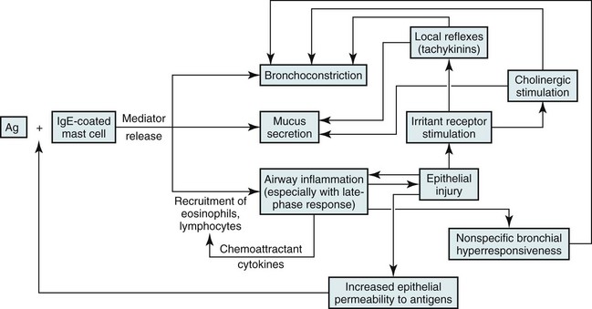
Mediators released from inflammatory cells may produce tissue damage that contributes to asthma pathogenesis. For example, when eosinophils degranulate, they release several toxic proteins from their granules, such as major basic protein and eosinophil cationic protein. These and other eosinophil products may contribute to the epithelial damage found in the asthmatic airway. Once the epithelium is injured or denuded, its barrier function is disrupted, allowing access of inhaled material to deeper layers of the mucosa. Additionally, the epithelial cells themselves may become actively involved in amplifying the inflammatory process (through production of cytokine and chemokine mediators) and in perpetuating airway edema (through vasodilation mediated by release of nitric oxide, leukotrienes, and prostaglandins). Finally, sensory nerve endings in the airway epithelial layer may become exposed, triggering a reflex arc and release of tachykinin mediators (e.g., substance P, neurokinin A), as shown in pathway 4 of Figure 4-3. These peptide mediators, released at bronchial smooth muscle, submucosal glands, and blood vessels, are capable of causing bronchoconstriction and airway edema.

Common Provocative Stimuli

A substantial amount is known about the sequence of events from the time of exposure to a stimulus until the clinical response of bronchoconstriction in asthmatic persons. Four specific types of stimuli that can result in bronchoconstriction are considered here: (1) allergen (antigen) exposure, (2) inhaled irritants, (3) respiratory tract infection, and (4) exercise.

Allergen Exposure

The pathogenetic mechanisms leading to bronchoconstriction are best defined for allergen-induced asthma. Allergens to which an asthmatic person may be sensitized are widespread throughout nature. Although patients and clinicians often first consider seasonal outdoor allergens such as pollen, many indoor allergens may play a more critical role. These allergens include antigens from house dust mites (Dermatophagoides and others), domestic animals, and cockroaches. Inhaled antigens are initially identified and processed by antigen-presenting cells called dendritic cells, which in turn present the antigenic material to T lymphocytes. Chemicals released by TH2 cells, especially IL-4 and IL-13, signal B lymphocytes to produce antigen-specific IgE antibodies. When an asthmatic person has IgE antibody against a particular antigen, the antibody binds to high-affinity IgE receptors on the surface of tissue mast cells and circulating basophils (see Fig. 5-1). If that particular antigen is inhaled, it binds to and cross-links IgE antibody (against the antigen) bound to the surface of mast cells in the bronchial lumen. The mast cell then is activated, leading to release of both preformed and newly synthesized mediators. Mediators released from the mast cell induce bronchoconstriction and increase airway epithelial permeability, allowing the antigen access to the much larger population of specific IgE-containing mast cells deeper within the epithelium. Binding of antigen to antibody on this larger population of mast cells again initiates a sequence of events leading to release of chemical mediators capable of inducing bronchoconstriction and inflammation. Several mediators have been recognized (Table 5-1), but the discussion here is limited to the few that have been primarily implicated in the pathogenesis of allergic asthma; major mediators include histamine and leukotrienes.

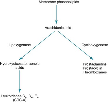
Leukotrienes: The leukotrienes include a series of compounds (LTC4, LTD4, and LTE4) that formerly were called slow-reacting substance of anaphylaxis (SRS-A). Unlike histamine, leukotrienes are not preformed in the mast cell but synthesized after antigen exposure and then released. To some extent, their actions are similar to those of histamine; they also have a direct bronchoconstrictor action on smooth muscle, increase vascular permeability, and stimulate excess production of airway mucus. Leukotrienes are synthesized from arachidonic acid (also the precursor for prostaglandins) but along a different pathway involving a lipoxygenase enzyme, as opposed to the cyclooxygenase enzyme used for prostaglandin synthesis (Fig. 5-2). LTC4 and LTD4 in particular are extraordinarily potent bronchoconstrictors and may have an important role in the pathogenesis of bronchial asthma. An interesting sidelight is provided by knowledge that some persons with asthma experience exacerbations of their disease after taking aspirin or other nonsteroidal antiinflammatory drugs (NSAIDs). These drugs are known inhibitors of the cyclooxygenase enzyme and may result in preferential shifting of the pathway shown in Figure 5-2 toward production of the bronchoconstrictor leukotrienes.

The role of other mediators listed in Table 5-1 in asthma pathogenesis is less clear. Platelet-activating factor has been proposed to play a role in recruiting eosinophils to the lung, and platelet-activating factor activates eosinophils, stimulating them to release proteins toxic to airway epithelial cells.

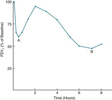
Late-Phase Asthmatic Response: The airway response to antigen challenge, as measured by changes in forced expiratory volume in 1 second (FEV1), appears to be more complicated and involves more than just the rapid mediator-induced bronchoconstriction seen within the first half hour following exposure. In many patients, the return of FEV1 to normal is followed by a secondary delayed fall in FEV1 occurring hours after antigen exposure (Fig. 5-3). This delayed fall in FEV1 is accompanied histologically by inflammatory changes in the airway wall. At the same time, increased bronchial hyperresponsiveness to nonspecific stimuli, such as histamine or methacholine, can be demonstrated and can last for days.

It now appears that this “late-phase response,” as it has been called, depends on the presence of antigen-specific IgE. Presumably, release of mediators after allergen binding to IgE-coated mast cells results in an influx of inflammatory cells, especially eosinophils, into the airway wall. Experimental data suggest that this heightened airway inflammation is responsible for the increased nonspecific bronchial hyperresponsiveness seen at the time of the late-phase response.

Exercise

Exercise can frequently provoke bronchoconstriction in patients with hyperreactive airways. The crucial factor in the pathogenesis appears to be heat movement from the airway wall, resulting in cooling of the airway. During exercise, individuals have a high minute ventilation, and the large amounts of relatively cool and dry inspired air must be warmed and humidified by the tracheobronchial mucosa. When the air is warmed and humidified, water evaporates from the epithelial surface, resulting in cooling and drying of the airway epithelium. The phenomenon of exercise-induced bronchoconstriction can be reproduced by having an asthmatic person voluntarily breathe cold dry air at a high minute ventilation. Inhalation of warm saturated air at the same minute ventilation does not produce a similar effect. The mechanism that links airway cooling and drying with bronchoconstriction is less clear. Alteration of the ionic environment after drying of the mucosa, mediator release, hyperemia of the mucosa following airway rewarming, and stimulation of irritant receptors all have been proposed as mechanisms, but none is universally accepted.

As might be expected from the description of exercise-induced bronchoconstriction, inhalation of cold air during the winter months can be responsible for asthma exacerbations or worsening of symptoms in selected patients. The mechanism of airway narrowing in these patients following inhalation of cold air is also believed to be due to airway cooling and drying and therefore is analogous to the mechanism of exercise-induced bronchoconstriction.

Pathology

Pathologic findings in asthma have often been described from autopsy studies and thus represent the consequences of particularly severe disease. In these cases, marked overdistention of the lungs is seen, and the airways are occluded by thick tenacious mucous plugs. However, information regarding the histologic appearance of airways in patients with stable mild disease is also available. Examination of the airways by microscopy demonstrates the following findings of variable severity that are apparent in both mild and more severe disease:

As described earlier in this chapter, the presence of histologic abnormalities presumably contributes to the nonspecific bronchial hyperresponsiveness in patients, even when they are free of an acute attack. In addition to the bronchial hyperresponsiveness that results from airway inflammation and remodeling, the more long-standing structural changes that characterize airway remodeling contribute to the component of persistent airflow obstruction that can be seen in some asthmatics.

Pathophysiology

The pathophysiologic features of asthma largely follow from the pathologic abnormalities. Contraction of smooth muscle in the bronchial walls, mucosal edema, and secretions within the airway lumen all contribute to decreased airway diameter, which increases airway resistance. Pathologic changes are present at many levels of the tracheobronchial tree, from large airways down to peripheral airways less than 2 mm in diameter.

As a result of narrowed airways with increased resistance, patients have difficulty with airflow during both inspiration and expiration. However, because intrathoracic airways are subjected to relatively negative external pressure (transmitted from negative pleural pressure) during inspiration, lumen size is larger during the inspiratory phase of the respiratory cycle. During expiration, relatively positive pleural pressure is transmitted to intrathoracic airways, thus decreasing their diameter. Therefore, greater difficulty with airflow on expiration than on inspiration is characteristic of asthma, as it is of any of the diseases that cause obstruction or narrowing of airways within the thorax. The greatest difficulty with expiration occurs when the patient is asked to perform a forced expiration (i.e., breathe out as hard and fast as possible). With forced expiration, pleural pressure becomes much more positive, thereby promoting airway narrowing and closure as well as air trapping.

The effects of increased airway resistance are readily seen by measuring pulmonary function in asthmatic persons. Pulmonary function studies performed during an attack show decreases in forced expiratory flow rates and evidence of air trapping. On the forced expiratory spirogram, patients generally exhibit a decrease in both forced vital capacity (FVC) and FEV1, with the decrease in FEV1 usually more pronounced than the decrease in FVC. Hence, the ratio FEV1/FVC, which reflects the proportion of FVC that can be exhaled during the first second, is decreased. In addition, the maximal midexpiratory flow rate (also called the forced expiratory flow between 25% and 75% of the vital capacity [FEF25–75]) is diminished.

Measurement of lung volumes during an exacerbation shows evidence of air trapping, with an increase in residual volume (RV) and functional residual capacity (FRC). The most impressive increase is seen in RV, the volume left in the lungs at the end of a maximal exhalation, which may be greater than 200% of the predicted value. The increase in RV is believed to be due to premature small airway closure as a result of smooth muscle constriction, mucous plugs, and inflammatory changes of the mucosa.

FRC, the resting point of the lungs after a normal expiration, may be increased for at least two reasons. First, because more time is required for expiration when airways are obstructed, patients may not have sufficient time before the next breath to fully exhale the volume from the previous breath. This phenomenon, sometimes called dynamic hyperinflation, is a particular problem when the asthmatic person is breathing at a rapid respiratory rate. Another reason for increase in FRC is related to persistent activity of the inspiratory muscles during expiration, maintaining lung volume at a level higher than expected throughout expiration. A physiologic advantage to breathing at a higher-than-normal FRC is having airways held open at a greater diameter. A disadvantage is increased work of breathing due to reduced compliance of the respiratory system at higher lung volumes (see Fig. 1-3, C) and a mechanical disadvantage for diaphragmatic function when the diaphragm is lower and flatter (see Mechanisms of Abnormal Gas Exchange in Chapter 6).

The focus so far has been on pulmonary function and physiologic abnormalities seen with a typical asthmatic attack. Between attacks, pulmonary function, as measured by FEV1 and FVC, often returns to normal. However, even when a person is not having an acute attack, subtle abnormalities in pulmonary function may be present, such as a decrease in maximal midexpiratory flow rate and a mild increase in RV. These abnormalities may reflect some residual disease in the small airways of the lung, frequently the last region to become normal after an attack.

A subgroup of asthmatic persons, generally those with long-standing disease, have pulmonary function that does not return to normal. Instead, they have easily demonstrable physiologic abnormalities (e.g., abnormal FEV1 and FVC) that persist between attacks. Even though asthma is generally characterized by reversible episodes of airflow obstruction, these persons also appear to have a component of irreversible disease. Nevertheless, they still generally experience episodes of reversible airflow obstruction and worsening of expiratory flow rates superimposed upon whatever irreversible disease is present.

The increased resistance to airflow in asthma exerts a toll on gas exchange, which is generally disturbed during acute attacks. The most common pattern of arterial blood gases consists of a low PO2 accompanied by a low PCO2 (respiratory alkalosis). The mechanism for the hypoxemia is ventilation-perfusion mismatch. The increased airway resistance in asthma is not evenly distributed, such that some airways are affected more than others. Therefore, inspired air is not distributed evenly but tends to go to less diseased areas. However, blood flow remains relatively preserved in the regions that are ventilating poorly. The regions of low ventilation-perfusion (image) ratio contribute blood with a low PO2 that cannot be compensated for by increases in the image ratio from other regions of the lung (see Chapter 1).

Patients are typically able to hyperventilate if an acute asthma attack is not too severe, and PCO2 usually is low. The stimulus or mechanism for the hyperventilation is not clear. During an acute attack, activation of irritant receptors may stimulate ventilation, or other reflexes originating in the airways, lung, or chest wall may stimulate ventilation. PCO2 that increases to either a normal or a frankly elevated level often means worsening airflow obstruction or fatigue of the respiratory muscles in a tiring individual who is no longer able to maintain normal or high minute ventilation in the face of significant airflow obstruction. Thus, the clinician should view a normal or high PCO2 as a serious warning sign.

Clinical Features

Onset of asthma occurs most frequently during childhood and young adulthood, although asthma can develop for the first time in older patients. In many patients, particularly those in whom asthma started before age 16 years, the disease eventually regresses, and patients are no longer subject to repeated episodes of reversible airway obstruction.

The symptoms most commonly noted by patients during an exacerbation of asthma are cough, dyspnea, wheezing, and chest tightness. Patients do not necessarily have a classic presentation with several or all of these complaints but may merely have an unexplained cough or breathlessness on exertion. In some cases, patients can clearly identify a precipitating factor for an attack, such as exposure to an allergen, respiratory tract infection, exercise, exposure to cold air, emotional stress, or exposure to irritating dusts, fumes, or odors. In other cases, no precipitant can be identified. Exposures in the workplace, related to proteins or other chemicals to which the patient may be sensitized, are important precipitants in a subgroup of patients who are said to have occupational asthma. Some asthmatic persons are particularly sensitive to ingestion of aspirin, which is believed to favor production of leukotrienes from arachidonic acid. Some patients with aspirin sensitivity also have nasal polyps, leading to a well-recognized triad of asthma, aspirin sensitivity, and nasal polyposis (sometimes referred to as triad asthma, Samter syndrome, or aspirin-exacerbated respiratory disease). Other NSAIDs (which also inhibit the cyclooxygenase enzyme) can also produce bronchoconstriction in patients who are aspirin sensitive.

On examination, patients experiencing an asthma attack usually have tachypnea and, on chest auscultation, prolonged expiration and evidence of wheezing. Wheezing is generally more prominent during expiration than inspiration and may be triggered by having the patient exhale forcefully. Although the tendency is to equate wheezing and asthma, the presence of wheezing does not necessarily indicate a diagnosis of asthma. Wheezing reflects only airflow through narrowed airways; it also can be seen in such diverse disorders as congestive heart failure and chronic obstructive pulmonary disease or in the case of a foreign body in the airway. On the other hand, not all asthmatic persons wheeze. It is a common observation that severe asthma may be associated with no wheeze at all if airflow is too impaired to generate an audible wheeze.

During a particularly severe attack that is refractory to treatment with bronchodilators, persons with asthma are said to be in status asthmaticus. These patients present difficult therapeutic challenges, may require assisted ventilation, and may even die as a result of the acute attack.

The overall severity of an individual’s asthma can be characterized on the basis of the frequency of exacerbations, nocturnal symptoms, and magnitude of abnormality and variability in pulmonary function. The features used to define four categories of severity (intermittent asthma, mild persistent asthma, moderate persistent asthma, and severe persistent asthma) are listed in Table 5-2.

Table 5-2

CLASSIFICATION OF ASTHMA BY SEVERITY: CLINICAL ASPECTS AND TREATMENT

image

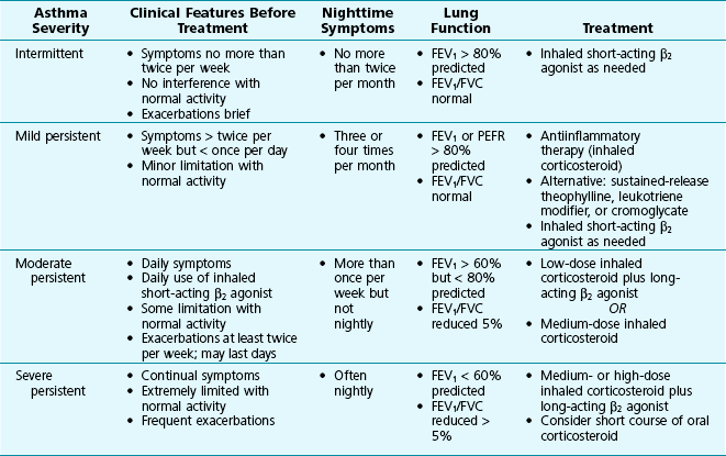
FEV1 = Forced expiratory volume in 1 second; PEFR = peak expiratory flow rate.

Adapted from National Asthma Education and Prevention Program: Guidelines for the diagnosis and management of asthma: Expert panel report 3, Bethesda, MD, 2007, National Institutes of Health, NIH publication 08-4051.

Diagnostic Approach

A clinical history of reversible episodes of bronchoconstriction brought on by characteristic triggers is often crucial to the diagnosis of asthma. Other helpful features in the history include other evidence for atopy (e.g., hay fever or eczema) or a family history of allergies or asthma. Physical examination demonstrating wheezes during an attack often provides confirmatory evidence for airflow obstruction.

The chest radiograph, although sometimes useful for ruling out other causes of wheezing or complications of asthma, is generally not particularly helpful in the diagnosis. It usually shows normal findings but may demonstrate a flattened diaphragm suggestive of air trapping.

If the patient is producing sputum, microscopic examination of the sputum frequently shows many eosinophils on the smear. An increased percentage of eosinophils in peripheral blood is also common, even when the asthma has no clear relationship to allergies.

The clinical usefulness of skin testing and inhalation testing with allergens in an attempt to identify antigens to which the patient is sensitized is controversial. Allergy skin testing with antigens and blood testing for specific IgE antibodies are available to help confirm or refute suspected allergic sensitivities to common aeroallergens. Unfortunately, neither is useful for the diagnosis of asthma, since persons with asthma may have no allergic sensitivities, and persons with allergic sensitivities may not have asthma but rather manifest with symptoms affecting the nose and conjunctivae.

Although the diagnosis of asthma is usually made on the basis of clinical features, spirometry, and response to therapy, provocation tests are sometimes used to make or confirm the diagnosis of asthma. These tests rely on the principle that asthmatic persons have hyperreactive airways. Therefore, when tested with inhalation of methacholine (a cholinergic agent) or histamine, persons with asthma respond with bronchoconstriction to comparatively small doses of either agent. Inhalation of cold air at high minute ventilations with PCO2 kept constant (termed isocapnic hyperpnea) also can be used as a challenge test to induce transient bronchoconstriction in patients in whom the diagnosis of asthma is uncertain.

Measurement of pulmonary function, especially FEV1 and FVC, is particularly useful in the patient with suspected or known asthma. Documentation of reversible airflow obstruction, either during attacks or with a challenge test, is frequently sufficient to make the diagnosis. In practice, the diagnosis of asthma is most commonly made by the history of episodic dyspnea, wheezing, or cough, with documentation of reversible airflow obstruction by pulmonary function testing.

Patients can conveniently test their own pulmonary function through measurement of the peak expiratory flow rate. Such testing is particularly useful for monitoring the course of the disease and alerting the patient to adjust the medication regimen, seek attention from a physician, or both. In addition, the efficacy of treatment or changes in the therapeutic regimen can readily be assessed by serial measurement of the peak expiratory flow rate.

Treatment

The major categories of drugs used to treat asthma are those that dilate smooth muscle of the bronchial wall and those that have an antiinflammatory action. Agents targeted at blocking production or activity of specific mediators are also used, and a monoclonal antibody against IgE has become available. The main categories of drugs used to treat asthma are listed in Table 5-3. Several of the drugs are also used for treatment of other types of pulmonary disease, particularly chronic obstructive pulmonary disease, and are mentioned in other chapters.
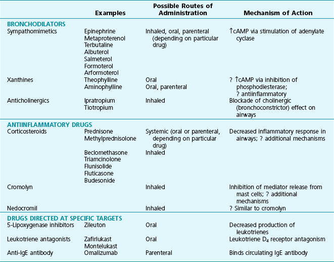
Bronchodilators

The most common bronchodilator agents used for treatment of asthma are the sympathomimetic agents, which act on β2 receptors to activate adenylate cyclase and increase intracellular cyclic adenosine monophosphate (cAMP). Increased levels of cAMP in bronchial smooth muscle, resulting specifically from stimulation of β2 receptors, activate protein kinase A, which phosphorylates several regulatory proteins that mediate bronchodilation. Additionally, β stimulation increases intracellular cAMP in mast cells, inhibiting release of chemical mediators that secondarily cause bronchoconstriction. Specific examples of available sympathomimetic drugs are listed in Table 5-3. To avoid some of the adverse cardiac effects induced by stimulation of β1 receptors, the preferred agents have action limited primarily to stimulation of β2 receptors. The β2-specific agents most commonly used are albuterol (short-acting β2 agonist) and salmeterol or formoterol (long-acting β2 agonists), and the typical route of administration is inhalation. Although some sympathomimetic agents can be given orally or parenterally, the inhaled route is preferred because it has fewer systemic side effects and provides direct delivery to the site of action in the airways.

Short-acting inhaled β2 agonists, such as albuterol, are typically used on an as-needed basis to reverse an acute episode of bronchoconstriction. They may be the only agents needed to control the patient’s asthma when episodes are infrequent. Effects of the long-acting β2 agonists salmeterol, formoterol, and arformoterol last for approximately 12 hours and are not appropriate for as-needed use to treat acute symptoms. Short-acting β2-agonist drugs can be used prophylactically before activities or exposure to stimuli known to precipitate bronchoconstriction. If asthma severity increases and more frequent or regular use of an inhaled β2 agonist is required, addition of an antiinflammatory agent (see following section) is important.

The second class of bronchodilator agents, the methylxanthines, is used much less frequently than β2 agonists. Theophylline, the prototype of this class, is generally believed to act by inhibiting the enzyme phosphodiesterase (PDE), which normally is responsible for metabolic degradation of cAMP. When degradation is inhibited, the levels of cAMP in smooth muscle and mast cells increase, resulting again in bronchodilation and decreased mediator release from mast cells. However, the serum levels of methylxanthines needed to inhibit PDE are higher than those actually achieved in patients, so whether PDE inhibition is the major or exclusive mechanism of action of theophylline as a bronchodilator is uncertain. In addition, theophylline may have a component of antiinflammatory activity, mediated by inhibition of the PDE-4 isozyme in inflammatory cells. Theophylline is available only for oral administration, whereas aminophylline (a water-soluble salt of theophylline) can be given either orally or intravenously. Because methylxanthines can be administered only systemically (as opposed to locally in the airway), systemic side effects (gastrointestinal, cardiac, neurologic) are more problematic than with inhaled sympathomimetic agents. This is an important reason why methylxanthines now are used relatively infrequently compared with inhaled β2 agonists.

The third class of bronchodilator agents, also used less frequently than β2 agonists in asthma patients, consists of drugs that have an anticholinergic action. Anticholinergic agents dilate bronchial smooth muscle by decreasing bronchoconstrictor cholinergic tone to airways. Ipratropium, available as an aerosol for inhalation, is the primary short-acting example of this class of agents. It is formally approved in the United States only for use in chronic obstructive lung disease (COPD; see Chapter 6), not in asthma. The major use of ipratropium for asthma has been as adjunctive therapy to inhaled β2 agonists in patients during an acute asthma attack. Tiotropium, a long-acting anticholinergic agent, is approved for and frequently used in patients with COPD, but it is neither approved for nor commonly used in patients with asthma.

Antiinflammatory Drugs

For years, the second major category of drugs used to treat asthma has included antiinflammatory agents: corticosteroids, disodium cromoglycate (cromolyn), and nedocromil. They suppress the inflammatory response by decreasing the number of eosinophils and lymphocytes infiltrating the airway and decrease production of a number of inflammatory mediators. Despite the general rationale for corticosteroid use, many aspects of their antiinflammatory action are unknown. Glucocorticoids are thought to bind to a cytoplasmic receptor present in nearly all cell types. After the receptor binds to its glucocorticoid ligand, it moves to the cell nucleus, where it interacts with transcription factors such as activator protein (AP)-1 and nuclear factor (NF)-κB, which regulate transcription of other target genes. Important target genes whose transcription is suppressed by the action of glucocorticoids include a variety of inflammatory cytokines (e.g., IL-1, IL-3, IL-4, IL-5, IL-6, and tumor necrosis factor [TNF]-α), the inducible form of nitric oxide synthase, and an inducible form of cyclooxygenase.

Because airway inflammation is believed to play an important role in asthma pathogenesis, particularly in the patient with more frequent attacks or more persistent airflow obstruction, corticosteroids have assumed a central role in the management of many cases of asthma. By decreasing airway inflammation, steroids are thought to ameliorate the underlying disease process in asthma, not just the bronchoconstriction resulting from airway inflammation.

Steroids have an important place in both management of acute asthma attacks and maintenance therapy for disease requiring more than just infrequent use of a β2-agonist bronchodilator. Frequently, steroids such as prednisone or methylprednisolone are started at high doses during an acute attack and then are tapered relatively rapidly. Because of the potential for significant adverse effects with long-term use of systemic (oral) steroids, chronic administration of oral steroids is avoided if the asthma can be managed with other modes of therapy. Foremost among these alternative forms of therapy are inhaled forms of corticosteroids that deliver the drug locally to the airway but have minimal systemic absorption and limited side effects. Inhaled steroids are now the preferred form of “controller” or preventive therapy for patients with asthma not adequately managed with infrequent use of a β-agonist inhaler.

Other antiinflammatory drugs (no longer available in the United States) are disodium cromoglycate (cromolyn) and nedocromil. Their mode of action traditionally was thought to be inhibition of mediator release from mast cells, but this mechanism has been disputed. Alternative mechanisms proposed include inhibitory effects on other types of inflammatory cells or on the action of tachykinins. Both cromolyn and nedocromil are given in inhaled form; neither drug is a bronchodilator, so neither is useful for treatment of acute attacks. Rather, they are generally given as ongoing medication, with the goal of preventing future exacerbations.

Agents with Specific Targeted Action

Agents are available that block the synthesis or action of a single type of mediator, specifically leukotrienes. Interestingly, these agents appear to be effective for only some patients with asthma. It is believed that underlying patient-related genetic factors affect the likelihood of a positive response. Specific agents that modify leukotrienes or leukotriene pathways include zafirlukast and montelukast, which antagonize the action of LTD4 at its receptor, and zileuton, which inhibits the enzyme 5-lipoxygenase and thus limits leukotriene production. On the basis of their mode of action, drugs that either block leukotriene synthesis or antagonize their action have a particularly important role in patients who are sensitive to aspirin or other NSAIDs.

The newest agent for treatment of asthma is omalizumab, a monoclonal antibody to IgE. On the basis of the principle that IgE is an important component of the pathobiology of allergic asthma, the drug was developed to prevent the binding of IgE to receptors on mast cells. Omalizumab is administered every 2 to 4 weeks by subcutaneous injection in a healthcare setting (e.g., outpatient medical office) and is extremely expensive. Its use has been limited to selected patients with particularly severe asthma who have elevated levels of IgE and continue to be symptomatic and prone to asthmatic attacks despite other treatment.

Management Strategy

At present, the overall strategy for management of asthma commonly proceeds in the following manner. A patient with relatively infrequent attacks, with symptom-free periods and normal pulmonary function between attacks, is managed with inhaled short-acting sympathomimetics (β2 agonists). These drugs are used on an as-needed basis, both for management of bronchospasm once it occurs and before exposure to stimuli often known to precipitate attacks (e.g., exercise or allergen exposure). These general guidelines are summarized in Table 5-2 according to the categories of clinical severity of disease.

When a patient’s asthma cannot be managed successfully with infrequent use of a β2-agonist inhaler, then an antiinflammatory agent is generally added as maintenance (ongoing) therapy to suppress the underlying airway inflammation. Inhaled corticosteroids are used most frequently and appear to be the most effective agents in this class, although leukotriene blockers are alternatives.

If therapy must be escalated beyond these measures because of inadequate control, addition of a regularly used long-acting inhaled β2 agonist (e.g., salmeterol or formoterol) is the preferred option. Other options are addition of an antileukotriene agent, escalation of the dose of inhaled corticosteroids, or addition of the methylxanthine theophylline. When patients have a significant acute attack or an attack that occurs despite adequate therapy as described, intensive bronchodilator therapy plus a short course of systemic steroids is generally effective. Particularly severe asthma exacerbations (i.e., status asthmaticus) often require high doses of systemic corticosteroids along with frequently administered bronchodilator therapy. Patients with respiratory failure may require intubation and mechanical ventilation.

For patients in whom allergen exposure is an exacerbating factor for their asthma, allergen avoidance is a fundamental component of the management regimen. Environmental control measures to minimize allergen exposure include removing carpets, encasing mattresses and pillows in allergen-impermeable covers (to minimize dust mite exposure), and removing pets from the home (to minimize exposure to animal antigens). Immunotherapy with repeated injections of antigen extract is sometimes used to desensitize the patient to the offending allergen. Although immunotherapy is effective in allergic rhinitis, its efficacy in patients with asthma is controversial and its role uncertain.

Because of the availability of effective forms of therapy, patients with asthma are generally capable of leading normal lives with relatively little or no alteration in their daily activities. However, not all patients with asthma are so fortunate. Refractory disease, persistent airflow obstruction, and rapid development of life-threatening attacks are some extreme examples of asthma that pose a continuing challenge to physicians caring for these patients.

References

Etiology and Pathogenesis

Barnes, PJ. Neurogenic inflammation in the airways. Respir Physiol. 2001;125:145–154.

Busse, WW, Lemanske, RF, Jr., Gern, JE. Role of viral respiratory infections in asthma and asthma exacerbations. Lancet. 2010;376:826–834.

Chung, KF. p38 mitogen-activated protein kinase pathways in asthma and COPD. Chest. 2011;139:1470–1479.

Fixman, ED, Stewart, A, Martin, JG. Basic mechanisms of development of airway structural changes in asthma. Eur Respir J. 2007;29:379–389.

Holgate, ST. Pathophysiology of asthma: what has our current understanding taught us about new therapeutic approaches? J Allergy Clin Immunol. 2011;128:495–505.

Holgate, ST. Innate and adaptive immune responses in asthma. Nat Med. 2012;18:673–683.

Holgate, ST, Yang, Y, Haitchi, HM, et al. The genetics of asthma. ADAM33 as an example of a susceptibility gene. Proc Am Thorac Soc. 2006;3:440–443.

Holt, PG, Sly, PD. Viral infections and atopy in asthma pathogenesis: new rationales for asthma prevention and treatment. Nat Med. 2012;18:726–735.

Holt, PG, Sly, PD. Interaction between adaptive and innate immune pathways in the pathogenesis of atopic asthma: operation of a lung/bone marrow axis. Chest. 2011;139:1165–1171.

Jacoby, DB. Virus-induced asthma attacks. JAMA. 2002;287:755–761.

Kaur, D, Brightling, C. OX40/OX40 ligand interactions in T-cell regulation and asthma. Chest. 2012;141:494–499.

Koppelman, GH, Sayers, I. Evidence of a genetic contribution to lung function decline in asthma. J Allergy Clin Immunol. 2011;128:479–484.

Martinez, FD. Genes, environments, development and asthma: a reappraisal. Eur Respir J. 2007;29:179–184.

Martinez, FD. New insights into the natural history of asthma: primary prevention on the horizon. J Allergy Clin Immunol. 2011;128:939–945.

Murphy, DM, O’Byrne, PM. Recent advances in the pathophysiology of asthma. Chest. 2010;137:1417–1426.

Paul, G, Brehm, JM, Alcorn, JF, et al. Vitamin D and asthma. Am J Respir Crit Care Med. 2012;185:124–132.

Postma, DS, Kerkhof, M, Boezen, HM, et al. Asthma and chronic obstructive pulmonary disease: common genes, common environments? Am J Respir Crit Care Med. 2011;183:1588–1594.

Robinson, DS. The role of the T cell in asthma. J Allergy Clin Immunol. 2010;126:1081–1091.

Thomson, NC, Chaudhuri, R, Livingston, E. Asthma and cigarette smoking. Eur Respir J. 2004;24:822–833.

Clinical Features and Diagnostic Approach

Anderson, SD. Indirect challenge tests: Airway hyperresponsiveness in asthma: its measurement and clinical significance. Chest. 2010;138:25S–30S.

Beach, J, Russell, K, Blitz, S, et al. A systematic review of the diagnosis of occupational asthma. Chest. 2007;131:569–578.

Corrao, WM, Braman, SS, Irwin, RS. Chronic cough as the sole presenting manifestation of bronchial asthma. N Engl J Med. 1979;300:633–637.

Jackson, DJ, Sykes, A, Mallia, P, et al. Asthma exacerbations: origin, effect, and prevention. J Allergy Clin Immunol. 2011;128:1165–1174.

Johnston, NW, Sears, MR. Asthma exacerbations. 1: Epidemiology. Thorax. 2006;61:722–728.

Katial, RK, Covar, RA. Bronchoprovocation testing in asthma. Immunol Allergy Clin North Am. 2012;32:413–431.

Martin, RJ. Nocturnal asthma: circadian rhythms and therapeutic interventions. Am Rev Respir Dis. 1993;147:S25–S28.

McFadden, ER, Jr., Gilbert, IA. Exercise-induced asthma. N Engl J Med. 1994;330:1362–1367.

McFadden, ER, Jr., Kiser, R, DeGroot, WJ. Acute bronchial asthma: relations between clinical and physiologic manifestations. N Engl J Med. 1973;288:221–225.

Naureckas, ET, Solway, J. Mild asthma. N Engl J Med. 2001;345:1257–1262.

Pratter, MR, Irwin, RS. The clinical value of pharmacologic bronchoprovocation challenge. Chest. 1984;85:260–265.

Singh, AM, Busse, WW. Asthma exacerbations. 2: Etiology. Thorax. 2006;61:809–816.

van den Berge, M, ten Hacken, NH, Cohen, J, et al. Small airway disease in asthma and COPD: clinical implications. Chest. 2011;139:412–423.

Wenzel, SE. Asthma phenotypes: the evolution from clinical to molecular approaches. Nat Med. 2012;18:716–725.

Treatment

Barnes, PJ. Severe asthma: advances in current management and future therapy. J Allergy Clin Immunol. 2012;129:48–59.

Barnes, PJ. Airway pharmacology. In: Mason RJ, Murray JF, Broaddus VC, et al, eds. Textbook of respiratory medicine. ed 4. Philadelphia: WB Saunders; 2005:235–279.

Barnes, PJ. Inhaled glucocorticoids for asthma. N Engl J Med. 1995;332:868–875.

Busse, WW. Long- and short-acting β2-adrenergic agonists. Arch Intern Med. 1996;156:1514–1520.

Chan, WW, Chiou, E, Obstein, KL, et al. The efficacy of proton pump inhibitors for the treatment of asthma in adults: a meta-analysis. Arch Intern Med. 2011;171:620–629.

Durrani, SR, Viswanathan, RK, Busse, WW. What effect does asthma treatment have on airway remodeling? Current perspectives. J Allergy Clin Immunol. 2011;128:439–448.

Fanta, CH. Drug therapy: asthma. N Engl J Med. 2009;360:1002–1014.

Glassroth, J. The role of long-acting β-agonists in the management of asthma: analysis, meta-analysis, and more analysis. Ann Intern Med. 2006;144:936–937.

Kazani, S, Wechsler, ME, Israel, E. The role of pharmacogenomics in improving the management of asthma. J Allergy Clin Immunol. 2010;125:295–302.

Lazarus, SC. Clinical practice. Emergency treatment of asthma. N Engl J Med. 2010;363:755–764.

O’ Byrne, PM, Parameswaran, K. Pharmacological management of mild or moderate asthma. Lancet. 2006;368:794–803.

Peters-Golden, M, Henderson, WR. Leukotrienes. N Engl J Med. 2007;357:1841–1854.

Price, D, Musgrave, SD, Shepstone, L, et al. Leukotriene antagonists as first-line or add-on asthma-controller therapy. N Engl J Med. 2011;364:1695–1707.

Strunk, RC, Bloomberg, GR. Omalizumab for asthma. N Engl J Med. 2006;354:2689–2695.

van den Berge, M, Hiemstra, PS, Postma, DS. Genetics of glucocorticoids in asthma. N Engl J Med. 2011;365:2434–2435.

Wahidi, MM, Kraft, M. Bronchial thermoplasty for severe asthma. Am J Respir Crit Care Med. 2012;185:709–714.

Weiss, ST. New approaches to personalized medicine for asthma: where are we? J Allergy Clin Immunol. 2012;129:327–334.