[level-membership-for-dermatology-category]
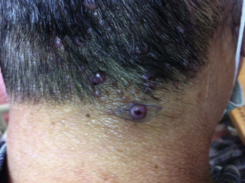
Angiolymphoid hyperplasia with eosinophilia


First-line therapies
 Surgery
 Laser therapy
 Corticosteroid, topical or intralesional
 Cryotherapy
Second-line therapies
 Imiquimod
 Tacrolimus
 Isotretinoin
 Radiofrequency ablation and sclerotherapy
Third-line therapies
 Interferon-α2b
 Anti-IL-5 antibody
 Suplatast tosilate
 Photodynamic therapy
 Radiotherapy
[/level-membership-for-dermatology-category][not-level-membership-for-dermatology-category]
Angiolymphoid hyperplasia with eosinophilia
