Anesthesia for the patient undergoing liver transplantation

Published on 13/02/2015 by admin

Filed under Anesthesiology

Last modified 22/04/2025

Print this page

rate 1 star rate 2 star rate 3 star rate 4 star rate 5 star
Your rating: none, Average: 0 (0 votes)

This article have been viewed 1295 times

Anesthesia for the patient undergoing liver transplantation

James Y. Findlay, MB, ChB, FRCA

Liver transplantation is an established therapy for end-stage liver disease, with the more than 6000 people who receive a liver transplant every year in the United States now having a 3-year survival rate of almost 80%. Despite recent advances in the use of living donors and of split-liver grafts for pediatric and adult recipients, the number of liver transplantations remains limited by the availability of suitable donors, with approximately 16,000 people waiting to receive a transplant. Liver transplantation presents a challenge to the anesthesia provider because, in addition to the operation being complex, most patients present for transplantation with greatly altered physiology because of their end-stage liver disease.

Preoperative evaluation

Table 176-1 lists some of the relevant physiologic consequences of liver failure and the consequences that may occur during liver transplantation. Prior to presenting for transplantation, candidates are screened for comorbid cardiopulmonary conditions: a resting echocardiogram assesses cardiac function and allows estimation of pulmonary artery pressures. A bubble test (injection of agitated saline while monitoring for echo-contrast in the right side of heart chambers) can also be performed; delayed appearance of the contrast agent suggests that the patient may have hepatopulmonary syndrome. If the patient has risk factors for coronary artery disease (∼40%-50% of adult patients), noninvasive testing is frequently performed, often by dobutamine stress echocardiography, because patients with significant coronary artery disease have poor peritransplant outcomes.

Table 176-1

Pathophysiologic Changes Associated with Liver Failure

Organ System Change Consequence(s)
Cardiovascular Hyperdynamic circulation (high cardiac output, low SVR)
Portal hypertension
Ascites
Pulmonary hypertension
Hypotension
Varices, splenomegaly
Bleeding (dilated vessels, thrombocytopenia)
Fluid shifts after drainage
High perioperative mortality rate (>80%) if severe
Respiratory Respiratory alkalosis
Restrictive physiology (ascites with or without pleural effusion)
Hepatopulmonary syndrome (intrapulmonary shunting)
Atelectasis; reduced compliance
Hypoxemia
Hematologic Decreased factor synthesis
Thrombocytopenia
Anemia
Bleeding potential
CNS Hepatic encephalopathy
Cerebral edema (in fulminant failure)
Delayed awakening
Raised ICP; consider ICP monitoring
Renal Hepatorenal syndrome
Hyponatremia
Renal failure—volume and electrolyte management concerns
Possibility of CPM if corrected intraoperatively

CNS, Central nervous system; CPM, central pontine myelinolysis; ICP, intracranial pressure; SVR, systemic vascular resistance.

Renal dysfunction often accompanies end-stage liver disease from hepatorenal syndrome, acute tubular necrosis, or a combination of both. In a patient requiring renal dialysis or continuous renal replacement therapy, consideration should be given to performing continuous dialysis or ultrafiltration in the operating room if problems managing volume or electrolytes are anticipated. Washing red blood cells prior to transfusion to reduce potassium may also be helpful.

The severity of end-stage liver disease is assessed by calculating the MELD (Model of End-stage Liver Disease) score, which incorporates the patient’s serum bilirubin, serum creatinine, and the international normalized ratio for prothrombin time to predict survival (Box 176-1). A higher MELD score is associated with more severe liver failure; higher MELD scores are also predictive of a greater rate of intraoperative blood product transfusion and need for vasopressors.

Intraoperative management

Anesthesia

Induction of anesthesia may be achieved using any of the commonly used agents. Maintenance is typically achieved using a balanced technique with an inhaled agent and opioid, often fentanyl. Cisatracurium may be preferred for maintenance of neuromuscular blockade because it is not dependent on hepatic metabolism for elimination, but other neuromuscular blocking agents can be used as long as redosing is guided by train-of-four monitoring.

Invasive monitoring is the norm; direct arterial pressure is best monitored by a brachial or femoral arterial catheter rather than a radial artery catheter because these sites allow for more accurate measurement of blood pressure at reperfusion. A pulmonary artery catheter is frequently placed; additionally or alternatively, transesophageal echocardiography provides valuable cardiac and hemodynamic information. A “stat lab” in close proximity to the operating room is useful for the rapid analysis of blood gases, electrolytes, glucose, and coagulation status. Many centers use thromboelastography to provide a rapid assessment of coagulation.

Adequate large-bore venous access, which is essential because of the potential for massive hemorrhage to occur, must be obtained in the upper body because the procedure involves partial or total clamping of the inferior vena cava (IVC). A dedicated peripheral or centrally placed 8F or larger catheter connected to a rapid infusion pump is used. If venovenous bypass is planned, a second dedicated large-bore catheter is centrally placed. Red blood cell salvage is typically used. The blood bank should be able to rapidly provide large quantities of blood and blood products.

The large surgical incision, prolonged operating times, and implantation of a cold graft make hypothermia a potential problem. The use of fluid warmers and forced-air convective warming blankets can help prevent or minimize perioperative hypothermia.

Transplantation procedure

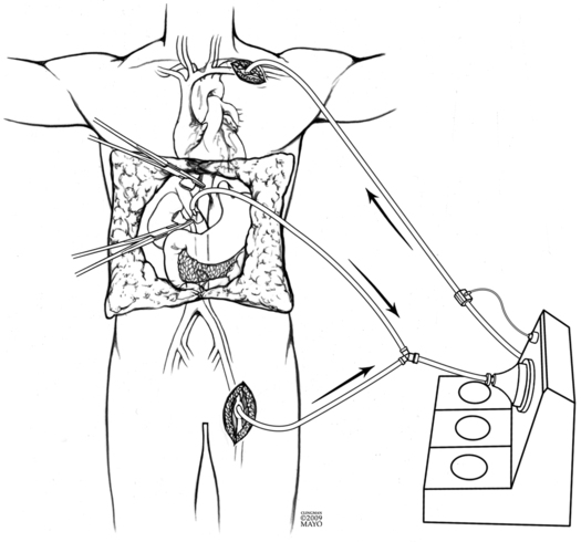
Initial dissection and hepatectomy can result in significant blood loss from friable dilated vessels in the abdominal wall, in the abdomen, and around the liver. Excision of the liver involves mobilization and then clamping and dividing the hepatic vasculature (hepatic artery, portal vein, and IVC). Venovenous bypass is occasionally used depending on surgeons’ experience and preference to overcome the loss of venous return to the heart, the lack of which can cause cardiovascular collapse. Cannulas are placed in the portal and femoral veins; blood drains by gravity to a centrifugal pump, which then returns the blood to the upper body via a large-bore cannula (Figure 176-1). An alternative surgical approach, and one more commonly used, is the “piggyback” technique, in which the surgeon separates the liver from the IVC using a side bite of the IVC (i.e., partial IVC occlusion), allowing some IVC flow to continue during surgery. With this approach, portal venous return is still lost.

Once vascular anastomoses to the graft are complete, recirculation occurs. Liver inflow is restored by opening the portal vein (with or without the hepatic artery), blood is flushed through the nearly complete anastomosis, and then the IVC clamp is released. This results in the abrupt delivery of cold, potassium-containing, acidic blood to the heart, along with, occasionally, microthrombi or even air. Hypotension is common, pulmonary artery pressures elevate, and cardiac arrhythmia or even cardiac arrest may occur. Intravenously administered calcium chloride antagonizes potassium-induced changes, and low-dose epinephrine is frequently used for immediate hemodynamic support. Prolonged hypotension after recirculation, termed the postreperfusion syndrome, is most often due to systemic vasodilation; however, myocardial depression is sometimes seen. Vasopressor, inotropic, or both vasopressor and inotropic support should be used, as indicated; resolution typically occurs within 30 min. The final stage of transplantation involves hepatic artery anastomosis (if not already performed) and a biliary drainage procedure.

Intraoperative coagulation and transfusion management is often challenging; although average volumes of transfusions of blood and blood products have decreased in recent years, catastrophic bleeding still occurs. In addition to having low levels of circulating clotting factors due to decreased synthesis, most patients are thrombocytopenic as a result of sequestration and destruction of platelets in the liver and spleen. After recirculation, tissue plasminogen activator activity rises, which can result in marked fibrinolysis. Heparinoids are also released from the reperfused liver. Treatment of coagulopathy with platelets, fresh frozen plasma, and cryoprecipitate should take into account both the results of coagulation testing and the clinical situation. In the absence of significant bleeding, complete correction of coagulation abnormalities (according to the results of laboratory tests) is not undertaken because correction may increase the risk of thrombosis, particularly of the hepatic artery. Prophylactic use of antifibrinolytic agents is not usual; their use may be considered if clinically significant fibrinolysis occurs, as can be demonstrated with thromboelastography. Should catastrophic coagulopathy and bleeding occur, the use of recombinant factor VIIa is a consideration, but several deaths due to complete intravascular thrombosis following this treatment have been reported. Therefore, the use of recombinant factor VIIa should be guided by thromboelastography to ensure that the patient is not hypercoagulable. When large volumes of blood and blood products are transfused, ionized hypocalcemia secondary to citrate chelation may occur. It should also be noted that the quantity of blood and blood products transfused are both independent predictors of poor outcome in liver transplantation, so overtransfusion should be avoided. The goal of maintaining a hemoglobin concentration in the 8-g/dL to 10-g/dL range is common.