Anesthesia for myotonic dystrophy

Published on 07/02/2015 by admin

Filed under Anesthesiology

Last modified 22/04/2025

Print this page

rate 1 star rate 2 star rate 3 star rate 4 star rate 5 star
Your rating: none, Average: 0 (0 votes)

This article have been viewed 1896 times

Anesthesia for myotonic dystrophy

Joseph J. Sandor, MD

Myotonia—the repetitive firing of muscle action potentials causing prolonged muscle contractions even after mechanical stimulations to the muscles have ceased—is a hallmark of myotonic dystrophy. The grip test is a quick and easy way to determine the presence of active myotonia. Patients with myotonic dystrophy are unable to relax the muscles of the hand after forming a fist.

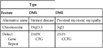
Myotonic dystrophies are autosomal dominantly transmitted disorders and classified as DM1 (Steinert disease) or DM2 (proximal myotonic myopathy), with DM1 further subdivided by age of onset. Prominent features of classic DM1, the most common myotonic dystrophy, include slowly progressive muscle weakness (dystrophy), cataracts, endocrine disturbances, and functional abnormalities of the cardiorespiratory system and gastrointestinal tract (Table 209-1).

Table 209-1

Genetics of Myotonic Dystrophy

  Type
Feature DM1 DM2
Alternative name Steinert disease Proximal myotonic myopathy
Chromosome 19q13.3 3q21
Defect
 Gene
 Repeat
DMPK
CTG
ZNF9
CCTG

image

An abnormality in the intracellular adenosine triphosphate (ATP) system that fails to return calcium to the sarcoplasmic reticulum is the theoretical pathologic mechanism of the myotonia. Contractions are not relieved by nondepolarizing neuromuscular blocking agents (NMBAs), regional anesthesia, or deep anesthesia; however, infiltration of local anesthetic agents into involved muscle may produce relaxation. Depression of rapid sodium flux into muscle cells by the use of phenytoin, procainamide, quinine, tocainide, or mexiletine may alleviate contractions by delaying membrane excitability.

Coexisting organ system dysfunction

Cardiac involvement is characterized by conduction system abnormalities, supraventricular and ventricular arrhythmias, and, less commonly, myocardial dysfunction and ischemic heart disease. Mitral valve prolapse occurs in 20% of affected individuals. Sudden death is usually related to abrupt onset of atrioventricular block. Cardiomyopathy is rare.

Pulmonary pathophysiology may be both structural and functional. Pulmonary function testing reveals a restrictive lung disease pattern due to contractures of intercostal muscles. Ventilatory responses to hypoxia and hypercarbia are impaired. Patients are predisposed to developing pneumonia as a result of reduced lung volume and ineffective cough mechanisms.

Gastrointestinal abnormalities that predispose patients with myotonic dystrophy to aspirating gastric content include dysphagia because of pharyngeal muscle weakness with impaired airway protection, gastric atony, and intestinal hypomotility. Additional gastrointestinal abnormalities include dysphagia, constipation, gallbladder stones, and pseudoobstruction.

Central nervous system manifestations include attention disorders, cognitive impairment, and mental retardation. Endocrine abnormalities include diabetes mellitus, thyroid dysfunction, adrenal dysfunction, and hypogonadism. Because insulin resistance is common in patients with myotonic dystrophy—likely related to reduced relative capacity of the myocyte insulin receptor in these patients—maintaining optimal glycemic control is often difficult.

Pregnancy exacerbates myotonic dystrophy, probably secondary to elevated progesterone levels. Women with myotonic dystrophy have a high incidence of obstetric complications, including polyhydramnios, premature onset of labor, breech presentation, impaired cervical dilation, uterine atony, retained placenta, and postpartum hemorrhage.

Anesthetic management

Patients with DM1 have an increased risk of developing complications related to anesthesia, with reported complication rates ranging from 10% to 42.9%; comparatively, the risk of serious complications occurring in patients with DM2 is low (0.6%), likely related to the general lack of impairment of respiratory muscles in DM2. Risk factors that appear to be related to a higher incidence of perioperative adverse events, at least in children, appear to be a high score on the muscular impairment rating scale, prolonged operative time (>1 h), perioperative morphine use, and lack of reversal of NMBAs.

The preoperative evaluation of a patient with myotonic dystrophy should focus on pulmonary function, the patient’s ability to protect the airway, cardiac conduction abnormalities, and cardiac reserve. Preoperative medications might include orally administered nonparticulate antacids and metoclopramide. Patients with DM1 have an increased sensitivity to preoperative sedative medications, and these agents should be avoided, if possible. These patients also have an increased sensitivity to intravenously administered induction agents; thus, these drugs should be used cautiously and titrated to effect. Etomidate may cause myoclonus and may precipitate fasciculations.

Depolarizing neuromuscular blockade is to be avoided because it may exacerbate fasciculations and contractures. Succinylcholine-induced fasciculation can lead to contractions severe enough to impair ventilation. Because patients with myotonic dystrophy often have impaired airway-protective mechanisms, placement of a cuffed tracheal tube is recommended.

Tracheal intubation can be performed while the patient is awake or as a rapid-sequence induction with cricoid pressure along with a reduced dose of an intravenously administered induction agent. Neuromuscular blockade might not be needed to facilitate intubation. Maintenance of anesthesia is best achieved with a balance of inhalation agent, opioids, and NMBA, all titrated to effect. Total intravenous anesthesia with propofol may also be a valuable and effective anesthetic technique for patients with myotonic dystrophy.

Myocardial depression may result with the use of any induction agents, but cautious use may minimize these effects and obviate the need for intravenously administered NMBAs. Exaggerated respiratory depressant effects from opioidergic agents used for anesthetic maintenance should be anticipated. The use of short-acting opioids (fentanyl, alfentanil, or remifentanil) for supplemental analgesia is recommended.

If neuromuscular blockade is needed, careful titration of shorter-acting agents (cisatracurium or mivacurium) is guided by the use of a peripheral nerve stimulator, the use of which is recommended throughout the procedure. Use of an anticholinesterase agent for reversal of neuromuscular blockade could theoretically precipitate myoclonus because of acetylcholine-facilitated depolarization at the neuromuscular junction. Use of short-acting NMBAs may eliminate the need for pharmacologic reversal.

Postoperative shivering should be avoided because shivering may precipitate fasciculation, myoclonus, or both. Patients’ core temperature should be maintained at 36.5° C or higher through the use of a body warmer and whatever other measures are appropriate.

Extubation should be performed when the patient can protect his or her airway.