Anatomy of the larynx
Description
The larynx connects the inferior pharynx with the trachea and, in so doing, serves three functions (Box 51-1): to maintain a patent airway, to guard against aspiration of liquids or solids into the trachea, and to permit vocalization. It is about 5 cm in length and, in adults, lies at the level of C4 to C5. In cross-section at the level of the laryngeal prominence (Adam’s apple), the larynx is triangular because of the shape of the thyroid cartilage. At the level of the cricoid cartilage, the larynx becomes more round. The larynx provides the area of greatest resistance to passage of air to the lungs.
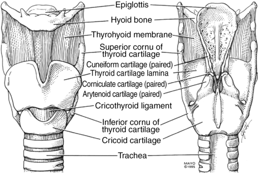
Laryngeal skeleton
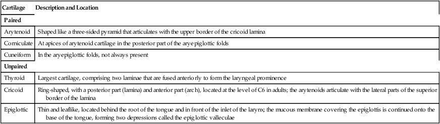
The laryngeal skeleton has a total of nine cartilages (Table 51-1): three sets of paired cartilages (arytenoids, corniculates, cuneiforms) and three unpaired cartilages (thyroid, cricoid, and epiglottic) (Figure 51-1).
Table 51-1
| Cartilage | Description and Location |
| Paired | |
| Arytenoid | Shaped like a three-sided pyramid that articulates with the upper border of the cricoid lamina |
| Corniculate | At apices of arytenoid cartilage in the posterior part of the aryepiglottic folds |
| Cuneiform | In the aryepiglottic folds, not always present |
| Unpaired | |
| Thyroid | Largest cartilage, comprising two laminae that are fused anteriorly to form the laryngeal prominence |
| Cricoid | Ring-shaped, with a posterior part (lamina) and anterior part (arch), located at the level of C6 in adults; the arytenoids articulate with the lateral parts of the superior border of the lamina |
| Epiglottic | Thin and leaflike, located behind the root of the tongue and in front of the inlet of the larynx; the mucous membrane covering the epiglottis is continued onto the base of the tongue, forming two depressions called the epiglottic valleculae |
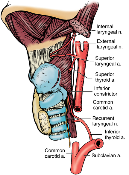
Innervation
Innervation to the larynx is supplied by the superior laryngeal nerve (Figure 51-2), a branch of the vagus nerve (X). This nerve has two terminal branches: the internal laryngeal nerve, which is purely sensory and has fibers from the mucosa of the tongue to the vocal folds (including the superior surface of these folds) and the external laryngeal nerve, which is purely motor and innervates the cricothyroid muscle. Another branch of cranial nerve X, the recurrent laryngeal nerve, provides branches to all of the muscles of the larynx, except the cricothyroid, and provides sensory innervation below the vocal cords. Its terminal branch, the inferior laryngeal nerve, has anterior and posterior branches.
Muscles
The extrinsic muscles comprise depressors and elevators (Box 51-2), which move the larynx as a whole. The intrinsic muscles, which are considered as functional groups, include the abductors and adductors. The abductors (the arytenoid muscle, aryepiglottic muscle, transverse arytenoid muscle, oblique arytenoid muscle, and thyroepiglottic muscle) open the inlet to the larynx and the adductor (lateral cricoarytenoid muscle) closes the vocal folds.