Published on 18/03/2015 by admin
Filed under Dermatology
Last modified 22/04/2025
This article have been viewed 1724 times
William Y-M Tang and Loi-yuen Chan
Evidence Levels: A Double-blind study B Clinical trial ≥ 20 subjects C Clinical trial < 20 subjects D Series ≥ 5 subjects E Anecdotal case reports
Amyloid is an altered, insoluble protein that can accumulate in one or many organs, causing dysfunction. Primary localized cutaneous amyloidosis is characterized by the deposition of amyloid in the skin without involving any internal organ. It occurs more commonly in Southeast Asian, Chinese, Middle Eastern, and South American people. There are three clinical forms: lichen, macular, and nodular. The co-occurrence of macular and lichen amyloidosis in a patient is known as biphasic amyloidosis. The amyloid in macular and lichen amyloidosis is derived from degenerated keratinocytes, whereas in nodular amyloidosis it is derived from immunoglobulin light chains from a local plasma cell clone.
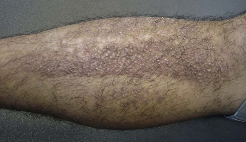
Lichen amyloidosis (see Figure) is a persistent eruption of multiple red-brown hyperkeratotic papules often affecting extensor aspects of extremities, especially the pretibial surfaces. It appears more commonly in males. Apart from its cosmetic nuisance, marked itching can occur. Although familial cases of lichen amyloidosis have been reported, most cases occur as isolated events having no association with systemic disease.
Macular amyloidosis is characterized by an eruption consisting of small, dusky-brown or grayish pigmented macules distributed symmetrically over the upper back and upper arm. It has a reticulated or rippled pattern. Itch is variable, and patients often seek medical advice for aesthetic issues and pruritus.
Nodular amyloidosis is the rarest subtype. It is characterized by single or multiple waxy, firm, brown or pink nodules involving the legs, head, trunk, arms, and genitalia. It is usually asymptomatic.
Lesions of localized cutaneous amyloidosis can produce considerable pruritus. Patients seek treatment to alleviate pruritus and the undesirable appearance. Currently there are no accepted standard treatments for the various types of cutaneous amyloidosis because of a lack of good clinical trials. As pruritus is a common symptom, antihistamines and topical corticosteroids are prescribed as first-line treatments.
Phototherapy (UVB or PUVA) has been used to treat lichen amyloidosis successfully for relief of pruritus. Acitretin may be added for combined therapy. Laser treatments reported to be successful in treating cutaneous amyloidosis include carbon dioxide, pulsed-dye (PDL) and neodymium:yttrium aluminum garnet (Nd:YAG).
Dermabrasion has been successful in treating lichen and nodular amyloidosis. This improves cosmesis and alleviates pruritus, but brings accompanying procedural pain and the development of skin atrophy. There is an anecdotal report that dermabrasion of lichen amyloidosis under tumescent anesthesia can result in remarkable pain reduction even though the total amount of local anesthetic required is low.
Other treatment choices include tacrolimus, transcutaneous electrical nerve stimulation (TENS) and tocoretinate. Topical dimethylsulfoxide (DMSO) also has been reported to benefit lichen and macular amyloidosis. However, a more recent study on 25 patients reported lack of efficacy.
Skin biopsy
All forms of amyloidosis have similar histological findings. On light microscopy, amyloid is characteristically a pink, amorphous material. Special stains, such as Congo red and crystal violet, can highlight the amyloid deposit. Amyloid can be metachromatically stained red by crystal violet staining of an aqueous mount of the specimen. Congo red staining of amyloid shows apple-green birefringence under polarized light microscopy.
Sedating antihistamines are commonly prescribed to relieve the pruritus. High-potency topical corticosteroids may provide symptom relief and thinning of lesions. Although there are no specific studies investigating the efficacy of antihistamines and topical corticosteroids in cutaneous amyloidosis, they are the first-line treatments and some patients respond well.
Jin AG, Por A, Wee LK, Kai CK, Leok GC. Photodermatol Photoimmunol Photomed 2001; 17: 42–3.
In this study, 14 patients with lichen amyloidosis were treated with either UVB (n = 9) or PUVA (n = 5) to half of the body, applying potent topical corticosteroids to the other half as a control. After 8 weeks of treatment, patients treated with phototherapy had more improvement in average roughness of lesions and itch than on the control side. Improvement of roughness in the UVB-treated lesions was significant. However, the difference in improvement in itch in the UVB- and PUVA-treated lesions compared to the control lesions was not significant.
Grimmer J, Weiss T, Weber L, Meixner D, Scharffetter-Kochanek K. Clin Exp Dermatol 2007; 32: 39–42.
Two male patients with lichen amyloidosis over the extensor surfaces of the lower legs were treated with bath PUVA three to four times per week for 11 weeks, plus oral administration of acitretin 0.5 mg/kg/day for 7 months. There was almost complete resolution of skin lesions in both patients. No relapse was observed for 8 months after discontinuation of treatment.
Hernandez-Nunez A, Dauden E, Moreno de Vega MJ, Fraga J, Aragues M, Garcia-Diez A. Clin Exp Dermatol 2001; 26: 256–9.
A 73-year-old man with a 15-year history of biphasic amyloidosis was treated with acitretin 35 mg once daily (0.5 mg/kg/day). Pruritus resolved completely after 2 weeks of treatment. Acitretin was continued for 6 months and then stopped. There was no recurrence during the subsequent 6 months of follow-up.
Hamzavi I, Lui H. Dermatol Surg 1999; 25: 726–8.
A 63-year-old woman with asymptomatic nodular amyloidosis on the nose was successfully treated with CO2 laser. Initially, the laser was set at a power of 8 W. During treatment it was noted that the tissue was highly friable, requiring an increase of the power to 15 W to achieve hemostasis. The lesion healed with an excellent cosmetic response 7 months after treatment.
The use of CO2 laser for nodular primary localized cutaneous amyloidosis was first reported by Truhan et al. in 1986. In their report the treatment produced a good cosmetic result, although post-treatment biopsies showed residual amyloid.
Sawamura D, Sato-Matsumura KC, Shibaki A, Akiyama M, Kikuchi T, Shimizu H. J Eur Acad Dermatol Venereol 2005; 19: 262–3.
A 59-year-old man with lichen amyloidosis for 5 years was treated with two sessions of 585 nm PDL. The treatment parameters were 7 mm spot size and a fluence of 6.0 J/cm2. Reassessment at 8 weeks showed great reduction of itch with decreasing size of papules, although complete clearance was not achieved. The improvement persisted for more than 15 months after treatment.
Ostovari N, Mohtasham N, Oadras MS, Malekzad F. J Eur Acad Dermatol Venereol 2008; 22: 442–6.
Twenty patients with histology confirmed macular amyloidosis were treated with Q-switched Nd:YAG laser: 532 nm in a part of their plaques and with 1064 nm in another part of their plaques. Assessment of efficiency was done by colorimetric scores and digital photographs before laser therapy and 8 weeks after treatment. Both lasers were effective in reducing pigmentation, with the 532 nm more effective. Ninety per cent of cases treated by 532 nm had good or very good response, and for the 1064 nm treated patches, 60% of cases had the good or very good response.
Lien MH, Railan D, Nelson BR. J Am Acad Dermatol 1997; 36: 315–16.
A 45-year-old white man with multiple nodular amyloidosis lesions on his chin for 2 years was treated by shave excision followed by superficial dermabrasion. The treatment area was first sprayed with fluoroethyl-free spray. Dermabrasion was carried out using a Bell hand engine with a regular wire brush followed by a smooth diamond fraise. There was no recurrence at 26 months of follow-up.
The largest study on efficacy of dermabrasion on nodular amyloidosis was reported by Wong CK et al. in 1982, where seven patients showed satisfactory improvement after a follow-up of at least 5 years.
Castanedo-Cazares JP, Lepe V, Moncada B. Dermatology 2002; 205: 420–1.
One patient with lichen amyloidosis diagnosed clinically was treated with tacrolimus 0.1% ointment twice daily. Resolution of pruritus was noted after 2 weeks of therapy, and marked improvement of plaque thickness was observed after 2 months.
This is the only report on the treatment of lichen amyloidosis using topical tacrolimus.
Yüksek J, Sezer E, Aksu M, Erkokmaz U. J Dermatol 2011; 38: 546–52.
Eight patients with macular amyloidosis and eight with lichen simplex were treated with high-frequency TENS thrice weekly for 4 weeks, 30 minutes duration to areas with most intense itching. All patients with macular amyloidosis and six (75%) with lichen simplex had relief of their pruritus.
Terao M, Nishida K, Murota H, Katayama I. J Dermatol 2011; 38: 179–84.
Tocoretinate is a synthetic esterified compound of retinoic acid and tocopherol that is used for treating skin ulcers and improving skin manifestations of scleroderma, morphea, and hypertrophic scars. Ten patients with lichen amyloidosis and macular amyloidosis were treated daily with topical tocoretinate ointment. The outcome was very good for four, good for two, moderate for two and poor for two. Normalization of epidermal differentiation shown in vivo is thought to be beneficial in the treatment of amyloidosis.
Treatment of Skin Disease Comprehensive Therapeutic Strategies 4e
WhatsApp us


 Sedating antihistamines
 Topical high-potency corticosteroids
 Phototherapy/photochemotherapy
 Oral retinoids
 Laser
Laser Dermabrasion
 Tacrolimus
 Excision
 Transcutaneous electrical nerve stimulation
 Tocoretinate