Alimentary system49
8.2 Respiratory system51
The alimentary and respiratory systems develop from the embryonic gut tube. Their common origin is reflected in both anatomy – they share a common upper portion (the pharynx) – and function, absorption of material from the external environment and excretion of waste products. They are associated with glands that secrete substances into the lumen of the tube for digestion or lubrication, and with numerous aggregations of lymphoid tissue in the walls that help repel foreign organisms.
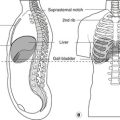
8.1. Alimentary system (Fig. 8.1)
The alimentary system consists of everything between the mouth and the anus together with all the tubes and secretory units (exocrine glands) that open into it.
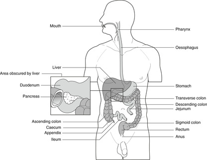 |
| Fig. 8.1 |
Exocrine glands
The glands which secrete substances into the lumen of the gut tube (exocrine glands) include:
• submandibular, sublingual, parotid and minor salivary glands secreting substances that begin the digestive process
• mucous glands secreting mucus for lubrication (particularly in the colon where the contents become progressively more solid)
• acid-secreting glands in the stomach for digestion
• glands secreting enzymes for digestion. These include exocrine pancreatic tissue and the bile-secreting cells of the liver. It is important to understand that these are both part of the alimentary system, as are all the smaller glands opening into the gut tube lumen.
Endocrine tissue
Endocrine cells secrete a variety of substances:
• insulin, glucagon and other digestive hormones
• amines such as catecholamines.
In some places endocrine cells are aggregated to form distinct endocrine organs, while in other places they remain isolated as single endocrine secretory units scattered throughout the epithelium.
Lymphoid tissue
The lumen of the gut tube is part of the external environment and gut epithelium is therefore a potential site of entry of foreign matter. Lymphoid material, which is concerned with the recognition of self/non-self and the defence of the organism from foreign invasion, is present in the wall of the gut tube to monitor potential trouble arising from this part of the outside world. It is particularly obvious in the mouth and pharynx where a ‘ring’ of lymphoid tissue (Waldeyer’s ring: tonsils, adenoids) encircles the oropharynx, the ileum (Peyer’s patches) and the appendix. From the lymph plexuses in the walls of the gut tube, lymph passes to nodes that drain, as for the rest of the body, to the thoracic duct.
Muscular coat of the gut tube
This involuntary muscle allows propulsion of contents through the gut tube and in the stomach is especially important for the churning movements of digestion. For the most part there are two muscular layers – inner circular and outer longitudinal – although there is an extra partial coat in the stomach. These muscles are supplied by the myenteric plexuses in and around the muscle coats of the gut tube.
Mobility of the gut tube: mesenteries
Those parts of the gut tube that are most mobile are in the abdomen and are surrounded by a serous membrane, the peritoneum, to allow friction-free movement. Figure 3.1A (p. 16) shows the original primitive state of the arrangement of the peritoneum with the mesentery of the gut tube connecting the gut tube to the body wall and allowing vessels and nerves access. For some parts of the intestines this persists (stomach, jejunum, ileum, transverse colon, sigmoid colon), even if somewhat modified, but for other parts of the gut tube, there is no apparent mesentery, it having been incorporated into the dorsal body wall, as described in Chapter 11.
8.2. Respiratory system (Fig. 8.2)
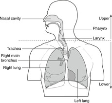 |
| Fig. 8.2 |
They are separated by the vocal cords.
Walls of the respiratory passages
The respiratory tubes must remain patent for the passage of air. They are held open by skeletal elements, notably the hyoid bone and laryngeal cartilages in the neck, and hyaline cartilage in the walls of the trachea and bronchi. Apart from the numerous small mucous glands, there are no important associated exocrine glands. As in the alimentary canal, endocrine cells are found in the walls and may (rarely) give rise to tumours. There is no significant lymphoid tissue in the walls of the respiratory system except for lymphatics and Waldeyer’s ring referred to earlier.