Disposition and origin53
9.2 Urinary tract53
9.3 Reproductive tract53
The urinary and genital components of the urogenital system are derived from common embryological sources. Their functions are at first sight dissimilar: the urinary system is mainly for excretion of waste products from the blood stream, and the genital system for reproduction. In mammals, organs of both systems are confined to the lower abdomen, pelvis and perineum.
9.1. Disposition and origin
The urogenital system is confined in mammals to the abdomen, pelvis and perineum. The two components, urinary and genital, have a common embryological origin.
• The gonads and kidneys (and adrenal cortex) are from embryonic tissue on the posterior coelomic wall (the coelom is in the early embryo what will, in this region, become the abdominal cavity). This common origin is reflected, despite subsequent movements during development, in the blood supply, lymph drainage and, for the adrenal cortex and the gonads, the secretion of steroid hormones.
• Much of the duct systems of the urogenital organs originate with the terminal portion of the gut: they are either cloacal derivatives, or they open into the cloaca (Latin: sewer). These structures include:
– in both sexes, the urethra, bladder, ureters, collecting ducts of the kidney
– in the female, the vagina, uterus and Fallopian tube
– in the male, the ductus (vas) deferens and seminal vesicles.
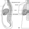
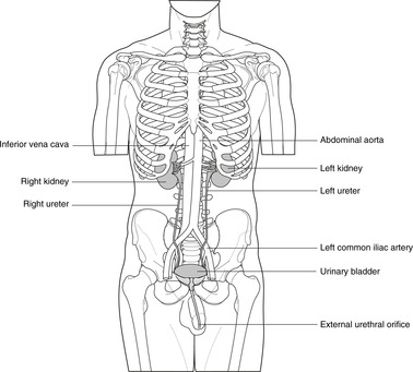
9.2. Urinary tract (Fig. 9.1)
Urine is produced in the right and left kidneys and passes through the right and left ureters to the bladder. It is expelled from the bladder through the (midline) urethra. This is short in the female and longer in the male, traversing the penis.
 |
| Fig. 9.1 |
9.3. Reproductive tract
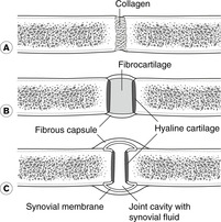
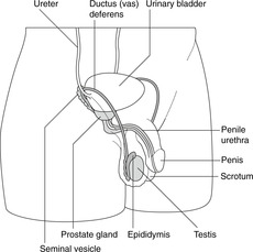
Male reproductive organs (Fig. 9.2)
The paired testes develop high on the posterior body wall and before birth descend to the scrotum. Spermatozoa are produced in the testis and at ejaculation are propelled along the ductus (vas) deferens on each side to join the (midline) urethra in the substance of the prostate gland. They travel to the tip of the penis in the urethra.
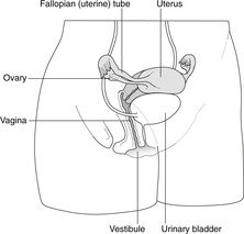
Female reproductive organs (Fig. 9.3)
The ovaries also develop high on the posterior body wall but do not descend as far as the testis so that the ovaries remain in the lower abdominal cavity. The uterus and Fallopian (uterine) tubes develop from a paired tubular system that partially unites. Those parts which remain paired are the Fallopian tubes; those that unite form the uterus in which the fetus develops, and the vagina which receives the penis at copulation and through which the baby is born.
Self-assessment on Section 1 (Chapter 1, Chapter 2, Chapter 3, Chapter 4, Chapter 5, Chapter 6, Chapter 7 and Chapter 8)
Single best answer questions
Questions 10–25
10. In the anatomical position, the palms of the hands face:
a. anteriorly.
b. inferiorly.
c. posteriorly.
d. superiorly.
11. Straightening the upper limb at the elbow joint is
a. abduction.
b. adduction.
c. extension.
d. flexion.
12. The tough fibrous membrane that encloses some compartments of the body is:
a. adipose tissue.
b. deep fascia.
c. loose connective tissue.
d. superficial fascia.
13. Which of the following bones is part of the axial skeleton:
a. humerus.
b. sacrum.
c. scapula.
d. tibia.
14. The equivalents in the foot of metacarpals of the hand are:
a. metatarsals.
b. phalanges.
c. talus.
d. tarsals.
15. The membranous outer covering of bone is:
a. cancellous bone.
b. cortical bone.
c. endosteum.
d. periosteum.
16. Which of the following is a disease of articular (hyaline) cartilage:
a. osteoarthritis.
b. rheumatoid arthritis.
c. synovitis.
d. tenosynovitis.
17. The suprasternal notch is at vertebral level:
a. C7.
b. T2.
c. T4.
d. T6.
18. Which is the correct order of components of the brain stem from below upwards:
a. medulla, midbrain, cerebellum.
b. medulla, pons, midbrain.
c. midbrain, pons, medulla.
d. pons, medulla, midbrain.
19. Cell bodies of primary sensory neurons are found in:
a. dorsal grey matter.
b. dorsal root ganglia.
c. ventral grey matter.
d. ventral root ganglia.
20. The spinal meningeal layer nearest the bone in the vertebral canal is the:
a. arachnoid mater.
b. dura mater.
c. filum terminale.
d. pia mater.
21. A venous system that begins and ends in capillary beds is called:
a. deep.
b. hepatic.
c. portal.
d. systemic.
22. The part of the gut tube that immediately succeeds the duodenum is the:
a. caecum.
b. ileum.
c. jejunum.
d. pylorus.
24. The gonads share a common embryological origin with the:
a. gut tube.
b. heart.
c. kidneys.
d. nervous tissue
25. Concerning the spinal cord and meninges:
a. the lower limit of the dural sac is at vertebral level S5.
b. the lower limit of the subarachnoid space is at vertebral level L3.
c. the sacral region of the spinal cord is in the region of vertebrae T12–L3.
d. the extra(epi)dural space contains CSF.
Matching item questions
Questions 26–30: cells of the nervous system
Match the numbered item to the lettered response. Each lettered response may be used once, more than once, or not at all.
a. ventral horn cells
b. lateral horn cells
c. dorsal columns
d. Schwann cells
e. oligodendrocytes
26. smooth muscle
27. skeletal muscle
28. preganglionic sympathetic neurons
29. sensation
30. myelin production in peripheral nerves
Questions 31–35: spinal canal contents
Match the numbered item to the lettered response. Each lettered response may be used once, more than once, or not at all.
a. extradural space
b. subdural space
c. subarachnoid space
d. cauda equina
e. filum terminale
31. contains a venous plexus
32. contains cerebrospinal fluid
33. consists of rootlets of spinal nerves
34. ends at vertebral level S2
35. begins at vertebral level L3/4 in the newborn child
Questions requiring short answers
36. Name the serous cavities of the body. What are the serous membranes, and how are they arranged? What is their function?
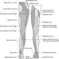
37. What are: (a) dermatomes, (b) myotomes, (c) Langer’s lines, (d) superficial fascia, (e) deep fascia?
38. What do the following terms mean in respect of veins: (a) superficial, (b) pulmonary, (c) portal, (d) systemic?
39. List the components of the alimentary system in the correct order from mouth to anus.
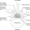
40. Draw a diagram of a typical vertebra naming the following parts: lamina, pedicle, transverse process, spinous process, body, vertebral canal. How do two typical vertebrae articulate with each other?
41. Describe or draw a cross-section through the mid-thoracic spinal cord labelling grey and white matter, central canal, and spinal nerve attachments. Indicate the position of cell bodies of motor neurons supplying voluntary muscle, cell bodies of motor neurons supplying involuntary muscle, and the dorsal white column. Which cells are attacked in poliomyelitis, and with what result?
43. List the structures that would be penetrated by a lumbar puncture needle. Where would you perform this, and why?
44. What is a dorsal root ganglion? Is it part of the peripheral or the central nervous system? What is its function? What disease may affect it?
Self-assessment on Section 1: answers
True/false answers
1.
b. False. Inferior means below (when standing in the anatomical position). Inferior is a term of position, not size or quality.
c. False. The division is into anterior and posterior portions, not right and left.
d. True.
e. True. Skeletal muscle is supplied by the somatic nervous system.
2.
a. True. These are synovial articulations. The head of a typical rib articulates with two vertebrae: that corresponding in number to the rib, and that above. The tubercle of a typical rib articulates with the transverse process of the corresponding vertebra.
b. False. The first costosternal is a primary cartilaginous joint.
c. True. There is also a small synovial cavity between the cartilage and the sternum.
d. True.
e. False. The second rib articulates at the sternal angle.
3.
a. True.
b. True.
c. False. These are symphyses (secondary cartilaginous).
d. False. This is also a symphysis.
e. True.
4.
a. True.
b. True.
c. False. It is only present in immature bone.
d. True.
e. True. This may have important consequences in the supply to the head of a bone in a child.
5.
a. False. This is the filum terminale, not cauda equina. The cauda equina is the name given to the collection of nerve rootlets.
b. True.
c. True. These veins are important in the spread of disease to the vertebral column.
d. True.
e. False. It is inside the dura mater.
6.
a. True.
b. True.
c. False. Hepatic veins drain blood from the liver to the inferior vena cava. Do not confuse the hepatic veins (liver–IVC) with the (hepatic) portal vein (intestines–liver).
d. True. These empty into the azygos vein which is a tributary of the superior vena cava.
e. False. Blood from the femoral veins reaches the heart in the IVC.
7.
a. True.
b. True.
c. False. It is a lower limb artery, at the back of the knee.
d. True.
e. False. It is an artery of the foot.
8.
a. True.
b. False. It is between L3/4 in an adult and lower in the newborn child.
c. False. It is taken from the subarachnoid space.
d. True.
e. True. Two or more basivertebral veins emerge from the posterior aspect of the vertebral body and drain into the plexus.
9.
a. False. This is white matter. Grey matter is predominantly made up of cell bodies.
b. True. It is made by oligodendrocytes in the central nervous system.
c. True. These cells are affected in poliomyelitis.
d. True. Cell bodies of preganglionic sympathetic neurons are here.
e. True.
Single best answers
10. a. See Fig. 2.1.
11. c. See Fig. 2.2.
12. b.
13. b. Axial means midline. The other bones belong to the appendicular skeleton (limbs).
14. a.
15. d. See Section 4.1. Cortical and cancellous are types of bone.
16. a. despite the name. See Section 4.2. Rheumatoid arthritis is a disease of synovium, and the other two terms signify, respectively, inflammation of synovial sheaths and the tendons within them.
17. b.
18. b. See Section 6.3.
19. b. See Sections 6.3 and 6.4. There is no such thing as a ventral root ganglion.
20. b. The arachnoid is ‘inside’ the dura, and the pia mater is on the surface of the spinal cord. The filum terminale is the inferior extension of the pia mater below the end of the spinal cord.
21. c. See Section 7.3. Hepatic veins convey blood from liver to inferior vena cava. The principal portal system is the hepatic portal system, but the word hepatic indicates association with the liver: it does not indicate beginning and ending in capillary beds. Deep veins are found in limbs. Systemic veins include everything except the portal system.
22. c. See Fig. 8.1.
23. b. See Fig. 9.1.
24. c.
25. c. Remember the cord is much shorter than the vertebral column.
Matching item answers
26. b. Lateral horn cells give rise to autonomic neurons supplying visceral smooth muscle.
27. a. Ventral horn cells give rise to somatic neurons supplying voluntary skeletal muscle.
28. b. See 1 above.
29. c. The dorsal columns are large tracts of white matter conveying sensory information up towards the brain.
30. d. Oligodendrocytes produce myelin in the central nervous system.
31. a. The extradural (epidural) space contains the internal vertebral venous plexus.
32. c.
33. d. The rootlets are present because the spinal cord is so much shorter than the vertebral column, so rootlets for the lumbar and sacral nerves have to pass for some distance in the subarachnoid space and vertebral canal before they reach the appropriate intervertebral foramen.
34. c.
35. e. See Figure 6.8 (p. 40).
Short answers
36. The serous cavities are the pleural (right and left), the pericardial and the peritoneal. They are lined by serous membranes: pleura, pericardium and peritoneum. These are arranged such that one layer – the parietal layer – is on the internal aspect of the body wall and one layer – the visceral layer – almost completely surrounds the internal organ (lungs, heart, guts). The parietal and visceral layers are continuous at the site where the internal organ is connected to the body wall (e.g. the hilum of the lung). See Figure 3.1 (p. 16).
37.
a. A dermatome is the area of skin supplied by one spinal nerve. See Figure 6.6 (p. 36).
b. A myotome is the group of skeletal muscle fibres supplied by one spinal nerve.
d. Superficial fascia is subcutaneous tissue of varying thickness; it includes fat, connective tissue, blood vessels and nerves.
e. Deep fascia is the fibrous layer, almost a membrane, which limits the superficial fascia; deep fascia often forms the sheath of a muscle.
38.
a. Superficial veins are those that run in the superficial fascia (see above) and are, if there is not too much fat, visible under the skin.
b. Deep veins accompany the principal arteries deep to the deep fascia. Deep and superficial veins communicate.
c. Portal veins are those which begin and end in capillary beds. The term portal veins usually means the hepatic portal vein conveying blood rich in nutrients to the liver from the alimentary canal between the lower oesophagus and anal canal. Its tributaries are the splenic, superior and inferior mesenteric veins (Fig. 7.3, p. 46).
d. Systemic veins are all other veins that convey blood from the body back to the heart through the superior and inferior vena cava. The term does not usually include the pulmonary veins conveying blood from the lungs to the heart.
39. Components of the alimentary system in order from mouth to anus are: mouth, pharynx, oesophagus, stomach, duodenum, jejunum, ileum, caecum, ascending colon, transverse colon, descending colon, sigmoid colon, rectum, anal canal.
40. See Figure 5.1. (p. 26).
41. See Figure 6.3 (p. 34). Cell bodies of motor neurons supplying voluntary muscle are in the ventral horn of grey matter. Cell bodies of motor neurons supplying involuntary muscle of the trunk are mostly in the lateral horn of grey matter. Ventral horn cells are attacked in poliomyelitis, leading to weakness or paralysis of the muscles supplied.
42. See Figure 6.9 (p. 41). The needle in Figure 6.9 is in the subarachnoid space containing cerebrospinal fluid. Anaesthetic agents injected here would give spinal anaesthesia. Anaesthetic agents injected into the extra(epi)dural space would give an epidural anaesthesia. When drawing the diagram, it would not be necessary to include the rootlets of spinal nerves, although it may be helpful if you let the examiner know that you knew they were there.
43. Assuming that the needle were inserted just to one side of the midline, it would pass through: skin, subcutaneous tissue (superficial fascia), postvertebral muscle, ligamentum flavum, extradural space (fat and vessels), dura mater, arachnoid mater. In an adult you would perform this between vertebrae L3 and L4, well below the termination of the spinal cord at L1/2. In a newborn child you would insert the needle between L4 and L5.
44. A dorsal root ganglion is a swelling on the dorsal root of a spinal nerve as it passes through the intervertebral foramen. It houses the cell bodies of all sensory (pseudounipolar) neurons within that nerve and so destruction of it would result in sensory changes in the dermatome concerned. These ganglia may be infected by microorganisms: the herpes zoster virus is an example. This may give rise to shingles in which there is a vesicular eruption in the skin over the affected dermatome, and dermatome pain.