Acute Respiratory Distress Syndrome

Published on 12/06/2015 by admin

Filed under Pulmolory and Respiratory

Last modified 22/04/2025

Print this page

rate 1 star rate 2 star rate 3 star rate 4 star rate 5 star
Your rating: none, Average: 0 (0 votes)

This article have been viewed 2566 times

28

Acute Respiratory Distress Syndrome

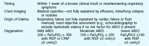
This chapter continues the discussion of respiratory failure with more detailed consideration of one important type of acute respiratory failure: acute respiratory distress syndrome (ARDS). This entity was initially called adult respiratory distress syndrome, but it is not limited to adults, so acute rather than adult is now used to describe it. ARDS represents a major form of hypoxemic respiratory failure. Its clinical and pathophysiologic features differ considerably from those noted for acute-on-chronic respiratory failure. ARDS is characterized by the presence of severe arterial hypoxemia and diffuse bilateral pulmonary infiltrates not due exclusively to cardiogenic or hydrostatic causes. The full criteria for establishing the diagnosis of ARDS are shown in Table 28-1. This chapter describes in detail each of these criteria and the associated pathology and pathophysiology.

Rather than a specific disease, ARDS truly is a syndrome resulting from any of a number of etiologic factors. It is perhaps simplest to consider this syndrome as the nonspecific result of acute injury to the lungs, characterized by breakdown of the normal barrier that prevents leakage of fluid out of the pulmonary capillaries and into the interstitium and alveolar spaces. Another term, acute lung injury, was formerly used to describe a similar process of lung injury in which the disturbance in oxygenation is less severe, whereas ARDS represented the more severe end of the spectrum. However, the current classification eliminates “acute lung injury” as a specific term and instead grades ARDS as mild, moderate, or severe based upon the degree of hypoxemia that is present. A number of other names have been used to describe ARDS, including noncardiogenic pulmonary edema, shock lung, and posttraumatic pulmonary insufficiency.

This chapter first considers the dynamics of fluid transfer between the pulmonary vessels and alveolar interstitium, because any alteration in this process is important in the pathogenesis of ARDS. Next is an outline of the many types of injury that can result in ARDS and some of the theories proposed to explain how such a diverse group of disorders can produce this syndrome. Following is a discussion of the pathologic, pathophysiologic, and clinical consequences. The chapter concludes with a general approach to treatment. More specific details about support of gas exchange are provided in Chapter 29.

Physiology of Fluid Movement in Alveolar Interstitium

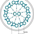
Despite the diverse group of disorders that can cause ARDS, the net result of the syndrome is the same: a disturbance in the normal barrier that limits leakage of fluid out of the pulmonary capillaries and into the pulmonary parenchyma. Before a discussion of some of the theories explaining how this barrier is damaged, a brief consideration of the determinants of fluid transport among the pulmonary vessels, interstitial space, and alveolar lumen may be helpful. The pulmonary parenchyma (Fig. 28-1) consists of (1) small vessels coursing through the alveolar walls, which for simplicity are referred to as the pulmonary capillaries; (2) pulmonary capillary endothelium, the lining cells that normally limit but do not completely prevent fluid movement out of the capillaries; (3) pulmonary interstitium, which is considered here as the alveolar wall exclusive of vessels and the epithelial cells lining the alveolar lumen; (4) lymphatic channels, which are found mainly in perivascular connective tissue in the lungs; (5) alveolar epithelial cells, which line the surface of the alveolar lumen; and (6) alveolar lumen or alveolar space.

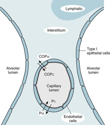
Movement of fluid out of the pulmonary capillaries and into the interstitial space is determined by a number of factors, including the hydrostatic pressures in the vessels and the pulmonary interstitium, the colloid osmotic pressures in these same two compartments, and the permeability of the endothelium. The effect of these factors in determining fluid transport is summarized in the Starling equation, examined in Chapter 15 with regard to fluid transport across the pleural space. The Starling equation is given as Equation 28-1:

image

where F = fluid movement; Pc and Pis = pulmonary capillary and interstitial hydrostatic pressure, respectively; COPc and COPis = pulmonary capillary and interstitial colloid osmotic (oncotic) pressure, respectively; K = filtration coefficient; and σ = reflection coefficient (measure of permeability of endothelium for protein).

If estimates of the actual numbers are substituted for normal hydrostatic and oncotic pressures in Equation 28-1, F is a positive number, indicating that fluid normally moves out of the pulmonary capillaries and into the interstitial space. Even though the rate of fluid movement out of the pulmonary capillaries is estimated to be approximately 20 mL/h, this fluid does not accumulate. The lymphatic vessels are quite effective in absorbing both protein and fluid that have left the vasculature and entered the interstitial space. However, if fluid movement into the interstitium increases substantially or if lymphatic drainage is impeded, fluid accumulates within the interstitial space, resulting in interstitial edema. When sufficient fluid accumulates or the alveolar epithelium is damaged, fluid also moves across the epithelial cell barrier and into the alveolar spaces, resulting in alveolar edema.

Two Mechanisms of Fluid Accumulation

In practice, the forces described in the Starling equation become altered in two main ways, producing interstitial and often alveolar edema (Table 28-2). The first occurs when hydrostatic pressure within the pulmonary capillaries (Pc) is increased, generally as a consequence of elevated left ventricular or left atrial pressure (e.g., in left ventricular failure or mitral stenosis). The resulting pulmonary edema is called cardiogenic or hydrostatic pulmonary edema, and the cause is essentially an imbalance between the hydrostatic and oncotic forces governing fluid movement. In this form of edema, the permeability barrier that limits movement of protein out of the intravascular space is intact, and the fluid that leaks out has a very low protein content.

Table 28-2

CATEGORIES OF PULMONARY EDEMA

Feature Cardiogenic Noncardiogenic
Major cause Left ventricular failure, mitral stenosis Acute respiratory distress syndrome
Pulmonary capillary pressure Increased Normal
Pulmonary capillary permeability Normal Increased
Protein content of edema fluid Low High

In the second mechanism by which fluid accumulates, hydrostatic pressures are normal, but the permeability of the capillary endothelial and alveolar epithelial barriers is increased as a result of damage to one or both of these cell populations. Movement of proteins out of the intravascular space occurs as a consequence of the increase in permeability. The fluid that leaks out has a relatively high protein content, often close to that found in plasma. This second mechanism is the one operative in ARDS. Because an elevation in pulmonary capillary pressure from cardiac disease is not involved, this form of edema is called noncardiogenic pulmonary edema.

Although cardiogenic and hydrostatic pulmonary edema are mentioned here, subsequent parts of this chapter focus on noncardiogenic edema (i.e., ARDS). However, it is important to remember that hydrostatic pressures have an important impact on fluid movement even when the primary problem is a defective permeability barrier. Specifically, higher pulmonary capillary hydrostatic pressures result in more fluid leaking through an abnormally permeable pulmonary capillary endothelium than do lower pressures. At the extreme, some patients with a permeability defect of the pulmonary capillary bed simultaneously have a grossly elevated pulmonary capillary pressure as a result of coincidental left ventricular failure. In these cases, the permeability defect and the elevated hydrostatic pressure work synergistically in contributing to leakage of fluid out of the pulmonary vasculature. Not only is the fluid leak compounded, but when both factors are involved, sorting out the relative importance of each and thus determining the optimal treatment priorities in a given patient can be difficult.

Etiology

Numerous and varied disorders are associated with the potential to produce ARDS (Table 28-3). What these diverse etiologic factors in ARDS have in common is their ability to cause diffuse injury to the pulmonary parenchyma. Beyond that, defining other features that link the underlying causes is difficult on the basis of our present knowledge. Even the route of injury varies. Some etiologic factors involve inhaled injurious agents; others appear to mediate their effects on the lungs via the circulation rather than the airway.

Inhaled Injurious Agents

Numerous injurious agents that reach the pulmonary parenchyma through the airway have been identified. In some cases, a liquid is responsible; examples include gastric contents, salt or fresh water, and hydrocarbons. With acid gastric contents, especially when pH is lower than 2.5, patients sustain a “chemical burn” to the pulmonary parenchyma, resulting in damage to the alveolar epithelium. In the case of near drowning in either fresh or salt water, not only does the inhaled water fill alveolar spaces, but secondary damage to the alveolar-capillary barrier causes fluid to leak from the pulmonary vasculature. Because salt water is hypertonic to plasma, it is capable of drawing fluid from the circulation as a result of an osmotic pressure gradient. Fresh water, on the other hand, is hypotonic to plasma and cellular contents and thus may enter pulmonary parenchymal cells, with resulting cellular edema. In addition, fresh water appears to inactivate surfactant, a complicating factor discussed in more detail under Pathophysiology. Finally, aspirated hydrocarbons can be toxic to the distal parenchyma, perhaps in part because they also inactivate surfactant and cause significant changes in surface tension.

A number of inhaled gases have been identified as potential acute toxins and precipitants of ARDS. Nitrogen dioxide is one example, as are some chemical products of combustion inhaled in smoke. High concentrations of oxygen, particularly when given for prolonged periods, have been considered to contribute to alveolar injury. The mechanism of O2 toxicity is believed to be generation of free radicals and superoxide anions, byproducts of oxidative metabolism that are toxic to pulmonary epithelial and endothelial cells. It is ironic that O2 can contribute to lung injury, given that it is so important in supportive treatment of ARDS. Chapter 29 discusses an additional way in which treatment of ARDS may worsen alveolar damage through overdistention and/or cyclic opening and closing of alveoli induced by mechanical ventilation.

Infectious agents may produce injury via airway access to the pulmonary parenchyma. Pneumonia is the most common underlying clinical problem associated with development of ARDS. Besides bacterial pneumonia, another important example in recent years was pneumonia due to Pneumocystis jiroveci, specifically in patients with AIDS. However, with common use of antiretroviral therapy and availability of effective prophylactic regimens against Pneumocystis, it now is an uncommon cause of ARDS. Another important cause of ARDS is viral pneumonia, which has the capability of damaging parenchymal cells and altering alveolar-capillary permeability.

Injury via Pulmonary Circulation

For causes of ARDS that do not involve inhaled agents or toxins, agents carried within the pulmonary circulation are proposed to initiate the injury in some way. However, in most cases a specific circulating factor has not been identified with certainty, even though several possibilities have been proposed. One of the most common precipitants for ARDS is sepsis, in which microorganisms or their products (especially endotoxin) circulating through the bloodstream initiate a sequence of events resulting in toxicity to parenchymal cells.

Although the term shock lung was used many years ago to describe what is now called ARDS, the presence of hypotensive shock alone is probably insufficient for development of ARDS. Patients in whom ARDS develops seemingly as a result of hypotension usually have complicating potential etiologic factors (e.g., trauma, sepsis) or have received therapy (e.g., blood transfusions) also capable of causing cellular damage.

Patients with the coagulation disorder known as disseminated intravascular coagulation (DIC) appear to have the potential for development of ARDS. In DIC, patients have ongoing activation of both the clotting mechanism and the protective fibrinolytic system that prevents clot formation and propagation. Like ARDS, DIC is a syndrome and can occur because of a variety of primary or underlying causes; although these two problems are frequently associated, whether and exactly how one causes the other is uncertain.

When fat or amniotic fluid enters the circulation, the material is transported to the lungs, resulting in the clinical problems of fat embolism and amniotic fluid embolism, respectively. Presumably these materials are directly toxic to endothelial cells of the pulmonary capillaries, and they certainly have been associated with development of ARDS.

A variety of drugs, many of which fall into the class of narcotics, are potential causes of ARDS. In most cases an overdose of the drug has been taken, although this is not always the situation. One of the agents most frequently recognized has been heroin, and the name “heroin pulmonary edema” sometimes is used. In addition to heroin and other narcotics, several other drugs have been described as occasionally causing ARDS, including even aspirin and thiazide diuretics. Although the problem of drug-induced pulmonary edema has been well described, the mechanism by which it occurs is not certain.

Some patients with acute pancreatitis develop a clinical picture consistent with noncardiogenic pulmonary edema. In this situation, enzymes released into the circulation from the damaged pancreas have been proposed to directly injure pulmonary parenchymal cells or initiate other indirect pathways, resulting in injury.

Certain disorders of the central nervous system, particularly trauma and intracerebral bleeding associated with increased intracranial pressure, are known to be associated with development of ARDS. Similarly, ARDS occasionally occurs after generalized seizures. An interesting and commonly accepted hypothesis to explain this so-called neurogenic pulmonary edema is that intense sympathetic nervous system discharge in response to intracranial hypertension produces extremely high pulmonary capillary pressures, resulting in mechanical damage to the endothelium and subsequent exudation of fluid out of the intravascular space.

Pathogenesis

How do these diverse clinical problems all result in the syndrome of increased pulmonary capillary permeability in ARDS? One important factor appears to be the ability to produce injury to pulmonary capillary endothelial and alveolar epithelial cells (primarily type I epithelial cells, the cytoplasmic processes of which provide most of the surface area lining the alveolar walls). Given the wide variety of insults that can damage these cell types, it seems unlikely that a single common mechanism is operative for all kinds of injury.

In the discussion of some specific causes of ARDS, brief mention was made of a few of the theories of pathogenesis for individual disorders. Here, the more generalized cellular and biochemical mechanisms that are operative during the course of injury to the pulmonary epithelial and capillary endothelial cells are considered. A particularly important component of the pathogenesis of acute lung injury and ARDS appears to be recruitment of inflammatory cells to the lungs, especially neutrophils. An early theory explaining recruitment of neutrophils to the lungs focused on the complement pathway. When complement is activated by sepsis, C5a is released and is responsible for aggregation of neutrophils within the pulmonary vasculature. These neutrophils may release a variety of substances that are potentially destructive to cellular and noncellular components of the alveolar wall. Superoxide radicals, other byproducts of oxidative metabolism, an array of cytokines, and various proteolytic enzymes all can be released by neutrophils and may be important pathogenetically in producing structural and functional injury to the alveolar wall. Examples of specific mediators include endotoxin, products of arachidonic acid metabolism, and cytokines such as tumor necrosis factor (TNF)-α, interleukin (IL)-8, endothelin, and transforming growth factor (TGF)-β. An inflammatory response also can be augmented by a reduction in antiinflammatory mediators, including cytokines such as IL-10 and IL-11.

Activation of complement likely is just one of multiple potential mechanisms for recruiting and sequestering neutrophils in the lungs. Other important factors include cytokines and other mediators that influence neutrophil trafficking in the lungs. Vascular endothelial cells, particularly in the pulmonary vascular system, also become activated, express leukocyte adhesion molecules, and lead to accumulation of neutrophils within the pulmonary vasculature.

Another important process appears to be activation of the coagulation system. Several factors are responsible for what has been called a “procoagulant state,” including release of procoagulant tissue factors, a decreased concentration of factors with anticoagulant activity (e.g., protein C and protein S), and increased activity of proteins that inhibit fibrinolysis (e.g., plasminogen activator inhibitor-1). The result is increased production of thrombin and fibrin as well as evidence of thrombosis within pulmonary capillaries.

Despite extensive research efforts over the past decades to elucidate the mechanisms of acute lung injury, a true understanding of ARDS is still a long way off. Such an understanding will be critical to the development of effective forms of prevention and therapy.

Pathology

Despite the number of etiologic factors in ARDS, the pathologic findings are relatively similar regardless of the underlying cause. As observed by the pathologist, this pattern of injury accompanying ARDS is frequently labeled diffuse alveolar damage.

Injury to type I alveolar epithelial cells and pulmonary capillary endothelial cells appears to be the primary factor in pathogenesis. Type I epithelial cells frequently appear necrotic and may slough from the surface of the alveolar wall. Damage to capillary endothelial cells is generally more difficult if not impossible to recognize with light microscopy; electron microscopy may be necessary to see the subtle ultrastructural changes.

Early in the course of ARDS, often called the exudative phase, fluid can be seen in the interstitial space of the alveolar septum as well as in the alveolar lumen. Scattered bleeding and regions of alveolar collapse, which are at least partly related to inactivation of surfactant (by protein-rich alveolar exudates) and decreased surfactant production resulting from injury to alveolar type II epithelial cells, may be seen. The lung parenchyma shows influx of inflammatory cells, both in the interstitial space and often in the alveolar lumen. The cellular response is relatively nonspecific, consisting of neutrophils and macrophages. Fibrin and cellular debris may be seen in or around alveoli.

A characteristic finding in the pathology of ARDS is the presence of hyaline membranes. These membranes are believed to represent the protein-rich edema fluid that has filled the alveoli. The membranes are composed of a combination of fibrin, cellular debris, and plasma proteins that are deposited on the alveolar surface. Although they are nonspecific, their presence suggests that alveolar injury and a permeability problem, rather than elevated hydrostatic pressures, are the cause of pulmonary edema.

After approximately 1 to 2 weeks, the exudative phase evolves into a proliferative phase. As an important part of the reparative process that occurs during the proliferative phase, alveolar type II epithelial cells replicate in an attempt to replace the damaged type I epithelial cells. These hyperplastic type II epithelial cells often figure quite prominently in the pathologic picture of ARDS.

Another component of the proliferative phase is accumulation of fibroblasts in the pulmonary parenchyma. In severe and prolonged cases of ARDS, this fibroblastic response becomes particularly important. In some cases, the damaged lung parenchyma is not repaired but goes on to develop significant scar tissue (fibrosis). Often accompanying the fibrosis are changes in the pulmonary vasculature, which include extensive remodeling and compromise of the lumen of small vessels by intimal and medial proliferation and by the formation of in situ thrombi.

Pathophysiology

Effects on Gas Exchange

Most of the clinical consequences of ARDS follow in reasonably logical fashion from the presence of interstitial and alveolar edema. The most striking problem is alveolar flooding, which effectively prevents ventilation of affected alveoli even though perfusion may be relatively preserved. These alveoli, perfused but not ventilated, act as regions where blood is shunted from the pulmonary arterial to pulmonary venous circulation without ever being oxygenated. This type of shunting is one of the mechanisms of hypoxemia (see Chapter 1), and there is perhaps no better example of intrapulmonary shunting than ARDS.

In ARDS there are regions of not only true shunting but also ventilation-perfusion mismatch. To some extent, this phenomenon results from a nonuniform distribution of the pathologic process within the lungs. In areas where the interstitium is more edematous or where more fluid is present in the alveoli, ventilation is more impaired (even though some ventilation remains) than in areas that have been relatively spared. Changes in blood flow do not necessarily follow the same distribution as changes in ventilation, and ventilation-perfusion mismatch therefore results.

In addition to the direct effects of interstitial and alveolar fluid on oxygenation, other changes appear to be secondary to alterations in the production and effectiveness of surfactant. Chapter 8 refers to surfactant as a phospholipid responsible for decreasing surface tension and maintaining alveolar patency. When surfactant is absent, as is seen in the respiratory distress syndrome of neonates, there is extensive collapse of alveoli. In ARDS, surfactant production may be adversely affected by injury to alveolar type II epithelial cells. Additionally, evidence suggests that as a result of extensive fluid within the alveoli, surfactant is inactivated and therefore ineffective in preventing alveolar collapse.

In terms of oxygenation, both ventilation-perfusion mismatch (with regions of low ventilation-perfusion ratio) and true shunting (ventilation-perfusion ratio = 0) are responsible for hypoxemia. Insofar as shunting is responsible for much of the drop in PO2, supplemental O2 alone may not be capable of restoring oxygenation to normal. In practice, PO2 does rise somewhat with administration of 100% O2, but not nearly to the level expected after such high concentrations of O2. Considering the nature of the problem of ARDS, this response to supplemental O2 should not be surprising. Oxygen improves whatever component of hypoxemia is due to ventilation-perfusion mismatch, but it is ineffective for true shunting.

On the other hand, the absolute level of ventilation in the patient with ARDS remains intact or even increases. As a result, the patient typically does not have difficulty with CO2 retention, except in terminal stages of the disease or in the presence of another underlying pulmonary process. Even though substantial amounts of what is effectively dead space may be present (as part of the overall ventilation-perfusion mismatch), the patient is able to increase total ventilation to compensate for the regions of maldistribution.

Changes in Pulmonary Vasculature

The pulmonary vasculature is subject to changes resulting from the overall pathologic process. Pulmonary vascular resistance increases, probably for a variety of reasons. Hypoxemia certainly produces vasoconstriction within the pulmonary arterial system, and fluid in the interstitium may increase interstitial pressure, resulting in a decrease in size and an increase in resistance of the small pulmonary vessels. The lumen of small vessels may be compromised by microthrombi and proliferative changes in vessel walls (discussed earlier under Pathogenesis and Pathology).

One consequence of the pulmonary vascular changes is alteration in the normal distribution of pulmonary blood flow. Blood flows preferentially to areas with lower resistance, which do not necessarily correspond to the regions receiving the most ventilation. Hence, ventilation-perfusion mismatch again results, with some areas having high and other areas low ventilation-perfusion ratios.

Effects on Mechanical Properties of the Lungs

When considering the mechanical properties of the lungs in ARDS, we must recognize that computed tomographic scanning has demonstrated the distribution of disease to be more heterogeneous than expected on the basis of the diffuse changes seen on chest radiograph. Whereas some regions have been damaged and are quite abnormal, others appear to have been spared from injury. As a result, the alveoli are not diffusely and relatively homogeneously stiffened. Rather, some regions of the lungs have significantly diseased alveoli that ventilate poorly or not at all, whereas others have relatively preserved and well-ventilated alveoli. The net result of having fewer effectively “functional” alveoli is that less volume enters the lungs for any given inflation pressure; by definition this means lung compliance is decreased.

The volume of gas contained within the lungs at functional residual capacity (FRC; i.e., resting end-expiratory position of the lungs) is also significantly decreased. Again, on the basis of the heterogeneity of the pathologic process, decreased FRC is not due to a uniform decrease in volume over all alveoli but rather to a group of alveoli containing little or no gas and another group containing a relatively normal volume of gas. The net result is that patients breathe at a much lower overall lung volume than normal, preferentially ventilating those alveoli that are relatively preserved. The typical breathing pattern resulting from these mechanical changes is characterized by rapid but shallow breaths. This type of breathing pattern is inefficient and demands increased energy expenditure by the patient, which probably contributes to the dyspnea so characteristic of ARDS patients.

Clinical Features

Because ARDS is a clinical syndrome with many different causes, the clinical picture reflects not only the presence of noncardiogenic pulmonary edema but also the presence of the underlying disease. Our concern here with the respiratory consequences of ARDS, irrespective of the cause, directs our focus to the clinical effects of the syndrome itself rather than to those of the underlying disorder.

After the initial insult, whatever it may be, there is generally a lag of several hours to a day or more before respiratory consequences ensue. In most cases, the first symptom experienced by the patient is dyspnea. As this develops, examination often shows the patient to be tachypneic, although the chest radiograph may not reveal significant findings. However, arterial blood gases reflect a disturbance of oxygenation, often with an increase in the alveolar-arterial difference in partial pressure of oxygen (AaDO2). Alveolar ventilation is either normal or (more frequently) increased, so PCO2 is generally below baseline. In the most severe cases, alveolar ventilation cannot be maintained, and PCO2 rises.

As fluid and protein continue to leak from the vasculature into the interstitial and alveolar spaces, clinical findings become even more florid. Patients may become extremely dyspneic and tachypneic, and rales may be heard on chest auscultation. Chest radiograph findings become grossly abnormal, revealing interstitial and alveolar edema that can be extensive. The radiographic aspects of ARDS are discussed under Diagnostic Approach.

Our improved ability over the past 40 years to provide respiratory support for these patients has now made death due to respiratory failure relatively uncommon. Rather, the high mortality seen with ARDS, currently estimated at 25% to 40%, is related to the underlying cause (particularly sepsis) or to failure of multiple organ systems in these critically ill patients. Patients fortunate enough to recover may have surprisingly few respiratory sequelae that are both serious and permanent. Pulmonary function may essentially return to normal, although sophisticated assessment frequently shows persistent subtle abnormalities. There is increasing recognition that a significant portion of survivors may suffer from impaired neurocognitive function, depression, anxiety, weakness, and posttraumatic stress disorder related to critical care.

Diagnostic Approach

The diagnosis of ARDS is generally based on a combination of clinical and radiographic information (assessment at a macroscopic level) and arterial blood gas values (assessment at a functional level). Although at one time, some clinicians and investigators advocated lung biopsy in patients with presumed ARDS, these procedures were performed primarily for research purposes and never achieved general clinical acceptance.

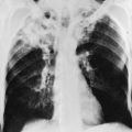
The chest radiograph in patients with incipient ARDS does not necessarily reveal abnormal findings at the onset of clinical presentation. However, within a short period of time, evidence of interstitial and alveolar edema generally develops, the latter being the most prominent finding on chest radiograph. Edema appears diffuse, affecting both lungs relatively symmetrically. As an indication that fluid is filling alveolar spaces, air bronchograms often appear within the diffuse infiltrates. Unless the patient has prior heart disease and cardiac enlargement unrelated to the present problem, heart size remains normal. A characteristic example of a chest radiograph in a patient with severe ARDS is shown in Figure 3-7.

Arterial blood gas values show hypoxemia and hypocapnia (respiratory alkalosis). Calculation of AaDO2 clearly shows that gas exchange is actually worse than it appears at first glance, with alveolar PO2 elevated as a result of hyperventilation. As the amount of interstitial and alveolar edema increases, oxygenation becomes progressively more abnormal, and severe hypoxemia results. Because true shunting of blood across unventilated alveoli is important in the pathogenesis of hypoxemia, PO2 may be relatively unresponsive to administration of supplemental O2. As a standardized method for interpreting PO2 in patients receiving different amounts of supplemental oxygen, a ratio of PaO2 to fractional concentration of inspired oxygen (PaO2/FIO2) less than 300 mm Hg is considered an appropriate gas exchange criterion for ARDS.

In many cases of ARDS, direct measurement of pressures within the central circulation is useful. This measurement is facilitated by use of either a central venous catheter (a catheter inserted into a systemic vein and then advanced to the superior vena cava) or a pulmonary artery catheter, which is advanced further and passed through the right atrium and right ventricle into the pulmonary artery. The relatively easy passage of the pulmonary artery catheter (commonly known as a Swan-Ganz catheter) results from a balloon at the tip, which can be inflated with air and then carried along with blood flow through the tricuspid and pulmonic valves into the pulmonary artery. The catheter is positioned at a point in the pulmonary artery where inflation of the balloon occludes the lumen and prevents forward flow. Consequently, if pressure is measured at the tip of the catheter when forward flow has been prevented, the measured pressure theoretically is a reflection of pressure in the left atrium, which corresponds to left ventricular preload. The pressure measured with the balloon inflated is commonly called the pulmonary artery occlusion pressure (PAOP) or pulmonary capillary wedge pressure. In recent years, central venous catheters are used more commonly than pulmonary artery catheters because they can be placed more quickly and easily, have fewer complications, and have been shown in large trials to produce similar patient outcomes to pulmonary artery catheters by using measurements of central venous pressure as a surrogate marker for left ventricular preload.

These measurements can distinguish whether the observed pulmonary edema is cardiogenic or noncardiogenic in origin. In cardiogenic pulmonary edema, the hydrostatic pressure within the pulmonary capillaries is high as a result of “back-pressure” from the pulmonary veins and left atrium. In noncardiogenic pulmonary edema or ARDS, the central venous pressure and the pressure within the left atrium (measured as the PAOP) are normal, indicating that the interstitial and alveolar fluid results from increased permeability of the pulmonary capillaries and not from high intravascular pressure.

Although use of these catheters for measurement of intravascular pressures is not essential to the diagnosis of ARDS, the information obtained may be useful for determining whether elevated hydrostatic pressures within the capillaries are contributing to the observed pulmonary edema. In addition, the catheters may provide helpful information during the course of the complicated management of these cases, even though their use has not been unequivocally demonstrated to improve mortality.

Treatment

Management of ARDS centers on three main issues: (1) treatment of the precipitating disorder, (2) interruption of or interference with the pathogenetic sequence of events involved in the development of capillary leak, and (3) support of gas exchange until the pulmonary process improves. Although treatment of the precipitating disorder is not always possible or successful, the principle is relatively simple: as long as the underlying problem persists, the pulmonary capillary leak may remain. In the case of a disorder such as sepsis, management of the infection with appropriate antibiotics (and drainage if necessary) is crucial to allowing the pulmonary vasculature to regenerate the normal permeability barrier for protein and fluid.

Meticulous supportive management, particularly support of gas exchange and avoidance of hypervolemia, is critical for patients with ARDS to survive the acute illness. Given the life-threatening nature of ARDS, patients typically are intubated, ventilated with a mechanical ventilator, and managed in an intensive care unit. Failure of other organ systems besides the respiratory system is common, and patients often present some of the most complex and challenging management problems handled in intensive care units. Because of the importance of mechanical ventilation and ventilatory support in the management of respiratory failure associated with ARDS and with other disorders, Chapter 29 is devoted to a more detailed consideration of mechanical ventilation in the management of respiratory failure.

In patients who are mechanically ventilated (as almost all patients with ARDS are), the most effective strategy involves applying lower tidal volumes than had been the traditional practice prior to the turn of the millennium. Currently this is the only intervention that has a significant mortality benefit documented in a large, well-designed randomized trial. However, the exact reasons why this approach is beneficial remain somewhat speculative. It is hypothesized that ventilating the lungs at lower lung volumes avoids overdistention of alveoli and a consequent deleterious release of inflammatory mediators.

Approaches aimed at altering the pathogenetic sequence of events in ARDS have focused on developing agents that block the effect of various cytokines or other initiating stimuli, such as endotoxin, in patients with septic shock. However, to date, this approach has been unsuccessful, and no agents blocking the effect of a particular mediator have been useful. A more nonspecific approach has been use of corticosteroids in an attempt to block a variety of mediators and control or reverse the capillary permeability defect allowing fluid and protein to leak into the interstitium and alveolar spaces. This approach is based in part on experimental evidence suggesting that corticosteroids inhibit aggregation of neutrophils induced by activated complement. However, because of disappointing clinical results and the potential for harmful effects, corticosteroids now are generally not considered appropriately indicated for treatment of ARDS, at least early in its course. Preliminary data suggested that steroids may be useful if administered during the later fibroproliferative phase of ARDS, but a subsequent controlled trial demonstrated no mortality benefit from corticosteroids used in this setting.

Because the mortality of ARDS remains considerable, a variety of newer and experimental forms of therapy have been tried. For example, one interesting approach has been the use of inhaled nitric oxide as a selective pulmonary vasodilator. By producing preferential vasodilation in areas of the lungs that are well ventilated (because these are the areas to which the gas is delivered), inhaled nitric oxide can facilitate better perfusion of well-ventilated areas, leading to better ventilation-perfusion matching and improved oxygenation. Unfortunately, however, beneficial physiologic effects on gas exchange have not been accompanied by documentation of improved survival in clinical trials conducted to date.

References

Treatment

Adhibari, N, Burns, KE, Meade, MO. Pharmacologic therapies for adults with acute lung injury and acute respiratory distress syndrome. Cochrane Database Syst Rev. (4):2004. CD004477

Brodie, D, Bacchetta, M. Extracorporeal membrane oxygenation for ARDS in adults. N Engl J Med. 2011;365:1905–1914.

Calfee, CS, Matthay, MA. Nonventilatory treatments for acute lung injury and ARDS. Chest. 2007;131:913–920.

Diaz, JV, Brower, R, Calfee, CS, et al. Therapeutic strategies for severe acute lung injury. Crit Care Med. 2010;38:1644–1650.

Esan, A, Hess, DR, Raoof, S, et al. Severe hypoxemic respiratory failure: part 1–ventilatory strategies. Chest. 2010;137:1203–1216.

Fan, E, Needham, DM, Stewart, TE. Ventilatory management of acute lung injury and acute respiratory distress syndrome. JAMA. 2005;294:2889–2896.

Malhotra, A. Low-tidal-volume ventilation in the acute respiratory distress syndrome. N Engl J Med. 2007;357:1113–1120.

Pipeling, MR, Fan, E. Therapies for refractory hypoxemia in acute respiratory distress syndrome. JAMA. 2010;304:2521–2527.

Randolph, AG. Management of acute lung injury and acute respiratory distress syndrome in children. Crit Care Med. 2009;37:2448–2454.

Raoof, S, Goulet, K, Esan, A, et al. Severe hypoxemic respiratory failure: part 2–nonventilatory strategies. Chest. 2010;137:1437–1448.

Steinberg, KP, Hudson, LD, Goodman, RB, et al. Efficacy and safety of corticosteroids for persistent acute respiratory distress syndrome. N Engl J Med. 2006;354:1671–1684.