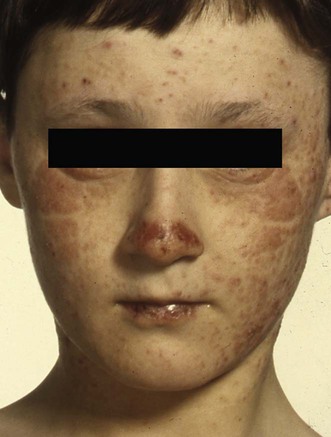
Published on 19/03/2015 by admin
Filed under Dermatology
Last modified 22/04/2025
This article have been viewed 1525 times
Robert S. Dawe
Evidence Levels: A Double-blind study B Clinical trial ≥ 20 subjects C Clinical trial < 20 subjects D Series ≥ 5 subjects E Anecdotal case reports
Actinic prurigo (AP) is a distinct photodermatosis, diagnosed on the basis of characteristic clinical features including perennial (albeit worse in summer) nature, vesiculopapular eruption during acute flares, persistent eroded nodules and/or dermatitic patches (sometimes affecting covered sites), scarring, dorsal nose involvement, cheilitis and conjunctivitis. Abnormal photosensitivity (UVA, UVB) is frequently severe, but generally gradually improves, especially when (as is usual) it presents before the age of 10 years.
Diagnosis is normally straightforward, but the differential diagnosis can include severe polymorphic light eruption and photoaggravated atopic dermatitis. Although differences between European and Amerindian forms of AP have been described, these are either closely related conditions or the same disease with some differences affected by population and environment. Phototesting and HLA typing may sometimes be helpful in cases of diagnostic uncertainty. Possible coexisting conditions such as sunscreen allergic contact dermatitis or photocontact reactions should be considered, as their presence will affect the recommended treatments.
Once the diagnosis is established, initial treatment consists of advice on sunlight avoidance measures (behavioral, clothing, and topical sunscreen), and the use of potent or very potent topical corticosteroids. This approach alone is often insufficient, and many patients require the addition of a springtime course of narrowband (TL-01) UVB or PUVA. When phototherapy is administered for this indication, only normally sunlight-exposed sites should be treated. It is helpful to apply a potent topical steroid to the treated areas immediately after each exposure, to reduce the risk of AP flares.
In Scotland, systemic treatment is rarely required, but is more often necessary where the availability of phototherapy is limited and in countries with more intense year-round sunlight exposure. Antimalarials and β-carotene are sometimes tried, but it remains uncertain whether they are truly of value. Thalidomide may be more useful, but its value is restricted by teratogenicity and the risk of irreversible peripheral neuropathy. Pentoxifylline has anti-TNF-α effects and, although listed as a third-line therapy here, may be worth considering before thalidomide because of its more attractive safety profile.
Phototesting
HLA typing
Histopathology of cheilitis
Addo HA, Frain-Bell W. Photodermatology 1984; 1: 119–28.
This study showed that almost 60% of AP patients have abnormal delayed erythemal responses on monochromator phototesting.
Phototest abnormalities tend to be more severe in AP than in polymorphic light eruption.
Bernal JE, Duran de Rueda MM, Ordonez CP, Duran C, de Brigard D. J Am Acad Dermatol 1990; 22: 1049–51.
In this population, the HLA class I antigen Cw4 was more frequent in AP patients than in controls.
Hojyo-Tomoka T, Vega-Memije E, Granados J, Flores O, Cortes-Franco R, Teixeira F, Dominguez-Soto L. Int J Dermatol 1995; 34: 380–4.
Menage H du P, Vaughan RW, Baker CS, Page G, Proby CM, Breathnach SM, Hawk JL. J Invest Dermatol 1996; 106: 362–7.
Dawe RS, Collins P, O’Sullivan A, Ferguson J. J Invest Dermatol 1997; 108: 233–4.
An even stronger association with the HLA class II antigen HLA-DR4 was shown and, more specifically, with HLA-DRB1*0407.
Suárex A, Valbuena MC, Rey M, de Porras Quintana L. Photodermatol Photoimmunol Photomed 2006; 22: 55–8.
These associations are not a feature of polymorphic light eruption. The absence of HLA-DR4 can help to rule out the diagnosis of AP, whereas the presence of HLA-DRB1*0407 helps to rule in the diagnosis.
Wiseman MC, Orr PH, MacDonald SM, Schroeder ML, Toole JW. J Am Acad Dermatol 2001; 44: 952–6.
No statistically significant association of AP with HLA-DR4 (frequent in the studied population) or HLA-DRB1*0407 was detected, and another HLA type (DRB1*14) was found more commonly than expected, although it was only present in 19 of 37 AP subjects. The authors acknowledge the possibility that they were studying a different condition from AP in other populations. Nevertheless, these findings suggest we should be cautious in attempting to use HLA typing as a diagnostic test, especially in populations in which strong HLA associations have not been confirmed. The diagnosis should still be based on the characteristic constellation of clinical features.
Mounsdon T, Kratochvil F, Auclair P, Neale J, Lee L. Oral Surg Oral Med Oral Pathol 1988; 65: 327–32.
Herrera-Geopfert R, Magana M. Am J Dermatopathol 1995; 17: 357–61.
Vega-Memije ME, Mosqueda-Taylor A, Irigoyen-Camacho ME, Hojyo-Tomoka MT, Dominguez-Soto L. Oral Surg Oral Med Oral Pathol Oral Radiol Endod 2002; 94: 83–91.
A ‘follicular cheilitis’ has been reported to be characteristic of AP. Thirty-two of 116 patients attending a dermatology clinic in Mexico City had cheilitis as their sole manifestation of disease.
Lip histopathology may be helpful diagnostically, especially in patients presenting with cheilitis without cutaneous features at presentation. Where other typical features (skin, eyes) are present it is arguable how much lip histopathology will contribute to diagnosis.
Farr PM, Diffey BL. Photochem Photobiol 1988; 47: 413–17.
Topical indomethacin was found to augment the erythemal response on phototesting. This study has not been replicated in the literature, and has not developed into a routine diagnostic test.
Fusaro RM, Johnson JA. J Am Acad Dermatol 1991; 24: 744–6.
The authors of this open study found 18 of 30 patients with hereditary polymorphic light eruption of American Indians (with described features indistinguishable from AP) to show ‘good to excellent results’ with use of a broad-spectrum sunscreen.
Although broad-spectrum sunscreens are useful, advice on other sunlight avoidance measures, including appropriate clothing and behavioral avoidance, is equally important.
Kerr AC, Ferguson J. Br J Dermatol 2007; 157: 619–20.
Window films that reduce the transmission of UV rays can be used as a method of environmental photoprotection. This case report describes an exacerbation of AP explained by increased transmission through such a window film as it aged.
Lane PR, Moreland AA, Hogan DJ. Arch Dermatol 1990; 126: 1211–13.
Seven out of eight patients treated with intermittent 3–14-day courses of topical 0.05% clobetasol 17-propionate cream or ointment cleared or markedly improved. All had previously found less potent topical steroids ineffective.
Collins P, Ferguson J. Br J Dermatol 1995; 132: 956–63.
Six patients with AP were included in this open study. All reported at least a sixfold increase in tolerable duration of sunlight exposure, which was sustained 4 months after treatment. In one patient, whose phototesting (severely abnormal before treatment) was repeated after treatment, the test results normalized.
Jong CT, Finlay AY, Pearse AD, Kerr AC, Ferguson J, Benton EC, et al. Br J Dermatol 2008; 159: 192–7.
Of the photodermatoses assessed in the British tertiary referral photodermatology centers participating in this study, AP was associated with the greatest health related quality of life impairment. We do not know whether measuring health related quality of life is useful in the day-to-day management of these patients, but important effects of AP on quality of life should be considered when planning treatments.
Schenck RR. JAMA 1960; 172: 1134–7.
This small (13 patients recruited, eight completed) placebo-controlled, crossover study showed no benefit. However, the psoralen dose was low (10 mg), the irradiation source was uncontrollable (the sun), and the study was conducted during summer.
Farr PM, Diffey BL. Br J Dermatol 1989; 120: 411–18.
Five patients were treated in this open study. Clinical improvement was accompanied by an increase of UVA minimal erythemal doses to within the normal range on phototesting. Corroboration that PUVA worked through a local effect was provided by before and after phototesting of areas kept covered during treatment. The UVA minimal erythema dose did not increase in these areas.
In the absence of any controlled study comparisons of PUVA and TL-01 phototherapy for this condition, PUVA should generally be reserved for those who fail to benefit from TL-01.
Londoño F. Int J Dermatol 1973; 12: 326–8.
Thirty-four patients were treated, with a starting dose of 300 mg thalidomide daily, gradually reducing to a minimum of 15 mg; 32 had good results while on the drug, but relapsed on stopping.
Lovell CR, Hawk JL, Calnan CD, Magnus IA. Br J Dermatol 1983; 108: 467–71.
Of 14 patients treated with thalidomide (adult starting dose 100–200 mg daily), 13 (one could not tolerate the drug owing to dizziness) reported improvement. This benefit was sustained in 11, of whom eight required maintenance doses of between 50 mg weekly and 100 mg daily.
Includes a review of earlier open studies reporting the use of thalidomide in AP.
Fusaro RM, Johnson JA. JAMA 1980; 244: 1456–9.
Seventeen of 54 patients who participated in this open study were reported to have achieved complete photoprotection, and 16 ‘marked improvement.’ The plasma carotene level tended to be higher in those who benefited. The authors also comment on their use of dihydroxyacetone and lawsone cream, with apparent benefit.
The clinical features of the study patients were not reported, but the authors’ introductory description of hereditary polymorphic light eruption suggests that they probably included AP.
Torres-Alvarez B, Castanedo-Cazares JP, Moncada B. Dermatology 2004; 208: 198–201.
Clinical improvement was reported in all 10 participants in this 6-month open-label uncontrolled study.
Duran MM, Ordonez CP, Prieto JC, Bernal J. Int J Dermatol 1996; 35: 413–16.
One group of eight patients was treated with tetracycline (1.5 g daily), and another eight with vitamin E (100 IU daily). On follow-up analysis comparing signs and itch, no difference was found between the groups. Both treatments were considered promising, and the possibility of using a tetracycline–vitamin E combination therapy raised.
These treatments need further investigation, but may be worth considering if first-line therapies are inadequate and thalidomide is contraindicated or not tolerated.
Lestarini D, Khoo LS, Goh CL. Photodermatol Photoimmunol Photomed 1999; 15: 183–7.
This is a review of the features and clinical course of 11 patients. Of three treated with systemic corticosteroids, one improved slightly. Intralesional steroids helped one. Three patients were treated with azathioprine: two appeared to benefit.
Hojyo-Tomoka MT, Dominguez-Soto L, Vargasocamp F. Int J Dermatol 1978; 17: 706–10.
In this review, the authors comment that chloroquine ‘seems to give temporary relief,’ and also suggest that antihistamines and tranquilizers may be of some benefit.
McCoombes JA, Hirst LW, Green WR. Am J Ophthalmol 2000; 130: 830–1.
Ortiz-Castillo JV, Boto-De-Los-Bueis A, De-Lucas-Laguna R, Pastor-Nieto B, Peláez-Restrepo N, Fonseca-Sandomingo A. Arch Soc Esp Oftalmol 2006; 81: 661–4.
In these two case reports topical 2% cyclosporine eye drops were associated with improvement in chronic AP conjunctivitis.
Treatment of Skin Disease Comprehensive Therapeutic Strategies 4e
WhatsApp us