[level-membership-for-dermatology-category]
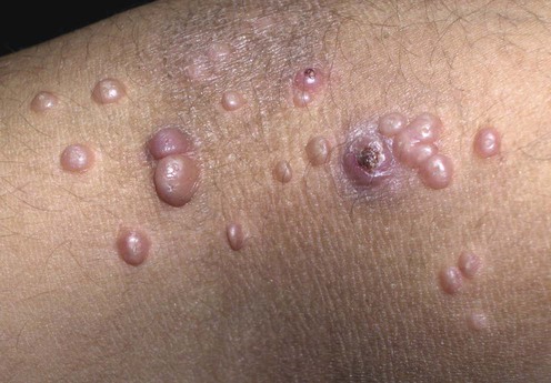
Molluscum contagiosum

 Await spontaneous resolution
 Manual extrusion with gloved fingers or fine forceps
Second-line therapies
 Topical 5% acidified nitrite co-applied with 5% salicylic acid
 Topical salicylic acid gel 12%
 Topical 10% povidone-iodine and 50% salicylic acid
 Topical 40% silver nitrate paste
 Topical 0.5% podophyllotoxin
 Cryotherapy
Third-line therapies
 Curettage with EMLA cream
 Pulsed dye laser
 Photodynamic therapy
 Application of duct tape
 10% potassium hydroxide solution
 Topical 1–5% imiquimod
 Cantharidin
 Diphencyprone
 Australian lemon myrtle
 Topical cidofovir
 Tretinoin
 Adapalene
 Melaleuca alternifolia and iodine
 Candida antigen – intralesional
 Cimetidine
 Interferon-α – subcutaneous
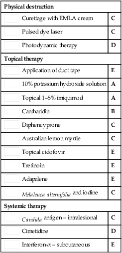
Curettage of molluscum contagiosum in children: analgesia by topical application of a lidocaine/prilocaine cream (EMLA).
Rosdahl L, Edmar B, Gisslen H, Nordin P, Lillieborgs S. Acta Derm Venereol 1988; 68: 149–53.
EMLA cream provided effective local anesthesia for the curettage of MC in 55 children.
[/level-membership-for-dermatology-category][not-level-membership-for-dermatology-category]
Molluscum contagiosum
Second-line therapies
Buy Membership for Dermatology Category to continue reading. Learn more here
[/not-level-membership-for-dermatology-category]
