Xeroderma pigmentosum

Published on 16/03/2015 by admin

Filed under Dermatology

Last modified 22/04/2025

Print this page

rate 1 star rate 2 star rate 3 star rate 4 star rate 5 star
Your rating: none, Average: 0 (0 votes)

This article have been viewed 1961 times

Xeroderma pigmentosum

Deborah Tamura, Kenneth H. Kraemer and John J. DiGiovanna

Evidence Levels:  A Double-blind study  B Clinical trial ≥ 20 subjects  C Clinical trial < 20 subjects  D Series ≥ 5 subjects  E Anecdotal case reports

image

Xeroderma pigmentosum (XP) is a rare autosomal recessive disorder characterized by cellular hypersensitivity to the damaging effects of ultraviolet radiation (UV) resulting in a 10 000-fold increased risk of skin cancers, that often develop in the first decade of life. About half of the patients present with acute photosensitivity with exaggerated reactions to minimal sun exposure: acute erythema and blistering. Burns can be so severe that child abuse or neglect is suspected. Patients who do not have these acute reactions often develop early freckle-like pigmentation before the age of 2 years. This may be accompanied by a dry ‘aged appearance’ to the skin, cheilitis, and photophobia. The eyes are particularly vulnerable and damage may include dry eye, ectropion, pterygia, pinguecula, corneal clouding, and scarring leading to blindness and cancer of the lids, sclera or cornea. Cancers can also occur on the lips and tip of the tongue.

The progressive skin and eye damage is due to inability to repair UV induced DNA damage in sun exposed tissues. XP patients have mutations in one of eight genes. Seven (XPA, XPB, XPC, XPD, XPE, XPF, or XPG) are in the nucleotide excision repair (NER) pathway and the other is DNA polymerase eta (XP variant). The NER pathway is critical for the identification, removal and repair of UV induced DNA damage while polymerase eta bypasses unrepaired DNA damage.

Management strategy

Diagnosis

The diagnosis of XP is based on clinical features, and once suspected UV protection should be initiated immediately. XP should be suspected in a child with a history of freckling on sun exposed skin before age 2 years, or severe sunburn after minimal sun exposure, photophobia, or cancer of the skin or eye. Disorders that should be excluded include porphyria, lupus, LEOPARD syndrome, Carney complex, familial melanoma, and nevoid basal cell carcinoma syndrome.

DNA testing for mutations in several XP genes is available (www.genetests.org). Since XP is a clinical diagnosis and mutation testing is not currently available for all the known XP genes, failure to identify a mutation does not rule out the diagnosis of XP. The cells of XP patients in culture are extremely sensitive to the killing effects of UV but there are currently no CLIA certified laboratories in the US performing this type of XP test.

Environmental management

The most critical management goal for XP is UV protection. While XP patients do not repair damage from UV in exposed tissues, they react normally to visible light. Eradicating all exposure to UV radiation may not be attainable; efforts should be directed towards providing a safer environment. Environments with high UV levels, such as outdoors during daylight hours, require the most rigorous protection and avoidance. However, long periods of lower levels of exposure also cause the accumulation of UV damage. Patients and their families can be taught to recognize and avoid UV sources and to institute protective measures in their home, school and other areas where the patient will spend long periods of time.

Protective measures should be a daily routine for XP patients. These include topically applied, broad spectrum, sun preparations with an SPF (sun protection factor) of at least 30 or higher, that protect against both UVB (short wavelength UV 290–320 nm) and UVA (long wavelength UV 320–400 nm), sun protective lip balms and make-up. The first application of the day should cover all exposed skin surface areas and applications should be reapplied to uncovered skin (usually face, ears, neck and hands) several times during the day. Finding the most acceptable sun block may take trial and error.

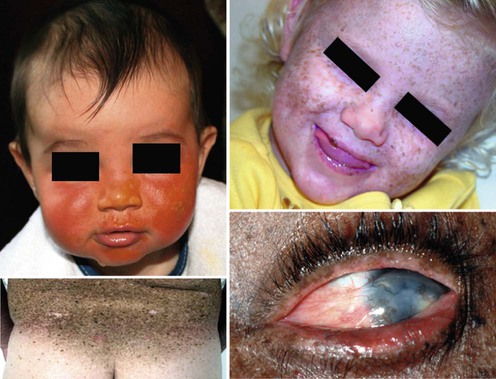
Clothing should cover as much skin surface as possible and should include long pants, long sleeved shirts, socks and closed toe shoes, hats which cover the ears or UV blocking face shields and hoods, and UV blocking sunglasses with side shields. For daytime outdoor exposure gloves or mittens can be worn to protect hands. Clothing material should be tightly woven (e.g., denim) or double layered. A simple test of effective UV protection by clothing is to hold the material up to a bright light. Material that permits visible light to pass will not block UV. Commercially available clothing made from sun blocking material is available but may be expensive. One alternative is a laundry additive called SunGuard (https://sunguardsunprotection.com/index.php), which is advertised to increase the ability of clothing to block UV. They recommend regular washing of outer clothing (pants, shirts, jackets, socks, gloves, and hats) to increase UV protection. The cornea and sclera are also at risk, and XP patients should wear UV blocking sunglasses in areas of potential exposure. The glasses should ‘wrap around’ the eyes to protect the sides of the eyes and large enough to fully protect both lids. Rigorous use of these measures should be initiated as soon as the diagnosis of XP is suspected, and need to be the mainstay of lifelong protection.

The major source of UV is the sun. Although window glass blocks most UVB wavelengths, UVA can pass through glass (along with visible light). Extremely sensitive XP patients have burned through window glass. The Americans with Disability Act and other laws and regulations mandate that children with XP be provided with a safe educational environment. Windows in rooms where XP patients will be spending substantial amounts of time, such as at home, in a car, in day care or school, should be shielded from UV radiation. This can be done with clear UV protective film, or, in some cases, by creating a consistently safe distance from windows. Schools should make accommodations during fire drills and physical education activities, since leaving the building during these activities is not safe for children with XP. An Individualized Educational Plan should be developed to keep them protected from UV sources but involved in these activities.

Medical alert bracelets (http://www.medicalert.org/home/Homegradient.aspx) and carrying an information card about the condition in a purse or wallet can be helpful in emergencies when affected individuals may not be able to communicate their UV sensitivity.

Unshielded fluorescent lighting and bare halogen bulbs are potential UV hazards. Replacing unshielded fluorescent bulbs with incandescent lights or placing plastic shielding over fluorescent bulbs can substantially reduce the ambient levels of UV. Halogen lights emit substantial levels of UVB and should be avoided.

Relatively inexpensive hand-held UV meters can measure UVB and UVA. While the UVB radiation is more damaging, UVA radiation can also produce DNA damage, so protection from all wavelengths of UV is desirable. Meters can be effective learning tools; since the level of UV cannot be easily estimated, directly measuring UV in the environment provides patients and families greater ability to assess the safety of surroundings.

Vitamin D is produced in the skin by UV exposure. Rigorous sun protection may lead to low levels of vitamin D in the blood and can eventually lead to increased risk of bone fracture. Vitamin D rich foods such as ‘fatty’ fish and vitamin D supplemented milk can help maintain normal vitamin D levels; however, patients should have periodic monitoring of vitamin D levels and oral supplementation if the serum levels of 25-OH vitamin D are low.

XP patients should not use any type of tobacco products and should be protected from second-hand smoke. The benzo[a]pyrene derivatives and other carcinogens found in tobacco products and smoke induce DNA damage repaired by the NER pathway, putting the XP patient at high risk for oral and respiratory tract cancers.

The substantial lifestyle adjustment necessary to provide a safer UV environment can be a challenge for the family. Outdoor activities should be avoided during daylight hours particularly between 10 am and 2 pm. Identifying appropriate indoor play and sports activities for the affected XP child, while continuing to meet the needs of the unaffected family members, can be difficult.

Social and psychological impact and effect on the family

The diagnosis of XP necessitates the need for adjustment in the entire family’s activities. Outdoor events will need to be adjusted. There will be a need for increased medical services that can place significant stressors on the family and developing child. The need for frequent painful procedures (surgeries, use of topical field agents, etc.) and hospitalizations for more extensive surgeries can lead in some instances to post-traumatic stress disorder type symptoms in some patients and families. This type of reaction occurs in patients of all ages and can result in avoidance of care. It is important to consider pain management and stress reduction appropriate for the developmental level of the XP patient when planning care. In addition, physical changes such as scarring from surgeries or progressive neurodegeneration, with the resulting disruption of normal childhood and adolescent psychosocial development, can adversely affect self-esteem. The emotional and economic stability of the family is often affected and, as with any chronic illness, the family and patient will need to go through a period of adjustment to the lifestyle limitations.

Obtaining health insurance and workplace accommodations may require considerable effort. The patients may be eligible for Social Security Disability Insurance or other medical assistance. XP support groups help patients connect with other affected families and minimize social isolation; these groups also sponsor UV safe family activities. (The XP Family Support Group, http://www.xpfamilysupport.org; The Xeroderma Pigmentosum Support Group UK, http://xpsupportgroup.org.uk; The Xeroderma Pigmentosum Society, http://www.xps.org). There are also support groups in Germany, Japan, and the Middle East. In situations with psychosocial pathologies (e.g., substance abuse or domestic violence), or if the family is having extreme difficulty coping with the diagnosis, referral to social services or a therapist may be advisable.

Since XP is a recessive disorder, parents are clinically normal carriers of the mutated gene. The risk of having another affected child is 1 in 4 for each subsequent pregnancy. Prenatal diagnosis is possible and consultation with a genetic counselor can address genetic risks in future pregnancies.

Medical and surgical management

For optimal management, the XP patient and family should partner with a dermatologist. Teaching patients and families to look for pre-malignant and malignant lesions can enable earlier identification and removal of small tumors. Depending on the rate of new tumor development, full skin exams should be scheduled regularly (every 3 to 6 months). For rapidly evolving lesions more frequent visits may be necessary. Baseline whole body photographs and close-up photos of lesions with a ruler in the image for size comparison can help track changes. Photos can be digitalized and given to the family for use at home and also kept in the medical record.

Pre-malignant lesions including actinic keratoses can be treated with cryotherapy, liquid nitrogen, topical 5-fluorouracil (5-FU), or topical imiquimod. Concern needs to be taken when using topical 5-FU or imiquimod as field treatments to insure that existing skin cancers are adequately treated. When using field treatments there is a risk of treating the superficial areas of skin cancer but leaving deeper areas of tumor untreated. Well controlled studies of topical field treatments in XP patients have not been published.

Dermabrasion or dermatome shaving has been used to remove the superficial photo-damaged epidermal layers. Theoretically, these procedures allow replacement of severely damaged epidermal cells with cells arising from the deeper, less UV damaged adnexal structures. For patients developing many new lesions chemoprevention with oral retinoids (isotretinoin, etretinate or acitretin) has been used. A longitudinal study of a small number of XP patients performed at the National Institutes of Health found oral isotretinoin was effective in decreasing the number of new non-melanoma skin cancers. However, there were many side effects in these patients especially at the higher drug dosages.

Early and adequate treatment of skin cancers is extremely important. All suspected tumors should be biopsied and removed. Standard techniques including curettage and desiccation, surgical excision or cryosurgical ablation can be used for small superficial lesions. Due to the extensive poikilodermatous changes and scarring in XP patients, it can be difficult to differentiate recurrent from new lesions. Mohs micrographic surgery is optimal for combining effective tumor removal with tissue sparing, which should be highly considered in the surgical plan. Areas developing skin cancers are usually in fields of severe actinic damage complicating the goal of ‘clear margins’. Movement of tissue with flaps can be complicated because of the presence of pre-cancers or small skin cancers in the severely damaged, surrounding skin. Despite their hypersensitivity to UV, many XP patients have demonstrated a normal response to X-ray therapy when it is used to treat recurrent skin and eye cancers, or brain and spinal cord neoplasms.

Ophthalmologic management

Ophthalmologic care is extremely important to preserve eyesight. Most ophthalmologic problems in XP patients occur in the surface structures of the eye and may begin in childhood as photophobia and conjunctivitis. Pre-cancerous and cancerous lesions can arise on the cornea, sclera, lids, and conjunctiva. UV damage on the cornea can lead to ‘dry eye’, keratitis, and conjunctival inflammation. Ectropion of the lids, resulting in defects of eye closure, can occur secondary to eyelid atrophy and surgical removal of malignancies from the periocular skin. Ectropion exacerbates eye dryness and, if left untreated, can lead to corneal ulceration, scarring and opacification of the cornea, and blindness. Corneal transplants have been performed but frequently have led to rejection from vascularization of the area. A thorough ophthalmologic exam at least yearly can include Schirmer’s test for dry eye and assessment of lid closure. Eye lubricants are recommended along with lubricating ointments at night, especially if lid closure is poor.

Neurologic management

Approximately 25% of XP patients develop progressive neurologic disease. The rate of symptom progression varies greatly among patients, with the most severe patients developing symptoms in early childhood (De Santis–Cacchione syndrome). Loss of deep tendon reflexes, most notably in the lower extremities, may be the first manifestation. This may be noted in early childhood as the only symptom for years. Routinely testing reflexes during skin examinations can help identify XP patients at risk for neurologic disease. Patients may have microcephaly. Progression includes cognitive decline, mobility difficulties with ataxia and falls, and eventually a wheelchair may be needed. MRI shows progressive dilation of the ventricles and loss of gray matter of the brain. Death may occur from aspiration pneumonia or other complications of severe debilitation. In an NIH study of 106 XP patients, the median age at death in XP patients with neurodegeneration (29 years) was significantly younger than those XP patients without neurodegeneration (37 years).

Progressive sensorineural hearing loss in XP patients may be diagnosed early, in the first decade of life, and may first be suspected secondary to inattentiveness in school. Audiology exams provide a sensitive method for early detection. The hearing loss can be helped with hearing aides and assisted hearing systems may be used in the classroom.

XP patients have a significantly increased risk for developing cancers on the lips and squamous cell carcinoma of the tip of the tongue, and these areas should be regularly evaluated. Telangiectasia on the tip of the tongue is an early UV induced change.

XP patients have an approximately 50-fold increased risk of developing primary central nervous system tumors. These may respond to full-dose X-ray therapy with normal skin reaction. However, since some XP cell lines have increased sensitivity to X-radiation, a small test dose might be advised prior to full-dose exposure.

With improved health care and better protective measures, XP patients are living longer, more active lives, marrying and having children. When contemplating a pregnancy, referral for pre-conceptual genetic counseling is recommended.

Optimal management of XP patients is a multi-disciplinary process involving several medical specialties and active cooperation and input from the patient and family. With early diagnosis, rigorous UV protection, and management of skin cancers, people with XP are living well into adulthood, working in the community and raising families.

Specific investigations

First-line therapies

imageUV protection C
imageRemoval of skin cancers C

Second-line therapies

imageTopical imiquimod B
imageTopical 5-fluorouracil B
imageOral retinoid therapy C

Third-line therapies

imageResurfacing and dermabrasion and chemical peels E