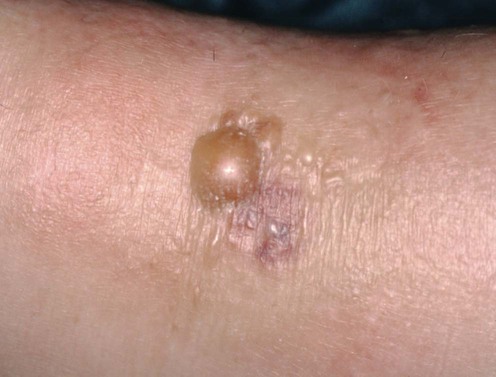
Wells syndrome

Published on 18/03/2015 by admin

Filed under Dermatology

Last modified 18/03/2015

Print this page

rate 1 star rate 2 star rate 3 star rate 4 star rate 5 star
Your rating: none, Average: 0 (0 votes)

This article have been viewed 1450 times

Wells syndrome

Pamela Fiandeiro, Emma Benton and Ian Coulson

Evidence Levels:  A Double-blind study  B Clinical trial ≥ 20 subjects  C Clinical trial < 20 subjects  D Series ≥ 5 subjects  E Anecdotal case reports

image

Wells syndrome or eosinophilic cellulitis is a rare condition resembling a bacterial cellulitis. It is of unknown etiology though there are several disease associations as well as drug-induced disease. The condition can be recurrent, and although thought to be sporadic, familial patterns have been reported. Erythematous patches and plaques are the most common clinical type, but papulonodular and bullous types have been described. Histologic features are characterized by edema, a marked eosinophilic dermal infiltrate, and flame figures. The usual course is of a pruritic sensation, followed rapidly by indurated, erythematous plaques of edema with violaceous edges and even blistering. The lesions progress over a few days, resolving without scarring within 8 weeks. The plaques can occur anywhere on the skin and may be solitary or multiple.

Management strategy

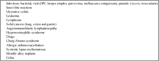
Although there is no known cause, several precipitating factors have been suggested (Table 247.1). Some of the associations are well reported, others anecdotal. The tumor necrosis factor antagonists may produce injection site or widespread lesions; etanercept and adalimumab may induce Wells syndrome localized to injection sites. Minocycline can also induce a Wells syndrome-like disorder.

image

Suspect culprit drugs should be withdrawn. If an underlying systemic disease is identified, this will require treatment in its own right.

The most frequently reported therapy is with systemic corticosteroids, used at moderate dose to gain control of symptoms, followed by tapering, although cases may resolve spontaneously. As with bullous pemphigoid, localized disease may respond to superpotent topical steroids. Topical tacrolimus has been successfully used. The H1 antihistamine with anti-eosinophil action cetirizine has been used successfully. Minocycline, dapsone, antimalarials, griseofulvin and cyclosporine are anecdotally beneficial. Sulfasalazine with oral corticosteroids proved effective in a case with ulcerative colitis and a case associated with HIV infection responded to interferon-α.