Vitiligo

Published on 16/03/2015 by admin

Filed under Dermatology

Last modified 16/03/2015

Print this page

rate 1 star rate 2 star rate 3 star rate 4 star rate 5 star
Your rating: none, Average: 0 (0 votes)

This article have been viewed 1637 times

Vitiligo

Suhail M. Hadi and James M. Spencer

Evidence Levels:  A Double-blind study  B Clinical trial ≥ 20 subjects  C Clinical trial < 20 subjects  D Series ≥ 5 subjects  E Anecdotal case reports

image

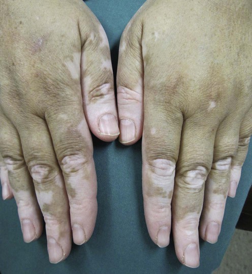
Vitiligo is an acquired idiopathic disorder in which localized areas of skin are devoid of melanocytes, resulting in depigmented macules that are often symmetrically distributed. The disease has 1–3% worldwide prevalence, with no predilection for age, sex, or race. About 50% of cases start before 20 years of age. Vitiligo can be quite psychologically and socially distressing, and in some cultures results in affected individuals being ostracized. This can lead to severe impairment of a patient’s life, particularly if the visible areas of the body (face, hands) are involved. Because of lack of melanin pigment there is an increased risk of sunburn and a theoretic increased risk of skin cancer in the amelanotic areas, and there is an association with ocular abnormalities, especially iritis.

There are many hypotheses to explain the etiology of vitiligo: autoimmune, autocytotoxic (toxic metabolites), neural, hereditary, and environmental factors. The autoimmune theory is supported by the association with a number of autoimmune diseases – thyroid disease, diabetes mellitus, Addison disease, and pernicious anemia – the finding of organ-specific autoantibodies and circulating antimelanocyte autoantibodies, and, in addition, evidence of cellular immunity by immunohistologic study of perilesional skin and T-cell analysis of peripheral blood. In generalized vitiligo reversal of helper T cells and suppressor T-cell ratio has been identified. Degenerative changes may be seen using electron microscopy in both melanocytes and keratinocytes in the involved and uninvolved skin. Oxidative stress may play a role in vitiligo and a free-radical-mediated injury may be the initial pathogenic finding in melanocyte degeneration. Vitiligo might be induced or exacerbated by drugs, with topical imiquimod being a recent example.

Recent studies have shown that genes play an important role in the pathogenesis of vitiligo. Vitiligo susceptibility genes have been discovered and need further study in the hope that this might help in disease management and prevention. Support groups can play an important role in alleviating patients’ suffering and improving their self-esteem. Lastly, reassurance and psychological therapy may have a positive impact on the progression of the disease.

Management strategy

Even though vitiligo does not cause physical impairment, the associated substantial disfigurement can cause serious emotional stress for the patient, which necessitates treatment. Protection of the vitiliginous areas with sun block is important because of the increased vulnerability of the lesional skin to the effects of ultraviolet light, which makes it more liable to sunburn. Cosmetic improvement can be achieved by camouflage products and self-tanning lotions, and might have psychosocial benefits.

Although vitiligo is a notoriously challenging disease to treat, there are several options. More than one treatment modality should be tried for many months before a patient is deemed resistant to therapy.

Associated autoimmune disease should be looked for. Recommended blood tests include thyroid studies, antinuclear antibodies (ANA), and screening for other organ-specific autoantibodies, fasting blood glucose levels, and complete blood count with indices for pernicious anemia. Patients with segmental vitiligo do not appear to have a higher risk of associated autoimmune disorders, although immunologic overlap between the various forms of vitiligo remains.

Treatment options include topical corticosteroids, topical calcineurin antagonists (which can be more effective if used under occlusion) and topical low-dose 8-methoxypsoralen (8-MOP) (0.1%) with UVA for small, localized patches. For widespread disease, systemic therapy with 8-MOP, 5-MOP, 4,5,8-trimethylpsoralen or a combination of these, along with sunlight or artificial UVA, may be beneficial. If the patients do not achieve visible repigmentation after 25–30 sessions with a given psoralen, an alternative therapy should be sought. Topical and systemic PUVA therapy may require 100–300 treatment sessions to achieve complete repigmentation. It has been shown that PUVA increases the number of active epidermal melanocytes in both the involved and uninvolved skin and also reduces the melanocyte and keratinocyte degeneration. Narrowband UVB radiation and 5-MOP with UVA have been shown to be effective alternatives to conventional PUVA, with fewer phototoxic effects. Studies have shown that narrowband UVB light stimulates the migration and proliferation of melanocytes, which might explain the beneficial effect of this treatment modality in vitiligo. Vitiligo patients with coexistent melasma show a significantly better repigmentation on the face and limbs with narrowband UVB compared with those without melasma.

Topical calcipotriene has been used in combination with topical corticosteroids, with narrowband UVB, and with PUVA, with some success. The most recent effective and approved therapy for vitiligo is the 308 nm excimer laser with or without topical calcineurin antagonists (tacrolimus and pimecrolimus). It has been shown that better repigmentation response can be achieved if the excimer laser is used at an earlier stage of the disease and for longer time (high cumulative UV dose). The pattern of repigmentation differs with different treatment modalities: it can be perifollicular, marginal, or diffuse.

For patients with refractory segmental or localized disease, thin split-thickness, punch and suction blister epidermal grafting have been reported to be the most effective surgical methods. Single hair grafting is effective for eyelid and eyebrow disease. Other surgical methods are autologous non-cultured melanocyte–keratinocyte cell transplantation and cultured pure melanocyte suspension. Non-cultured melanocyte transplantation can be improved significantly by suspending the melanocytes in the patients’ autologous serum instead of normal saline.

Cosmetic tattooing is used for localized stable vitiligo, especially of the mucosal type.

Patients with extensive disease (>50% body area) who desire permanent matching of skin color but for whom repigmentation is not possible can be depigmented with 20% monobenzyl ether of hydroquinone, twice daily for 9 to 12 months. The results are excellent but irreversible. For vitiligo universalis, treatment with topical 4-methoxyphenol and the Q-switched ruby laser has been effective. The psychological impact of the disease being a stigmatizing condition and the effect on the patient’s quality of life should be addressed.

Specific investigations

New insights into the pathogenesis of vitiligo: imbalance of epidermal cytokines at sites of lesions.

Moretti S, Spallanzani A, Amato L, Hautmann G, Gallerani I, Fabiani M, et al. Pigment Cell Res 2002; 15: 87–92.

In vitiligo the expression of epidermal cytokines may be modified compared to normal skin. Fifteen patients with active non-segmental vitiligo were evaluated. Compared to perilesional, non-lesional, and healthy skin, in vitiligo skin there was significantly lower expression of keratinocyte-derived cytokines with stimulatory activity on melanocytes, e.g., granulocyte–monocyte colony-stimulating factor, and significantly higher expression of keratinocyte-derived cytokines with inhibitory activity on melanocytes, e.g., interleukin-6 and tumor necrosis factor-α.

The majority of patients with vitiligo do not have an accompanying systemic disease, but directed medical history and blood tests are helpful to identify high-risk individuals who require further evaluation.

First-line therapies

Buy Membership for Dermatology Category to continue reading. Learn more here
imageTopical corticosteroids A
imageTopical photochemotherapy B
imageSystemic photochemotherapy A
imageNarrowband UVB (NB-UVB) A