Published on 19/03/2015 by admin
Filed under Dermatology
Last modified 22/04/2025
This article have been viewed 4139 times
Michael Romano, Jeffrey Mailhot and Karen Wiss
Evidence Levels: A Double-blind study B Clinical trial ≥ 20 subjects C Clinical trial < 20 subjects D Series ≥ 5 subjects E Anecdotal case reports
Rubella (German measles, 3-day measles) is usually a mild disease of low-grade fever, generalized erythematous macules and papules, and generalized lymphadenopathy. It is caused by an enveloped RNA virus of the Togaviridae family.
In children, there is typically no prodrome. In adolescents and adults, a prodrome of fever, malaise, sore throat, nausea, anorexia, and generalized lymphadenopathy is often seen. The erythematous pink macules and papules start on the face and neck and spread down and out in a centrifugal fashion over 1–2 days. These lesions disappear in 2–3 days. Forschheimer spots, an enanthem consisting of petechiae on the hard palate, may accompany the rash.
Rubella is a self-limited illness. The treatment is generally supportive. Teenagers and adults may experience transient polyarthralgia and polyarthritis. Thrombocytopenia and encephalitis are extremely rare complications. During the first trimester of pregnancy, maternal rubella can result in fetal death or congenital rubella syndrome. The main fetal anomalies include ophthalmic disease (cataracts, glaucoma, microphthalmia, and chorioretinitis), sensorineural deafness, cardiac abnormalities (patent ductus arteriosus, atrial septal defects, ventricular septal defects), pulmonic stenosis, and blueberry muffin lesions (extramedullary hematopoiesis).
It can be difficult to distinguish rubella from other viral exanthems, in particular enteroviruses. Rubella also mimics measles, parvovirus B19, human herpesvirus (HHV)-6, and arboviruses. It is essential to differentiate infection between these viruses during pregnancy. Virus identification by culture is available. In congenital infections, rubella can be isolated from the blood, urine, cerebrospinal fluid, and the posterior pharynx. In postnatal infections, the virus is harbored in the nasopharynx. Amplification is accomplished by reverse transcription polymerase chain reaction (RT-PCR). Viral serology is also available for diagnosis. Both serum IgM and seroconversion of convalescent IgG with a fourfold increase in titer suggest recent infection. A more reliable diagnosis is obtained by both RT-PCR and serology, which can detect virus on day 0 of rash eruption. Microarray technology is a cheap and promising future methodology for diagnosis of acute infection. Children with rubella should be excluded from school for 7 days after onset of the rash. Rubella vaccine is recommended in combination with the measles and mumps vaccine, with or without the varicella vaccine (MMR or MMRV), at 12–15 months of age with a second dose at 4–6 years. However, an increased risk of febrile seizures has been reported with the combination MMRV vaccine. Post-pubertal females can be tested for rubella IgG and vaccinated if necessary. The vaccine contains live virus and should not be given to pregnant women.
Viral culture
Serology (acute IgM or acute and convalescent IgG)
PCR
Confirmation of rubella within 4 days of rash onset: comparison of rubella virus RNA detection in oral fluid with immunoglobulin M detection in serum or oral fluid.
Abernathy E, Cabezas C, Sun H, Zheng Q, Chen M, Castillo-Solorzano C, et al. J Clin Microbiol 2009; 47: 182–8.
On days 1 and 2, rubella infection was confirmed best by RT-PCR. On days 0, 3, and 4, serum IgM detected rubella infection. The most sensitive and specific testing for acute rubella infection would be to combine these methodologies within 1 week after the exanthem eruption.
A protein microarray immunoassay for the serological evaluation of the antibody response in vertically transmitted infections.
Ardizzoni A, Capuccini B, Baschieri M, Orsi C, Rumpianesi F, Peppoloni S, et al. Eur J Clin Microbiol Infect Dis 2009; 28: 1067–75.
Enzyme immunoassays are recommended as the ‘gold standard’ for rubella IgM and IgG detection. This study suggests that microarray technology offers a cheaper and more reliable diagnostic test which may be applicable for multiple congenital diseases.
The epidemiological profile of rubella and congenital rubella syndrome in the United States, 1998–2004: the evidence for absence of endemic transmission.
Reef SE, Redd SB, Abernathy E, Zimmerman L, Icenogle JP. Clin Infect Dis 2006; 43: S126–32.
Since the advent of the national rubella vaccination program, epidemiologic evidence strongly supports that rubella virus is no longer endemic in the US.
Observational safety study of febrile convulsion following first dose MMRV vaccination in a managed care setting.
Jacobsen SJ, Ackerson BK, Sy LS, Tran TN, Jones TL, Yao JF, et al. Vaccine 2009; 27: 4656–61.
Though MMRV and MMR + V have similar immunogenicity, MMRV has demonstrated increased side effects, most notably febrile convulsions in children on days 5–12 after vaccination.
Roseola infantum (exanthem subitum, sixth disease) is a disease of high fever in a well-appearing child with an exanthem of pink macules and papules upon defervescence. It is caused by infection with HHV-6 or -7.
Roseola is an illness of children between 6 and 36 months of age. The first sign of illness is a high fever (>39.5°C) that persists for 3 to 7 days, followed by an exanthem that spreads centrifugally from the neck, lasting hours to days. Typically, no treatment is necessary and the illness resolves in a few days. Febrile seizures are common in infants during the febrile phase, usually requiring emergency room care.
Identification of HHV-6 or -7 is difficult because most infections are asymptomatic. Culture from peripheral blood is available in specialized facilities but is of limited use due to slow turn-over. High population seroprevalence (~95%) also renders serology less useful. Seroconversion of convalescent IgG with a fourfold increase in titer is more indicative of acute infection. However, there is considerable antibody cross-reactivity between HHV-6, -7, and cytomegalovirus (CMV). Viral DNA detection by nucleic acid amplification of whole blood, serum, or plasma is the current method of diagnosis, though standardized methods of measurement are yet to be determined. Recent studies suggest that PCR alone cannot reliably distinguish between active and latent infection; a multiple assay approach is more sensitive and specific.
Most individuals harbor HHV-6 and -7 in their saliva, while only 1% of the population carries chromosomally integrated viral DNA. In the immunocompromised, viral reactivation frequently causes severe disease such as fever, bone marrow suppression, hepatitis, pneumonia, lymphoproliferative disorders, and encephalitis. In these patients, ganciclovir and foscarnet are first-line treatments. Foscarnet has shown prophylactic efficacy in stem-cell transplant patients. In vitro studies show that ganciclovir, cidofovir, and foscarnet inhibit HHV-6 and -7 replication. Individual case reports have suggested benefit from these agents in immunocompetent patients with end-organ disease. Infection with HHV-6 can be associated with a severe course of drug-induced hypersensitivity syndrome.
None
HHV-6 serology (acute IgM or acute and convalescent IgG)
Diagnostic assays for active infection with human herpesvirus 6 (HHV-6).
Caserta MT, Hall CB, Schnabel K, Lofthus G, Marino A, Shelley L, et al. J Clin Virol 2010; 48: 55–7.
Qualitative or quantitative PCR alone is not capable of differentiating active HHV-6 infection from latent or chromosomally integrated virus; a multiple assay approach (RQ-PCR, RT-PCR, qualitative PCR) is more reliable.
Association of human herpesvirus 6 reactivation with the flaring and severity of drug-induced hypersensitivity syndrome (DIHS).
Tohyama M, Hashimoto K, Yasukawa M, Kimura H, Horikawa T, Nakajima K, et al. Br J Dermatol 2007; 157: 934–40.
The combination of HHV-6 reactivation and the hypersensitivity response to medication may lead to a more severe course of DIHS with more organ involvement and a prolonged clinical course. Diagnosing HHV-6 reactivation may be a useful diagnostic and prognostic marker in this setting.
High-dose ganciclovir in HHV-6 encephalitis of an immunocompetent child.
Olli-Lähdesmäki T, Haataja L, Parkkola R, Waris M, Bleyzac N, Ruuskanen O. Pediatr Neurol 2010; 43: 53–6.
Though antiviral treatment is usually reserved for immunocompromised patients, high-dose ganciclovir (18 mg/kg/day) was safe and efficacious in this 15-month-old with HHV-6 encephalitis.
Human herpesvirus type 6 and human herpesvirus type 7 infections of the central nervous system.
Dewhurst S. Herpes 2004; 11: 105A–11A.
Ganciclovir and foscarnet may be used for managing HHV-6-related disease. Ganciclovir, but not foscarnet, may be useful for HHV-7-related illness.
Safety of pre-engraftment prophylactic foscarnet administration after allogeneic stem cell transplantation.
Ishiyama K, Katagiri T, Ohata K, Hosokawa K, Kondo Y, Yamazaki H, et al. Transpl Infect Dis 2012; 14: 33–9.
In 10 stem cell transplant patients, prophylactic foscarnet reduced the risk of HHV-6 or -7 associated encephalitis but was unable to prevent HHV-6 reactivation.
Rubeola (measles) is a systemic illness with fever, cough, coryza, conjunctivitis, erythematous macules and papules, and Koplik spots. It is caused by infection with the measles virus, an enveloped RNA virus of the Paramyxoviridae family.
The measles rash begins along the hairline and behind the ears and spreads down the body. It is the prototype of the ‘morbilliform eruption.’ Koplik spots, the pathognomonic enanthem of clustered white dots on the buccal mucosa, precede the rash by approximately 2 days. It is important to distinguish the measles rash from drug eruptions, Kawasaki disease, and other viral infections, in particular exanthems from enteroviruses and adenoviruses. Complications include pneumonia, croup, diarrhea, encephalitis, and death. Subacute sclerosing panencephalitis due to persistent measles infection is a rare degenerative neurologic disease that can occur years after original infection.
Rapid diagnosis by immunofluorescence of desquamated nasal mucosal cells is available. Both serum IgM at the onset of rash and seroconversion of convalescent IgG with a fourfold increase in titer suggest recent infection. RT-PCR of nasopharyngeal secretions or peripheral blood mononuclear cells (PBMC) provides the most reliable diagnostic method of acute infection. Oral fluid assays display similar diagnostic efficacy and may represent the future of measles detection.
Children with measles should be isolated for 4 days after rash eruption, and physicians should notify the appropriate monitoring authorities. Measles vaccine contains live virus and is recommended as part of the MMR or MMRV regimen at 12–15 months with a second dose at 4–6 years of age. It can also be given as a measles-only formulation.
Individuals with poor nutritional status are at greatest risk for complications from measles. Dietary supplementation with vitamin A may reduce the morbidity and mortality of the disease. Supplementation should be given to children 6–24 months of age who are hospitalized for measles complications. Any child with measles and compromised immune function, malnutrition, vitamin A deficiency, or recent travel to high measles mortality areas is also a candidate for treatment. The World Health Organization recommends two doses of 50 000 IU for infants <6 months, 100 000 IU for children 6–12 months, and 200 000 IU for individuals >1 year. A recent meta-analysis demonstrated that at least two doses of 200 000 IU for children >1 year and 100 000 IU for infants was found to reduce measles mortality by approximately 60%. Immunoglobulin prophylaxis can prevent or modify disease within 6 days of exposure. It is recommended for susceptible household contacts, especially for infants, pregnant women, or immunocompromised individuals.
Measles virus is susceptible to ribavirin in vitro, though its clinical efficacy is questionable. It has been given intravenously and intranasally to treat immunocompromised children with severe illness. Novel non-nucleoside inhibitors of measles RNA polymerase are in development and show promise.
Immunofluorescence
Oral fluid for the serological and molecular diagnosis of measles.
Hutse V, Van Hecke K, De Bruyn R, Samu O, Lernout T, Muyembe JJ, et al. Int J Infect Dis 2010; 14: e991–7.
Oral fluid demonstrates equal diagnostic ability compared to the gold standards of serum ELISA and nasopharyngeal RT-PCR swabs. In addition, oral fluid collection is painless and cost-effective.
Reevaluation of laboratory methods for diagnosis of measles.
Akiyoshi K, Suga T, Nukuzuma S, Kon-no M, Shibata M, Itoh M, et al. Jpn J Infect Dis 2010; 63: 225–8.
RT-PCR of PBMCs was the most effective method to diagnosis measles infection (virus isolation via immunofluorescence versus RT-PCR versus IgM level.)
Progress in global measles control, 2000–2010.
Centers for Disease Control and Prevention. MMWR 2012; 61: 73–8.
Prior to widespread vaccination (1980), there were an estimated 2.6 million measles-related deaths. WHO and UNICEF started an initiative in 2001 to increase vaccine coverage, which resulted in a dramatic decrease in disease-related mortality: 733 000 in 2000 and 164 000 in 2008. The number of measles cases remained stable in 2009 and increased in 2010.
Measles in the 21st century.
Mulholland EK, Griffiths UK, Biellik R. N Engl J Med 2012; 366: 1755–7.
Maintaining high levels of vaccination is essential in preventing measles transmission, as the risk for imported disease and outbreaks remains. Ninety percent of the 222 cases reported in the US in 2011 were imported.
Effectiveness of measles vaccination for control of exposed children.
Barrabeig I, Rovira A, Rius C, Muñoz P, Soldevila N, Batalla J, et al. Pediatr Infect Dis J 2011; 30: 78–80.
There is considerable debate about the efficacy of post-exposure prophylaxis with measles vaccine. This study demonstrated that vaccination within 72 hours of exposure prevented 90.5% of disease.
Effectiveness of measles vaccination and vitamin A treatment.
Sudfeld CR, Navar AM, Halsey NA. Int J Epidemiol 2010; 39(Suppl 1): i48–55.
One dose of measles vaccine is 85% effective in preventing measles disease while a second dose is estimated at 98.1–100% effective. Vitamin A treatment demonstrated a 62% reduction in measles mortality with two doses of 200,000 IU in children >1 year and 100 000 IU for infants.
Measles control – can measles virus inhibitors make a difference?
Plemper RK, Snyder JP. Curr Opin Investig Drugs 2009; 10: 811–20.
Ribavirin and IFN-α are used clinically for severe cases of measles infection, though their efficacy and side effect profiles are unfavorable. A novel antiviral agent is greatly needed and novel non-nucleoside inhibitors of measles RNA polymerase show potential.
Enteroviruses are a group of non-enveloped RNA viruses that include coxsackieviruses A and B, enteroviruses 68–71, echoviruses, and polioviruses. As polio has been eliminated in the US, non-polioviruses are the main concern. There is a wide spectrum of clinical disease, including mild fever, upper respiratory tract infections, aseptic meningitis, myocarditis, and encephalitis. Enteroviruses also cause more specific syndromes, such as hand, foot, and mouth disease, herpangina, hemorrhagic conjunctivitis, and pleurodynia.
The majority of enterovirus infections are mild and self-limited, peaking in summer and fall seasons. The viruses are transmitted by the orofecal route and distribution is worldwide. Young children are most frequently affected and commonly present with fever, malaise, diarrhea, vomiting, and upper respiratory symptoms. The exanthem is typically a non-pruritic, morbilliform eruption often with a petechial component.
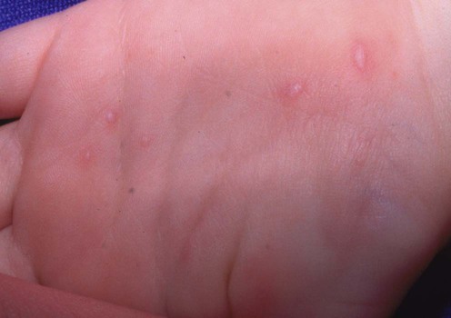
Hand, foot, and mouth disease is typically caused by coxsackievirus A16. There is a prodrome of fever, malaise, and sore throat followed by painful red macules, which become vesicles, on the tongue, soft palate, uvula, and tonsillar pillars. Ovoid, opaque vesicles with a surrounding rim of erythema may be noted on the hands, feet, and buttocks. The disease is usually self-limited. Epidemics in Taiwan, caused by enterovirus 71, led to deaths due to pulmonary hemorrhage/edema, meningitis, encephalitis, myocarditis, and flaccid paralysis.
Herpangina may present with a similar prodrome. White-gray vesicles with a rim of erythema are later observed on the soft palate, uvula, and tonsillar pillars. Typically fewer than 20 lesions are noted.
Enteroviral infections may be diagnosed by rapid or traditional viral culture of the oropharynx or stool and occasionally of the blood, CSF, urine, or tissue. The specific serotype can usually be identified. Disadvantages to viral culture include low sensitivity and slow turn-over. Serology is not routinely used but is commercially available. Because all enteroviruses share common genomic sequences, PCR detects almost all enterovirus serotypes and is particularly reliable in the CSF.
Treatment of most enteroviral infections, including hand, foot, and mouth disease and herpangina, consists of hydration and pain control. These infections are of particular concern in newborns and immunosuppressed patients, who have been treated with immunoglobulin for severe life-threatening illness. Pleconaril is an antiviral drug that prevents viral attachment and fusion, and has the potential to improve morbidity and mortality. Presently, its use is limited to severe life-threatening infections, specifically meningitis. Small interfering RNA (siRNA) shows promise as a future treatment option.
Enteroviral PCR
A convenient rapid culture assay for the detection of enteroviruses in clinical samples: comparison with conventional cell culture and RT-PCR.
Terletskaia-Ladwig E, Meier S, Hahn R, Leinmüller M, Schneider F, Enders M. J Med Microbiol 2008; 57: 1000–6.
Rapid culture assay should be used on stool samples. RT-PCR should be used on CSF samples.
Enterovirus infections of the central nervous system review.
Rhoades RE, Tabor-Godwin JM, Tsueng G, Feuer R. Virology 2011; 411: 288–305.
Pleconaril and immunoglobulin remain the gold standard for enteroviral infections of the CNS, though immunoglobulin therapy has not been properly demonstrated in clinical trials. siRNA may become a future therapeutic option.
Treatment of Skin Disease Comprehensive Therapeutic Strategies 4e
WhatsApp us


 Antipyretics: acetaminophen (paracetamol), ibuprofen
 Analgesics: non-steroidal anti-inflammatory drugs
 School avoidance for 7 days
 Immunization


 Antipyretics – acetaminophen, ibuprofen
 Ganciclovir
 Foscarnet


 Antipyretics: acetaminophen, ibuprofen
 Report to local or state health department
 Measles vaccine (prevention and after exposure)
 Oral vitamin A
 Immunoglobulin prophylaxis
 Ribavirin and IFN-α

 Antipyretics such as acetaminophen or ibuprofen
 Analgesics
 Hydration
 Immunoglobulin
 Pleconaril