Upper extremity blocks

Published on 07/02/2015 by admin

Filed under Anesthesiology

Last modified 22/04/2025

Print this page

rate 1 star rate 2 star rate 3 star rate 4 star rate 5 star
Your rating: none, Average: 0 (0 votes)

This article have been viewed 1348 times

Upper extremity blocks

Sandra L. Kopp, MD, Susan M. Moeschler, MD and Denise J. Wedel, MD

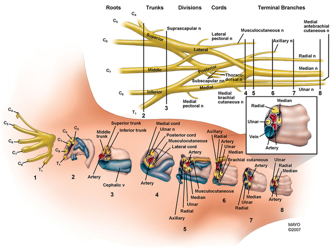
Successful neural blockade of the upper extremity requires extensive anatomic knowledge of the brachial plexus, from its origin as the roots emerge from the intervertebral foramina and of the nerves of the arm and forearm (Figure 127-1). Also important is knowledge of the side effects and complications of peripheral nerve blocks in the upper extremity, as well as the clinical application of available local anesthetic agents for these blocks. Finally, one must not underestimate the role of appropriate sedation during placement of the nerve block and during the surgical procedure (Table 127-1).

Table 127-1

Regional Anesthetic Techniques for Upper Extremity Operations

Brachial Plexus Technique Level of Blockade Peripheral Nerves Blocked Surgical Applications Comments
Axillary Peripheral nerves Radial, ulnar, median; musculocutaneous unreliably blocked Operations of the forearm and hand Unsuitable for proximal humerus or shoulder surgeryRequires patient to abduct the arm
Supraclavicular Distal trunk–proximal cord Radial, ulnar, median, musculocutaneous, axillary Operations of the midhumerus, elbow, forearm, and hand Risk of pneumothorax requires caution in ambulatory patientsPhrenic nerve paresis in 30% of cases
Interscalene Upper and middle trunks Entire brachial plexus, although inferior trunk (ulnar nerve) is inconsistently blocked Surgery to shoulder, proximal and mid humerus Phrenic nerve paresis in 100% of patients for duration of the blockUnsuitable for patients unable to tolerate a 25% reduction in pulmonary function

image

Adapted from Kopp SL, Horlocker TT. Regional anaesthesia in day-stay and short-stay surgery. Anaesthesia. 2010;65(Suppl 1):84-96.

Interscalene block

The interscalene approach to the brachial plexus, at the level of the trunks, is best suited to operations on the shoulder, when a block of the cervical plexus is also desirable. Blockade of the inferior trunk (C8-T1) is often incomplete, requiring supplementation of the ulnar nerve for adequate surgical anesthesia in that distribution. Advantages of this block include technical ease because of easily palpated landmarks and the ability to perform the block with the patient’s arm in any position, which is especially important for cases involving upper extremity trauma or other painful conditions. Use of a nerve stimulator or ultrasound guidance is recommended with this technique to place the local anesthetic solution accurately.

Technique

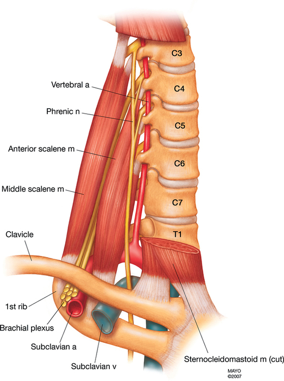
With the patient in the supine position, the patient’s head is turned away from the side to be anesthetized. The lateral border of the sternocleidomastoid muscle is palpated and marked; identification of the muscle is facilitated by having the patient briefly lift his or her head. The interscalene groove may be palpated by rolling the fingers posterolaterally from the muscle border, over the belly of the anterior scalene muscle. A line is extended laterally from the cricoid cartilage to intersect the vertical line of the interscalene groove; this represents the level of the C6 transverse process. The external jugular vein often crosses at this level but is not a reliable anatomic landmark (Figure 127-2).

A 22-gauge, 4-cm, short-bevel needle is inserted perpendicular to the skin with a 45-degree caudad and slightly posterior angle. The needle is advanced until the patient exhibits paresthesia or, if a nerve stimulator is being used, a motor response is observed in the forearm or hand. The brachial plexus is usually quite superficial in the interscalene area (1 to 2 cm). A “click” may be felt as the blunt needle penetrates the prevertebral fascia, giving another confirmation of accurate needle location. If the needle encounters bone within 2 cm of the skin surface, this is likely the transverse process, and the needle should be gently “walked off” anteriorly. After a test dose of local anesthetic agent is given, 10 to 30 mL of the agent is injected incrementally, with frequent aspiration. Caudad spread of the local anesthetic may be facilitated by maintaining digital pressure proximal to the injection site and placing the patient in a head-up position during or following blockade.

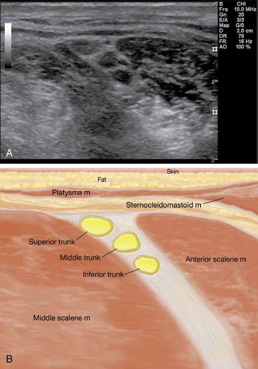
This block is very suited to the use of ultrasound guidance. It is often easiest to obtain a supraclavicular view (see description later) of the subclavian artery and brachial plexus and then trace the plexus up the neck with the ultrasound probe until the plexus trunks are visualized as hypoechoic structures between the anterior and medial scalene muscles (Figure 127-3, A and B). The needle can then be advanced in an out-of-plane or an in-plane approach to a depth of 1 to 3 cm in most patients. After negative aspiration, a test dose is administered to confirm appropriate placement of the needle.

Side effects and complications

Nerve damage or neuritis can occur secondary to needle trauma or pharmacologic toxicity but is uncommon and usually self-limited. Local anesthetic toxicity as a result of intravascular injection should be guarded against by careful aspiration and incremental injection. The phrenic nerve is frequently blocked, probably because of its anatomic proximity on the anterior surface of the anterior scalene muscle. The patient may complain of subjective shortness of breath. The risk of pneumothorax is low when the needle is correctly placed at the C5 or C6 level because of the distance from the dome of the pleura. Blockade of the vagus, recurrent laryngeal, and cervical sympathetic nerves, as well as epidural and intrathecal injections, have been reported to have occurred during interscalene block. Reports of catastrophic nerve damage resulting from cord injection or high-dose spinal injections underscore that this block should not be performed in a heavily sedated or anesthetized patient.

Supraclavicular block

Because of the compact arrangement of the trunks of the brachial plexus at the level of the first rib, the supraclavicular approach is extremely efficient; relatively small volumes of local anesthetic agent result in rapid and profound neural blockade when injected accurately. The supraclavicular approach provides excellent surgical anesthesia for the elbow, forearm, and hand. The use of ultrasound has led to a resurgence of this block.

Technique

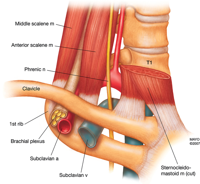
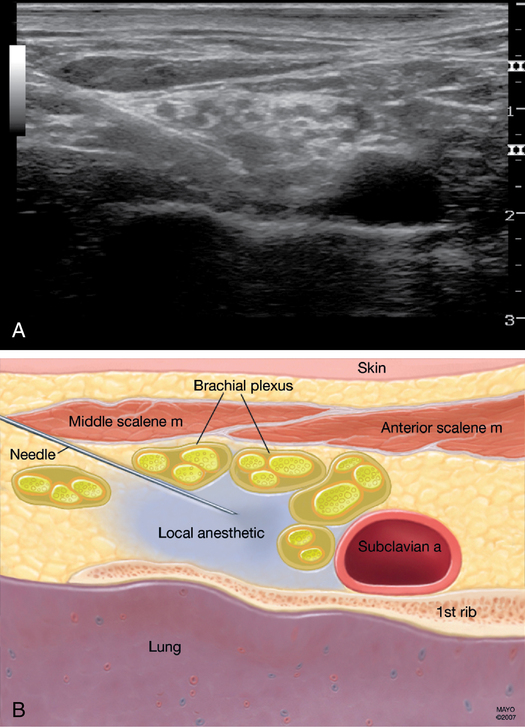
The three trunks of the brachial plexus are compactly arranged cephaloposterior to and surrounding the subclavian artery at the level of the first rib, inferior to the clavicle at approximately its midpoint (Figure 127-4).

The patient is positioned in the supine position with the head turned away from the side to be blocked and the arm adducted and stretched as far as possible toward the ipsilateral knee. In the classic description, the midpoint of the clavicle is marked. The lateral border of the sternocleidomastoid muscle is identified (aided by the patient lifting the head), and the interscalene groove is palpated by rolling the fingers back from the muscle border over the anterior scalene muscle. A mark is then made 1.5 to 2.0 cm posterior to the clavicle at its midpoint within the interscalene groove. Palpation of the subclavian artery, if possible, provides further verification of the correct needle placement.

A 22-gauge, 4-cm, short-bevel needle is directed in a caudad, slightly medial, and posterior direction until the patient exhibits a paresthesia or the first rib is encountered. This needle orientation lies in a plane parallel to a line joining the skin-entry site and the patient’s ear. If the first rib is encountered before a paresthesia is elicited, then the needle can be walked anteriorly and posteriorly along the rib until a paresthesia is elicited or the subclavian artery is encountered. If the artery is located, the needle should be redirected in a more posterolateral direction, a maneuver that usually results in elicitation of a paresthesia. A nerve stimulator can also be used to aid needle placement.

The use of ultrasound for the supraclavicular block allows the practitioner to see the brachial plexus structures to be blocked, as well as the subclavian artery and pleura, just below the first rib, which are to be avoided. The patient is positioned as described previously, and the ultrasound probe is placed just cephalad and parallel to the clavicle. The probe is moved medially and laterally until the plexus is viewed just lateral to the subclavian artery. The needle is advanced in plane, lateral to medial, toward the plexus. Following negative aspiration, 20 to 40 mL of local anesthetic agent is injected around the plexus; spread around the neural structures can be seen on the ultrasound (Figure 127-5).
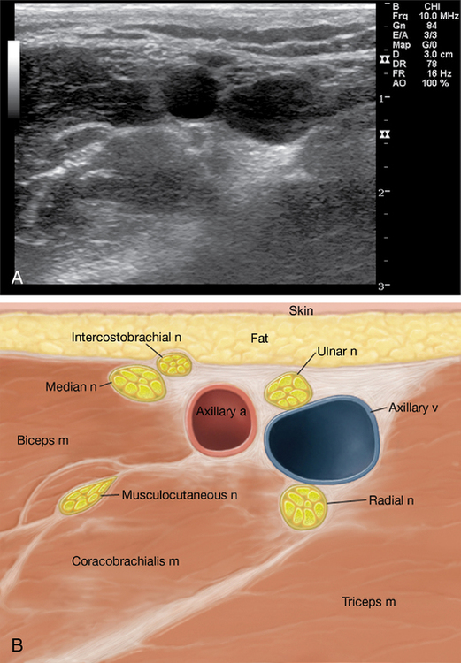
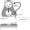
Axillary block

The axillary approach to the brachial plexus is used because of its ease of performance, safety, and reliability, particularly for hand and forearm surgery. A variety of approaches to the axillary block have been described, including elicitation of paresthesias, transarterial injection, sheath blocks, use of a nerve stimulator, and the use of ultrasound guidance. In experienced hands, all seem to result in a reasonable success rate. Use of this technique is confined to patients who are able to abduct their arms sufficiently to allow access to the neurovascular bundle within the axilla. The musculocutaneous nerve may not always be blocked with this approach but can be supplemented either at the level of the coracobrachialis muscle or as it courses superficially above the interepicondylar line at the elbow.

Technique

For all approaches to the axillary block, the patient is positioned supine with the arm to be anesthetized abducted at right angles with the body and the elbow flexed to 90 degrees. The axillary artery is palpated as close to the axillary crease as possible, and a line is drawn tracing the course of the artery distally. The artery is fixed against the humerus at the level of the axillary crease by the index and middle fingers of the nondominant hand. Placement of the needle proximal to the fingers, along with maintenance of distal pressure, encourages proximal spread of the local anesthetic solution, increasing the likelihood of blocking the musculocutaneous nerve.

Paresthesia techniques involve elicitation of single or multiple paresthesias with a small-gauge (22G-25G), 2-cm needle. A minimum volume of 10 mL of local anesthetic agent is carefully injected at each paresthesia.

Nerve stimulators can be used with a Teflon-coated (insulated) needle or commercially available nerve stimulators. This technique avoids sensory paresthesias but requires additional equipment.

Transarterial techniques have been described involving placement of a sharp needle through the axillary artery and injecting a local anesthetic agent (40 to 50 mL) behind the artery or, in some descriptions, dividing the total volume of agent and injecting it behind and superior to the artery. Obviously, great care must be taken to avoid intravascular injection with this technique.

The ultrasound approach to the axillary block involves visualizing the axillary artery and surrounding distinct neural structures at various positions relative to the artery. The block requires several needle redirections to adequately deposit local anesthetic agent around each neural structure. The patient is positioned as described previously. The ultrasound probe is placed just distal and parallel to the axillary crease at a point that best identifies the artery in close proximity to the median, ulnar, and radial nerves (Figure 127-6). The needle is advanced in an in-plane approach to individually block each nerve. Finally, the musculocutaneous nerve can be identified by scanning further laterally within the coracobrachialis muscle. It should be blocked via a separate needle-insertion site.