Ultrasound training in critical care medicine fellowships
Overview
Critical care ultrasound (CCU) is a noninvasive tool used for diagnostic evaluation and for guiding procedures in critical care patients. Ultrasound improves success and lowers complication rates when used to guide procedures such as vascular access and fluid drainage.1 Despite these advantages, formal training in CCU has not been incorporated into many fellowship programs, and training in this skill remains heterogeneous. The American Board of Internal Medicine (ABIM) has recommended that fellowship training in critical care medicine include training in the use of ultrasound to guide thoracentesis and central venous access.2 The definition of competence in CCU, suggested training guidelines, the prevalence of fellowship training in this skill, and current barriers to ultrasound training are considered in this chapter.
Definition of competence in critical care ultrasound
A consensus statement sponsored jointly by the American College of Chest Physicians (ACCP) and La Société de Réanimation de Langue Française (SRLF) outlined specific competency guidelines for achieving proficiency in CCU.3 The statement divides CCU into general ultrasound (pleural, lung, abdominal, vascular access, and vascular diagnostic) and echocardiography (basic and advanced). For each, the panel defines a reasonable minimum standard of specific skills required to achieve proficiency in CCU. Importantly, competence is distinguished from certification, which refers to the recognition of competence by an external agency. In the United States, no formal certification process for CCU is currently available. The specific competency skills outlined in the ACCP/SRLF document will be briefly summarized here.
Skills for achieving competency in the basic principles of critical care ultrasound
Knowledge of basic ultrasound physics is required to acquire images and recognize artifacts. Also important is knowledge of the machine controls and how to manipulate the transducer to obtain images. Intensivists should be aware of when the examination required exceeds the scope of their capabilities and seek appropriate assistance when necessary (see Chapters 1 and 57).
Skills for achieving competency in diagnostic vascular ultrasound
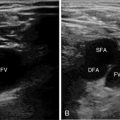
Ultrasound can be used by intensivists to diagnose venous thrombosis at the bedside. Clinicians must be able to correctly locate the relevant vein and associated artery and should be able to identify venous thrombosis. They should be familiar with the proper technique of performing a compression study and be aware that a compression study should not be performed in patients with a visible thrombus.
Skills for achieving competency in critical care echocardiography
According to the ACCP/SRLF consensus statement, competence in critical care echocardiography is divided into basic and advanced levels. Because competence in advanced echocardiography requires a high degree of skill and more extensive training than most critical care fellowship training programs would reasonably be able to provide, a discussion of competency in advanced echocardiography is beyond the scope of this chapter (see Chapter 61).
Suggested fellowship training guidelines
The European Society of Intensive Care Medicine (ESICM) has recently proposed training guidelines designed around achieving proficiency in the skills outlined by the ACCP/SRLF competence statement.4 The ESICM document proposes that general CCU and basic echocardiography training should be a mandatory component of intensivists’ training and that advanced echocardiography should be an optional component. Furthermore, the authors suggested that to achieve competency in general CCU and basic echocardiography, intensivists should receive at least 10 hours of training in each, divided between lectures and didactics (including image acquisition and interpretation training).
Current prevalence of training in critical care ultrasound
A recent survey of critical care medicine and pulmonary/critical care medicine fellowship program directors in the United States examined the prevalence of fellowship training in CCU.1 This survey examined ultrasound training in five specific areas: vascular access, lung/pleural, cardiac, abdominal, and vascular diagnostic. Training in using ultrasound for vascular access was offered by 98% of responding programs. Training in the use of ultrasound in other areas was less prevalent: lung and pleural, 74%; cardiac, 55%; abdominal, 37%; and vascular diagnostic, 33%.1 This survey demonstrated that even though nearly all programs train fellows in ultrasound-guided vascular access, training in other aspects of ultrasound is not universal.
Barriers to training
This same survey of program directors examining the prevalence of CCU training also examined barriers to training in this skill. The most commonly cited barriers to ultrasound training included fellow turnover, lack of faculty trained in ultrasonography, financial considerations, and lack of access to an ultrasound machine.1
References
1. Eisen, LA, Leung, S, Gallagher, AE, Kvetan, V, Barriers to ultrasound training in critical care medicine fellowships: a survey of program directors. Crit Care Me. 2010; 38:1978–1983.
2. American Board of Internal Medicine. Critical care medicine certification policies. Available at http://www.abim.org/certification/policies/imss/ccm.aspx#tpr, April 29, 2013. [Accessed].
3. Mayo, PH, Beaulieu, Y, Doelken, P, et al. American College of Chest Physicians/La Société de Réanimation de Langue Française statement on competence in critical care ultrasonography. Chest. 2009; 135:1050–1060.
4. Expert Round Table on Ultrasound in ICU. International expert statement on training standards for critical care ultrasonography. Intensive Care Med. 2011; 37:1077–1083.


