Published on 16/03/2015 by admin
Filed under Dermatology
Last modified 22/04/2025
This article have been viewed 4937 times
Murtaza Khan and John Berth-Jones
Evidence Levels: A Double-blind study B Clinical trial ≥ 20 subjects C Clinical trial < 20 subjects D Series ≥ 5 subjects E Anecdotal case reports
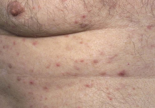
Grover disease is an acquired pruritic, papulovesicular eruption characterized histologically by focal acantholytic dyskeratosis. It is predominantly self-limiting. It is more common in middle-aged and elderly people, especially men, and involves mainly the trunk. The evolution is acute or chronic. The etiology is unknown, but excessive UV exposure, heat, sweating, and ionizing radiation are linked to the disease. Drugs, chemotherapeutic agents and cancers are also known triggers. Other skin disorders such as psoriasis or eczema of various types may coexist.
Grover disease is an uncommon disorder characterized by discrete erythematous, edematous papulovesicles or keratotic papules. The duration of the eruption may be weeks to months and it may be persistent or recurrent. Pruritus of variable intensity is experienced by most patients and may be out of proportion to the clinical signs. Constitutional symptoms are usually absent.
Treatment is difficult. There have been no large clinical trials and reports are based on small numbers.
Patients should be advised to avoid excessive sun exposure, strenuous exercise, heat, and occlusive fabrics. In mild cases, simple antipruritic measures such as avoidance of soap, simple emollients, and soothing baths with bath oils or colloidal oatmeal may be of benefit. Wet compresses with zinc oxide, calamine, or topical corticosteroids may help to relieve the itching.
Topical calcipotriol (ointment) twice daily 50 µg/g may be helpful after 3 to 4 weeks of treatment. Topical vitamin A acid (retinoic acid) is of limited use owing to skin irritation.
Systemic therapy may be indicated in more extensive and persistent disease. Oral vitamin A has been recommended in the past. The aromatic retinoid acitretin been used successfully in doses of 0.5 mg/kg daily. Isotretinoin 40 mg daily has been used for periods ranging from 2 to 12 weeks. It may be administered on a reducing regimen if the initial response is rapid, with a maintenance dose of 10 mg daily. Side effects include dry skin, cheilitis, teratogenicity, and elevation of cholesterol and triglycerides.
Systemic corticosteroids have been used to suppress inflammation and pruritus, but relapses frequently occur on drug withdrawal.
Psoralen with UVA (PUVA) may be useful, but an initial exacerbation may occur. There are anecdotal reports of the success of narrowband UVB and of medium-dose UVA1 phototherapy.
Topical 5-fluorouracil, dapsone, antibiotics, and cryotherapy are ineffective. Recently rituximab and etanercept have been reported to be useful.
Skin biopsy
Acantholysis is the characteristic epidermal change. The histologic changes may mimic Darier disease, pemphigus, and Hailey–Hailey disease. Hyperkeratosis, parakeratosis, and spongiosis are other common epidermal changes. Immunofluorescence does not aid in making the diagnosis.
Heenan PJ, Quirk CJ. Br J Dermatol 1980; 102: 515–20.
This study looked at a series of 24 cases of transient acantholytic dermatosis. Most of them required topical fluorinated corticosteroids to control the pruritus, and two required intermittent courses of oral corticosteroids. Antihistamines were of limited value in controlling the pruritus.
French LE, Piletta PA, Etienne A, Salomon D, Saurat JH. Dermatology 1999; 198: 410–1.
A prospective study of 28 hospital inpatients diagnosed with Grover disease. In over 80% of cases the duration of hospitalization exceeded 2 weeks and was associated with strict bed rest. The authors suggested a sweat-related pathogenesis.
Hu H, Michel B, Farber EM. Arch Dermatol 1985; 121: 1439–41.
Seven cases of Grover disease are presented in this article, which demonstrated a causal association with heat and sweating. Five of the cases are reported to have responded to topical corticosteroids.
Dodd HJ, Sarkany I. Clin Exp Dermatol 1984; 9: 431–4.
A case report of a 41-year-old man with a 5-year history of an itchy truncal and lower limb rash consistent with persistent acantholytic dermatosis. Bath emollients and aqueous cream BP afforded minor relief. Etretinate (50 mg daily) cleared the skin lesions and reduced the itching.
Etretinate has been replaced by its active metabolite acitretin. Lower doses of the latter may be effective.
Helfman RJ, Gables C. J Am Acad Dermatol 1985; 12: 981–4.
Four patients with biopsy-proven Grover disease responded to 40 mg daily of isotretinoin for 2 to 4 months. In two patients most lesions had cleared after 3 to 4 weeks of treatment. Their dose was reduced by 10 mg daily for a further 8 weeks. One patient required 40 mg daily for 8 weeks. These patients remained in remission for up to 10 months after treatment. The final patient obtained partial relief and then discontinued treatment because of elevated triglycerides.
Mancuso A, Cohen EH. Int J Dermatol 1989; 28: 58–9.
A 48-year-old man with severe pruritus had a 2-month history of Grover disease. Systemic corticosteroids initially relieved his symptoms. On discontinuation of prednisone he relapsed. Isotretinoin was commenced at an initial dose of 1 mg/kg daily (40 mg twice daily) and then reduced to 40 mg daily due to adverse effects. He had improved by 6 weeks. Lesions and pruritus resolved after 3 months of therapy.
Paul BS, Arndt KA. Arch Dermatol 1984; 120: 121–2.
A 59-year-old man with persistent Grover disease was unresponsive to oral prednisone and vitamin A (300 000 units daily). PUVA was initiated with 50 mg (0.6 mg/kg) methoxsalen and 2 J/cm2 of UVA. Treatment was twice weekly and the UVA dosage was increased by 0.5 J/cm2 with each treatment. The patient experienced a flare after four treatments, but improved by week 6, with maximal improvement by week 8. Therapy was then tapered off over the following 4 weeks, with complete clearing. No recurrence had occurred 25 months after therapy.
Honig B, Morison WL, Karp D. J Am Acad Dermatol 1994; 31: 775–90.
The authors comment that Grover disease may occur in patients receiving PUVA for other skin conditions. Continuation of PUVA clears the rash, with the pruritus resolving within 10 treatments and the eruption clearing within 20–30 treatments. No numbers are supplied.
Reports show that approximately 10 treatments are required for resolution of pruritus and 20–30 treatments may be needed to clear the eruption. Paradoxically, Grover disease may complicate PUVA prescribed for other conditions.
Rohr JR, Quirk CJ. Arch Dermatol 1979; 115: 1033–4.
Eight patients were treated with vitamin A 50 000 units three times a day for up to 2 weeks; all patients responded. Once initial improvement was noted the dose was reduced to 50 000 units daily as maintenance or for several weeks. No signs of toxicity were noted. One patient required re-institution of the drug owing to recurrence on cessation of treatment.
Keohane SG, Cork MJ. Br J Dermatol 1995; 132: 832–3.
A 50-year-old man had a 13-month history of Grover disease. He responded poorly to oxytetracycline, topical corticosteroids, dapsone, and etretinate. Lesions cleared following hospitalization and prednisone 100 mg daily, but he relapsed with any reduction in dose. Oral corticosteroids were stopped and he was commenced on an alternating regimen of calcipotriol ointment and 0.025% betamethasone valerate ointment. There was complete clearance of lesions after 1 month of treatment, but the disease relapsed when treatment was stopped.
Mota AV, Correia TM, Lopes JM, Guimaraes JM. Eur J Dermatol 1998; 8: 33–5.
Case report of an 84-year-old man with a 2-year history of Grover disease. He improved significantly despite initial moderate irritation following a 3-week course of calcipotriol 50 µg/g twice daily. Lesions did not recur during a 6-month follow-up.
Hayashi H. Clin Exp Dermatol 2002; 27: 160–1.
A 31-year-old man with a 2-month history of Grover disease that failed to respond to topical corticosteroid was commenced on tacalcitol ointment twice daily. He improved dramatically within 1 week and was in remission after 1 month.
Kouba DJ, Dasgeb B, Deng AC, Gaspari AA. Dermatol Surg 2006; 32: 1083–8.
A 46-year-old woman had a 6-month history of progressive Grover disease with intractable pruritus. She failed to respond to topical calcipotriene (Dovonex). She was treated with an even, light application of 40% (w/v) TCA – single-pass strokes with TCA-dampened gauze. Individual lesions of Grover disease were identified and were re-treated with 40% TCA using a cotton-tipped applicator. Three weeks post procedure she had re-epithelialized and was disease free, and was still in remission 8 months later.
The authors stress that practitioners not accustomed to using TCA in office applications should use low-strength formulations such as 20–30% to avoid scarring.
Breuckmann F, Appelhans C, Altmeyer P, Kreuter A. J Am Acad Dermatol 2005; 52: 169–70.
A 78-year-old man with persistent Grover disease had failed to respond to topical and oral corticosteroids. He was treated with a medium-dose UVA1 cold light monophototherapy containing a special filtering and cooling system (21°C). Irradiation (50 J/cm, 1.9 J/cm/minute; 26 minutes), six times weekly for 3 weeks, then three times weekly for 3 weeks. A total of 24 treatments with a cumulative dose of 1200 J/cm2. Complete remission was achieved after 4 weeks, with no subsequent relapse.
Ishibashi M, Nagasaka T, Chen KR. Clin Exp Dermatol 2008; 33: 206–7.
An 80-year old woman developed Grover disease simultaneously with a relapse of follicular lymphoma. Six years prior to this episode her lymphoma had been treated with rituximab. She was given rituximab again and her skin lesions cleared within a week. The lymphoma improved initially but after nine cycles of treatment she suffered another relapse. However, her skin lesions did not recur.
Norman R, Chau V. J Am Acad Dermatol 2011; 64: 796–8.
A 55-year old man had a 2-year history of Grover disease that did not respond to topical or oral steroids and antihistamines. Isotretinoin given for 2 months resulted in improvement of his pruritus, but was discontinued after he had a myocardial infarction. He was prescribed etanercept 50 mg subcutaneously, twice weekly for 6 weeks which reduced the pruritus by 98%. Over the next 4 months he had very mild symptoms and did not develop any new lesions.
Treatment of Skin Disease Comprehensive Therapeutic Strategies 4e
WhatsApp us


 Emollients
 Avoid heat/sweating
 Topical corticosteroids
 Antihistamines
 Isotretinoin/acitretin
 Systemic corticosteroids
 PUVA
PUVA Vitamin A
 Calcipotriol
 Tacalcitol
 Trichloroacetic acid
 UVA1
UVA1 Rituximab
 Etanercept