Chapter 53 Transfusion Reactions to Blood and Cell Therapy Products
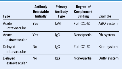
Table 53-1 Types of Acute Transfusion Reactions
| Reaction Type | Presenting Signs and Symptoms |
|---|---|
| Acute intravascular hemolytic | Fever, chills, dyspnea, hypotension, tachycardia, flushing, vomiting, back pain, hemoglobinuria, hemoglobinemia, shock |
| Acute extravascular hemolytic | Fever, indirect hyperbilirubinemia, posttransfusion hematocrit increment lower than expected |
| Febrile reaction | Fever, chills |
| Allergic (mild) | Urticaria, pruritus, rash |
| Anaphylactic | Dyspnea, bronchospasm, hypotension, tachycardia, shock |
| Hypervolemic | Dyspnea, tachycardia, hypertension, headache, jugular venous distention |
| Septic | Fever, chills, hypotension, tachycardia, vomiting, shock |
| Transfusion-related acute lung injury | Dyspnea, decreased oxygen saturation, fever, hypotension |
Workup of an Acute Intravascular Hemolytic Transfusion Reaction
If an acute transfusion reaction occurs:
1. Stop blood component infusion immediately.
2. Maintain intravenous access with a suitable crystalloid or colloid solution.
3. Monitor/maintain blood pressure and heart rate.
4. Maintain an adequate airway.
5. Give a diuretic or institute fluid diuresis, or both.
6. Obtain blood and urine studies for the transfusion reaction workup.
If intravascular hemolytic reaction is confirmed:
8. Monitor renal status (BUN, creatinine).
9. Monitor coagulation status (prothrombin time, partial thromboplastin time, fibrinogen).
10. Monitor for signs of hemolysis (lactate dehydrogenase, bilirubin-total/direct, haptoglobin).
Table 53-3 Diagnosis of Transfusion-Related Acute Lung Injury
| Onset | Within 6 hours of start of transfusion |
| Frequency | 1 : 1000 to 1 : 4500 transfusions (probably underreported) |
| Signs and symptoms | Decreased O2 saturation, fever, hypotension, tachypnea, dyspnea, diffuse pulmonary infiltrates, normal cardiac pressures (requires Swan-Ganz catheter), copious amounts of pulmonary edema fluid |
| Pathogenesis | HLA/granulocyte-specific antibodies (usually of donor origin) reacting with recipient leukocytes. Activates recipient neutrophils; stimulates complement activation; generates CD11/18 on the polymorphonuclear neutrophil surface, resulting in pulmonary capillary adherence and diapedesis and eventual pulmonary capillary leak syndrome. The latter is associated with generation of proteolytic enzymes and toxic O2 metabolites, which cause endothelial cell damage. Neutrophil priming lipids may also play a role. |
| Diagnosis | Chest radiograph; blood gases; blood for HLA or antineutrophil antibodies |
| Differential diagnosis | Fluid overload; septic transfusion; anaphylaxis |
| Treatment | STOP TRANSFUSION! Provide ventilatory support (administer O2, intubate as needed), support blood pressure, administer steroids; diuretics are of no value. |
Risk Groups for Transfusion-Associated Graft-Versus-Host Disease
Risk Well Defined
Congenital T-cell defects (known or suspected)
Immunologic immaturity (fetus or premature infant)
Neonates undergoing intrauterine exchange transfusion or extracorporeal membrane oxygenation
Bone marrow or peripheral blood stem cell transplant recipients (allogeneic or autologous)
Haplotype sharing between donor and recipient

