[level-membership-for-dermatology-category]
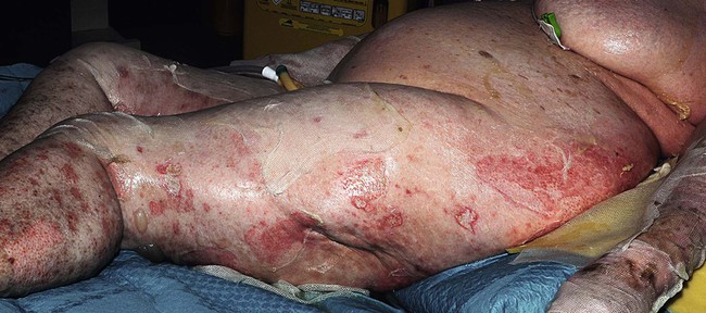
Toxic epidermal necrolysis and Stevens–Johnson syndrome
Specific investigations


Incidence of Stevens–Johnson syndrome and toxic epidermal necrolysis in patients with the acquired immunodeficiency syndrome in Germany.
Rzany B, Mockenhaupt M, Stocker U, Hamouda O, Schöpf E. Arch Dermatol 1993; 129: 1059.
HIV testing should be considered for high-risk patients presenting with TEN/SJS.
First-line therapies
 Supportive measures
 Withdrawal of culprit drug
 Analgesia
 Bioengineered skin substitute
Toxic epidermal necrolysis and Stevens–Johnson syndrome: does early withdrawal of causative drugs decrease the risk of death?
Garcia-Doval I, LeCleach L, Bocquet H, Otero XL, Roujeau JC. Arch Dermatol 2000; 136: 323–7.
Early discontinuation of the causative drug improved prognosis in this study of 113 patients.
ALDEN, an algorithm for assessment of drug causality in Stevens–Johnson syndrome and toxic epidermal necrolysis: comparison with case-control analysis.
Sassolas B, Haddad C, Mockenhaupt M, Dunant A, Liss Y, et al. Clin Pharmacol Ther 2010; 88: 60–8.
A validated algorithm for assessing likelihood of causality of drugs in cases of SJS and TEN.
Second-line therapies
 Cyclosporine
 Systemic corticosteroids
 Intravenous immunoglobulin
Third-line therapies
 Plasmapheresis
 Pentoxifylline
[/level-membership-for-dermatology-category][not-level-membership-for-dermatology-category]
Toxic epidermal necrolysis and Stevens–Johnson syndrome
Specific investigations
Incidence of Stevens–Johnson syndrome and toxic epidermal necrolysis in patients with the acquired immunodeficiency syndrome in Germany.
Rzany B, Mockenhaupt M, Stocker U, Hamouda O, Schöpf E. Arch Dermatol 1993; 129: 1059.
HIV testing should be considered for high-risk patients presenting with TEN/SJS.
