[level-membership-for-dermatology-category]
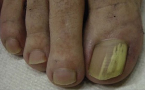
Tinea unguium
Specific investigations


First-line therapies – SYSTEMIC

Second-line therapies


Third-line therapies


Topical Therapies
 Topical amorolfine
 Topical ciclopiroxolamine
 Topical terbinafine
[/level-membership-for-dermatology-category][not-level-membership-for-dermatology-category]
Tinea unguium
