[level-membership-for-dermatology-category]
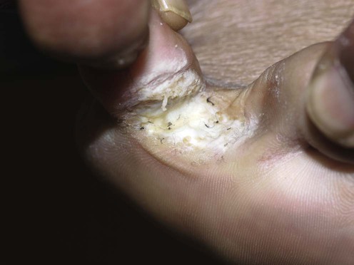
Tinea pedis and skin dermatophytosis
Specific investigations


First-line therapies


Second-line therapies


Third-line therapies
 40% urea cream
 Photodynamic therapy
 Terbinafine
 Itraconazole
 Fluconazole
 Griseofulvin
[/level-membership-for-dermatology-category][not-level-membership-for-dermatology-category]
Tinea pedis and skin dermatophytosis
