Published on 19/03/2015 by admin
Filed under Dermatology
Last modified 22/04/2025
This article have been viewed 3657 times
Eirini E. Merika and L. Claire Fuller
Evidence Levels: A Double-blind study B Clinical trial ≥ 20 subjects C Clinical trial < 20 subjects D Series ≥ 5 subjects E Anecdotal case reports
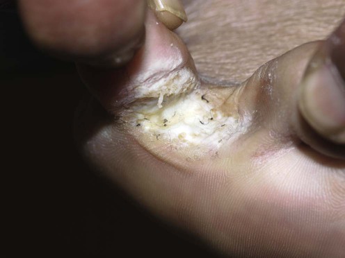
Superficial fungal infections are the commonest of all mucocutaneous infections and appear to be on the increase. Dermatophytes infect keratinized epithelium, the hair and nails. Tinea pedis (athlete’s foot) describes a dermatophyte infection of the soles of the feet and interdigital spaces; tinea cruris, an infection of the groin; tinea facei, the face; and tinea corporis the rest of the skin.
Vena GA, Chieco P, Posa F, Garofalo A, Bosco A, Cassano N. New Microbiol 2012; 35: 207–13.
A retrospective analysis involving 6133 patients with 20.4% suffering with tinea pedis with a male predominance showing that over the last decades tinea pedis has becoming increasingly common.
Skin dermatophytosis rarely causes significant morbidity and certainly not mortality, but there is some evidence that, especially tinea pedis, it acts as a portal of entry for bacteria that produce impetiginization, lymphangitis, and bacterial cellulitis.
Topical antifungal treatment (with topical azoles or allylamines) of tinea pedis is generally adequate, as it is for small areas of tinea corporis and cruris, but for extensive infections, and especially those in immunosuppressed patients, oral therapy may be required. Several antifungal topical therapies are available without prescription, and the vast majority of the disease burden is likely to be managed without intervention from medical personnel. Treatment schedules vary but it is generally accepted that treatment should be applied once to twice daily for 2 to 4 weeks, continuing for at least 1 week after the lesions have cleared. Eradication of tinea elsewhere in the body, such as tinea pedis in the context of tinea cruris or tinea ungium in tinea pedis, is paramount for effective treatment and prevention of relapse.
Extensive forms of infection, failure of local treatment or relapse from inadequate therapy may require oral therapy with systemic antifungals such as terbinafine, fluconazole or itraconazole. Oral therapy remains the most effective agent against extensive or non-responsive forms of tinea. British prescribing guidelines suggest the following regimens: terbinafine 250 mg daily for 14 days, fluconazole 50 mg daily for 2 to 4 weeks (up to 6 weeks in tinea pedis), and itraconazole 100 mg daily for 15 days or 200 mg daily for 7 days (longer for tinea pedis and manuum). Severe macerated forms of tinea pedis may well be superinfected with bacteria justifying concomitant antibiotic therapy, although some topical antifungal agents have in vitro antibacterial activity, e.g., ciclopiroxolamine. Other than general hygiene, there is only anecdotal evidence to support prevention strategies to minimize re-infection, such as using antifungal powder to areas prone to fungal infections after showering, and wearing shower shoes in public bathing facilities.
Roujeau JC, Sigurgeirsson B, Korting HC, Kerl H, Paul C. Dermatology 2004; 209: 301–7.
Two hundred and forty-three patients with acute bacterial cellulitis and 467 age- and gender-matched controls were investigated, and mycology-proven tinea pedis was shown to be a significant risk factor for cellulitis (odds ratio [OR] 2.4; p<0.001). Interdigital tinea pedis conferred the highest risk (OR 3.2, p<0.001) followed by onychomycosis (OR 2.2, p<0.001) and then plantar-type tinea pedis (OR 1.2, p=0.005). A previous history of cellulitis, venous insufficiency, and leg edema was also reported to increase the risk.
Skin scrapings for mycological microscopy and culture
Skin swabs for bacteriology
The lesions should be scraped carefully, harvesting surface scale with a no. 15 blade or banana-shaped knife. The active edges of large lesions are likely to yield more scale. Avoiding the application of emollients prior to sampling aids sample collection and analysis. Blister and pustule tops may be ruptured and the contents swabbed and placed directly onto agar plates.
Marks R, Dykes P, Motley R. London: Taylor & Francis, 1993.
The principal organisms responsible for tinea pedis are Trichophyton rubrum, T. mentagrophytes and Epidermophyton floccosum. Not all scaly eruptions are due to tinea, so confirming the presence of a dermatophyte enables the instigation of relevant, targeted therapy and avoids causing unwanted side effects in non-fungal causes.
Havlickova B, Czaika VA, Friedrich M. Mycoses 2008; 51(Suppl 4): 2–15.
Trichophyton, Microsporum and Epidermophyton are the pathogens responsible for most skin dermatophytoses with considerably variable incidence globally mainly due to local socio-economic conditions and cultural practices. There seems to be a predominance of tinea pedis in developed countries with Trichophyton rubrum the leading pathogen.
Fuchs A, Fiedler J, Lebwohl M, Sapadin A, Rudikoff D, Lefkovits A, et al. Am J Med Sci 2004; 327: 77–8.
Culture-positive rates in 874 patients suspected of having tinea pedis were only 32%.
Many clinicians will scrape a scaly rash or an acral eruption to rule out a tinea contribution, so this is probably an appropriately low positive result. Macerated tinea pedis may well be superinfected with bacteria.
Leyden JJ, Kligman AM. Arch Dermatol 1978; 114: 1466–72.
Quantitative cultures of 140 cases of interdigital ‘athlete’s foot’ showed that as the clinical disease became more macerated, increasing numbers of resident aerobic organisms, such as large colony diphtheroids, were present.
Sweeney SM, Wiss K, Mallory SB. Arch Pediatr Adolesc Med 2002; 156: 1149–52.
Dermatophyte infections, especially in children, may be difficult to diagnose and often mimic bacterial infections. Scraping microscopy can help discriminate before final culture results are available.
Topical
ClotrimazoleA
MiconazoleA
Hart R, Bell-Syer SE, Crawford F, Torgerson DJ, Young P, Russell I. Br Med J 1999; 319: 79–82.
This rigorous review found 17 placebo-controlled trials for azoles with an estimated pooled risk of failure to cure of 0.54, versus 12 trials of allylamines against placebo with a rate of 0.3 of failure to cure. There are 12 randomized controlled trials of azoles against allylamines, and although allylamines are slightly more effective than azoles and undecanoic acid, they are more expensive, so the authors recommended first to treat with azoles topically and to use allylamines only in treatment failures. This saves £1.94 (sterling) for each cured patient.
Rotta I, Otuki MF, Sanches AC, Correr CJ. Rev Assoc Med Bras 2012; 58: 308–18.
Forty-nine studies met the selection criteria and were included in this meta-analysis. The grouped efficacy data evidenced the superiority of antifungal drugs compared to placebo, regardless of the dermatomycosis under evaluation, with odds ratio values ranging from 2.05 (95% CI 1.18–3.54) to 67.53 (95% CI 11.43–398.86). Allylamines were better than azoles only for the outcome sustained cure (OR 0.52, 95% CI 0.31–0.89).
Croxtall JD, Plosker G. Drugs 2009; 69: 339–59.
A placebo-controlled trial showing that topical 2% sertaconazole cream once or twice daily for tinea pedis led to mycological cure and was more effective compared to 2% miconazole cream twice daily. Non-inferiority trial data has shown that 2% solution is as effective as 2% cream. Note that sertaconazole is not yet available in the UK.
Terbinafine 1% (allylamine) as either cream, solution, gel or film forming solutionA
Cicolpirox (hydroxyopiridone)A
Korting HC, Kiencke P, Nelles S, Rychlik R. Am J Clin Dermatol 2007; 8: 357–64.
An efficacy analysis from 19 eligible studies involving nearly 3000 patients demonstrated that mycological cure rates with terbinafine were significantly superior to those of placebo, but no significant differences were shown between the different formulations, durations of therapy, or frequencies of application. Regimens varied from a single application of the film forming solution to 4 weeks once or twice daily of the other formulations. Ten randomized controlled studies compared terbinafine with an active control, and although clinical and mycological cure rates were slightly higher with terbinafine, this did not reach statistical significance.
Aly R, Fisher G, Katz I, Levine N, Lookingbill DP, Lowe N, et al. Int J Dermatol 2003; 42: 29–35.
Three hundred and seventy-four subjects with mild to moderate tinea pedis were enrolled and randomized to one of two treatment groups: ciclopirox gel 0.77%, or ciclopirox gel vehicle, applied twice daily for 28 days. Eighty-five percent of ciclopirox subjects were mycologically cured, compared to only 16% of vehicle subjects 2 weeks post treatment.
Subissi A, Monti D, Togni G, Mailland F. Drugs 2010; 70: 2133–52.
Ciclopirox has a broader spectrum of action compared to most other antimycotics and is active against azole-resistant Candida species.
Elewski BE, Haley HR, Robbins CM. Cutis 2004; 73: 355–7.
Twelve patients with moccasin-type tinea pedis were treated with 40% urea cream once daily as an adjunct to the topical antifungal ciclopirox cream twice daily. At 3 weeks a 100% cure rate was shown.
Qiao J, Li R, Ding Y, Fang H. Mycopathologia 2010; 170: 339–43.
A review of published cases showing that 6 out of 10 patients with tinea pedis and six out of nine with foot-interdigital mycoses responded to PDT after 1–4 treatments but at 8 weeks the cure rate was only 30% and 22% respectively. It is difficult to recommend this as a treatment unless there has been intolerance to all the previous treatments mentioned.
Tausch I, Decroix J, Gwiezdzinski Z, Urbanowski S, Baran E, Ziarkiewicz M, et al. Int J Dermatol 1998; 37: 140–2.
A double-blind randomized multicenter phase III study involving 300 patients with palmar-type tinea pedis (moccasin) or manuum using 200 mg itraconazole twice daily for 7 days versus 250 mg terbinafine once daily for 14 days, showed no difference in the mycological cure rates of 79% vs 80%. Side effects were reported in 14% of the itraconazole group and 19% of the terbinafine group, with no clinically relevant changes in laboratory variables evaluated.
Although this is a high dose of itraconazole this is shown both to be efficacious and safe in the treatment of tinea pedis.
Faergemann J, Mork NJ, Haglund A, Odegard T. Br J Dermatol 1997; 13: 575–7.
Two hundred and thirty patients were entered into a double-blind, parallel-group study comparing fluconazole 150 mg once weekly with griseofulvin 500 mg once daily for 4 to 6 weeks in the treatment tinea corporis or tinea cruris. Mycological cure was achieved in 78% of the fluconazole group compared to 80% in the griseofulvin group.
Svejgaard E, Avnstorp C, Wanscher B, Nilsson J, Heremans A. Dermatology 1998; 197: 368–72.
Seventy-two patients with moccasin-type tinea pedis were randomized to receive either itraconazole 200 mg twice daily for 1 week or placebo. After 8 weeks of follow-up mycological cure rates were 56% vs 8% (p<0.001) in favor of itraconazole, and clinical cure rates 75% vs 11% (p<0.001).
Short-term treatment showed that itraconazole is significantly more effective than placebo in treating tinea pedis.
Montero-Gei F, Perera A. Clin Infect Dis 1992; 14: S77–81.
Twenty patients each with tinea corporis and/or tinea cruris and 20 patients with tinea pedis were treated with a single dose of 150 mg fluconazole and followed weekly. A further dose was administered each week in the persisting presence of clinical evidence of infection, to a maximum of four doses. In tinea corporis/cruris 70% required two doses, 20% three doses, and 10% four doses. At 30 days mycological clearance was observed in 95%. In tinea pedis 20% needed two doses, 20% three, and 60% four doses, with mycological clearance at 30 days observed in 75%.
Rich P, Houpt KR, LaMarca A, Loven KH, Marbury TC, Matheson R, et al; Tinea Corporis/Tinea Cruris Research Group. Cutis 2001; 68: 15–22.
A small prospective open-label study randomly treated six HIV-positive and eight diabetic adult patients with tinea corporis or tinea cruris using oral terbinafine 250 mg once daily for either 1 or 2 weeks. At 6 weeks, of the 11 patients that had full mycological results available for analysis, all the HIV-positive patients and 83% of diabetic patients achieved mycological clearance. There was no difference observed between 1 or 2 weeks’ treatment, and the drug was well tolerated.
Treatment of Skin Disease Comprehensive Therapeutic Strategies 4e
WhatsApp us


 40% urea cream
 Photodynamic therapy
 Terbinafine
 Itraconazole
 Fluconazole
 Griseofulvin