The Metabolism of Cell Growth and Proliferation

Published on 09/04/2015 by admin

Filed under Hematology, Oncology and Palliative Medicine

Last modified 22/04/2025

Print this page

rate 1 star rate 2 star rate 3 star rate 4 star rate 5 star
Your rating: none, Average: 0 (0 votes)

This article have been viewed 7542 times

Figure 13-1 Mitochondria efficiently degrade metabolic fuels to generate ATP Metabolic fuels—carbohydrates, fats, and proteins—are broken down into their constituent units: glucose, fatty acids, and amino acids. These molecules are further degraded into metabolic intermediates that enter the tricarboxylic acid (TCA) cycle. Glucose-derived pyruvate, fatty acids, and many amino acids enter the TCA as acetyl-CoA, which condenses with oxaloacetate to form citrate. A series of biochemical reactions oxidize citrate back to oxaloacetate, while using the high-energy electrons released by these oxidation reactions to reduce the electron carriers NAD+ and FAD to NADH and FADH2, respectively. NADH and FADH2 carry these electrons to complex I and complex II of the electron transport chain, regenerating NAD+ and FAD. The high-energy electrons are passed through the electron transport chain (complex I-III-IV or complex II-III-IV), and their energy is used to pump protons from the mitochondrial matrix into the inner membrane space, generating an electrochemical gradient known as the mitochondrial membrane potential. Ultimately, the electrons reduce molecular oxygen to form water. The energy released by protons flowing down their electrochemical gradient through complex V/ATP synthase is harnessed to synthesize ATP from ADP and Pi. Reactive oxygen species (ROS) are formed when electrons leak out of the electron transport chain prematurely, combining with oxygen to form the superoxide radical (O2 . ). Although ROS is an inevitable by-product of oxidative phosphorylation, its production can be increased whenever flow through the electron transport chain is slowed, whether by heightened membrane potential or reduced oxygen concentrations. Metabolites are shown in blue. Electron carriers are shown in green.

Regulation of Cell Metabolism

During conditions of nutrient scarcity, anabolic programs are suppressed in favor of energy-generating catabolic pathways. Conversely, multiple cellular metabolic pathways must be coordinately rewired to support cell proliferation. Elevated nutrient uptake—particularly of glucose and glutamine—provides the substrates for cell growth. Rapid catabolism of glucose via glycolysis produces sufficient ATP and NADPH to support energy-dependent anabolic reactions while generating the metabolic intermediates that will be critical for macromolecular biosynthesis. Similarly, catabolism of glutamine will help maintain bioenergetics while ensuring adequate substrates for cell growth. Multiple regulatory mechanisms ensure that energy production and macromolecular biosynthesis are appropriately balanced with the metabolic needs of the cell.
Unicellular organisms are directly exposed to the environment and any fluctuations in nutrient availability that may occur. Consequently, these organisms have elegant mechanisms to sense the available nutrients and rewire their metabolism accordingly. Thus, for unicellular organisms, nutrient availability directly controls the signals that regulate growth and proliferation. If a unicellular organism is in an environment with abundant nutrients, these nutrients will directly activate signaling pathways that instruct the cell to engage anabolic metabolic pathways and to undergo cell division. Conversely, conditions of low nutrient availability will halt cell growth and division. Cells will engage catabolic pathways in order to produce energy to survive through the period of scarcity (Figure 13-2 ).
In contrast, metazoans have complex organ systems that maintain a relatively constant level of extracellular nutrients throughout the body. Cell-autonomous metabolic regulation would be catastrophic, as well-fed cells might undergo aberrant growth and proliferation. Thus, nutrient availability alone cannot determine whether cells engage anabolic or catabolic pathways; the metabolism of individual cells must be aligned with the needs of the organism as a whole. This coordination is largely achieved through extracellular growth factors that regulate nutrient uptake and utilization. Binding to receptors on the cell surface, growth factors stimulate intracellular signal-transduction cascades that regulate many facets of nutrient uptake and metabolism. In particular, growth factor signaling enables cells to take up high levels of nutrients such as glucose and glutamine and to engage in anabolic pathways supporting cell growth (see Figure 13-2). 9 In this manner, systemic signals can target individual cells (expressing the proper growth factor receptor) to induce specific activities, thereby ensuring that the behavior of individual cells is tailored to the needs of the entire organism. In the absence of growth factor signaling, metazoan cells are largely quiescent, maintaining homeostasis by the efficient degradation of the limited nutrients they are directed to take up. By engaging oxidative, catabolic pathways that preserve intracellular energy levels, these cells are able to survive and fulfill their allotted functions.
Most cells have the capacity to engage both anabolic and catabolic pathways, and the balance between the two—the balance that guards against inappropriate cell proliferation while maintaining cell survival—is carefully regulated by a number of factors. External signals such as growth factors instruct the cell to grow, activating anabolic reactions and driving nutrient uptake accordingly. The activities of key metabolic enzymes that determine whether metabolites enter anabolic or catabolic pathways are regulated by posttranslational modifications, most commonly phosphorylation triggered by growth factor signaling. Metabolites themselves function as critical allosteric regulators of metabolic enzyme activity, increasing or inhibiting flux through metabolic pathways according to the needs of the cell. For example, signals of abundant energy stores such as ATP and NADH can allosterically inhibit multiple enzymes that otherwise promote the channeling of metabolites into catabolic, energy-producing pathways. This allows for the diversion of metabolites toward anabolic processes when energy supply is plentiful. Similarly, both metabolic cues and growth factor signaling pathways can regulate the expression levels of metabolic enzymes. Through these mechanisms, cells carefully control the activation of anabolic or catabolic metabolic programs and ensure that during cell proliferation, metabolic pathways are coordinately rewired to support cell growth.
image
Figure 13-2 Proliferative metabolism is differentially regulated in single-cellular and multicellular organisms Quiescent cells in both single-cellular organisms and multicellular organisms engage in oxidative metabolism, efficiently converting available nutrients to ATP and carbon dioxide. In the presence of abundant nutrients, single-cellular organisms will engage in proliferative metabolism, consuming high levels of nutrients to produce biomass while excreting large amounts of lactate or other anaerobic fermentation products, such as ethanol. In contrast, the presence of abundant nutrients is insufficient to trigger proliferative metabolism in multicellular organisms, which require additional growth factor signals in order to engage proliferative metabolism. Once stimulated, these cells will undergo high rates of glycolysis to fuel cell growth.
A major hallmark of cancer is the development of cell-autonomous regulation of cell growth. 10 Through multiple genetic events, including activation of growth factor signal transduction pathways (oncogenes) and loss of inhibitory signals (tumor suppressors), cancer cells circumvent dependence on external growth factor signaling. Constitutive activation of growth factor signaling pathways ensures that cancer cells are not subject to the normal regulation of metazoan cells. Consequently, cancer cells exhibit metabolic transformation, taking up high levels of nutrients and engaging in proliferative metabolism to support unchecked cell growth.

The Metabolic Profile of Cancer Cells

It is increasingly clear that cancer cells exhibit metabolic phenotypes that are similar to rapidly proliferating normal cells, with the major difference that the metabolic alterations in cancer cells stem from oncogenic cell-autonomous signaling, rather than the appropriate result of specific growth signals originating outside the cell. The characteristic metabolic features of rapidly proliferating tumor cells include elevated glucose uptake and glycolysis, increased glutamine uptake and utilization, and enhanced lipid and nucleotide biosynthesis. This section discusses the common metabolic signatures of tumor cells and how these metabolic alterations may support tumor growth.

Aerobic Glycolysis: The Warburg Effect

The most well-known and prevalent metabolic change associated with cancer cells is the enhanced uptake and metabolism of glucose, often referred to as the Warburg effect. In 1926, Otto Warburg noted that rapidly proliferating ascites cancer cells take up high levels of glucose and produce large amounts of lactate, even in the presence of oxygen. This finding was not intuitive: Work begun by Louis Pasteur had shown a clear inverse relationship between oxygen availability and the rate of glucose fermentation to lactate. The ability of eukaryotic cells to switch between anaerobic energy production through fermentation to aerobic energy production through oxidative phosphorylation depending on the presence of oxygen is known as the Pasteur effect (Figure 13-3 ). In cancer cells in the presence of oxygen, one might expect glucose to be metabolized to pyruvate, which would then be completely oxidized in the mitochondrion to produce ATP through the oxygen-dependent process of oxidative phosphorylation (see Figure 13-1). The surprising observation that cancer cells converted pyruvate to lactate despite abundant oxygen availability—the process of aerobic glycolysis—led Warburg to speculate that mitochondrial function is impaired in tumor cells, forcing a reliance on glycolytic metabolism. 11
We now know that most cancers do not exhibit impaired mitochondrial energy production. 12 Moreover, research increasingly suggests that mitochondrial metabolic pathways are not simply catabolic and energy-producing; they may also play a critical role supporting anabolic biosynthetic pathways, as discussed later. These findings indicate that high aerobic glycolysis is not the secondary result of a metabolic failure, but rather a specific adaptation that promotes cell growth. Despite the centrality of the Warburg effect to cancer cell metabolism, there is still some debate as to how aerobic glycolysis confers a proliferative advantage to tumor cells.
image
Figure 13-3 Normal, nonproliferating cells coordinate the balance between glycolysis and oxidative phosphorylation based on oxygen and glucose availability The Crabtree effect describes the inverse relationship between glucose availability and oxidative phosphorylation. In normal cells, abundant glucose promotes glycolytic metabolism and inhibits oxidative metabolism; conversely, low glucose availability stimulates mitochondrial oxidative phosphorylation to maximize ATP production from the available glucose. Similarly, the Pasteur effect summarizes the influence of oxygen tension on cellular metabolism: as cells adapt to low oxygen, they reduce their reliance on oxygen-dependent oxidative phosphorylation and increase their glycolytic rate to compensate. The Warburg effect appears to contradict these relationships, as highly proliferative cells undergo glycolytic metabolism even in the presence of abundant oxygen.

The Paradox of the Warburg Effect

Glycolysis comprises a series of reactions that convert 1 molecule of glucose to 2 molecules of pyruvate, generating 2 molecules each of ATP and NADH (Figure 13-4 ). If oxygen is present, pyruvate is converted to acetyl-CoA in the mitochondrion, and acetyl-CoA is oxidized by the TCA cycle, producing 1 molecule of GTP and four pairs of high-energy electrons that will be used to fuel OXPHOS (3 molecules of NADH and 1 molecule of FADH2). As the glycolytic reactions occur in the cytosol, the reducing equivalents of the NADH generated by glyceraldehyde-3-phosphate dehydrogenase (GAPDH) must be transferred to the mitochondrion in order for their energy to be harnessed into ATP. The malate-aspartate shuttle and the glycerol-3-phosphate shuttle transfer the electrons into the mitochondrion for oxidation by the electron transport chain. All together, this process of complete glucose oxidation produces 38 molecules of ATP. Thus, when oxygen is present, glucose can be efficiently converted to ATP while regenerating the NAD+ required to maintain glycolysis.
If NADH is not oxidized to NAD+, the subsequent depletion of NAD+ will inhibit GAPDH and block glycolysis. To avoid this, under anaerobic conditions lactate dehydrogenase (LDH) reduces pyruvate to lactate, consuming NADH and producing NAD+. In total, therefore, anaerobic glycolysis yields two molecules of ATP and two molecules of lactate that are secreted from the cell.
The paradox of the Warburg effect is that cells convert pyruvate to lactate even in the presence of oxygen. If glucose oxidation produces 19 times more ATP than anaerobic glycolysis, what benefit could a cell gain from choosing the less efficient route? And why would a cell that is growing rapidly throw away valuable carbon units in the form of lactate? Warburg’s studies described an astonishing rate of lactate production in ascites tumor cells—cells produced up to 30% of their dry weight in lactic acid per hour. 13 Although it is now known that this is a much higher rate than for most tumors, it is clear that aerobic glycolysis occurs at a high rate in many tumor cells. There are several potential explanations for how tumors could sustain such apparently wasteful metabolism and the benefits that rapid aerobic glycolysis might bestow:

1. Aerobic glycolysis is a source of rapid ATP generation. According to Warburg’s calculations, the high rate of glucose consumption enabled cells to produce approximately the same amount of ATP through fermentation as through respiration. 11 Similarly, a series of studies demonstrated that stimulating cells to proliferate increased ATP turnover, as expected; however, the increased ATP demand was met entirely by increased glycolytic flux and not by any increase in ATP production by oxidative phosphorylation. 14 Thus, the relative inefficiency of ATP production may be counterbalanced by the high rate of glucose consumption. Furthermore, tumor cells may extract more than two molecules of ATP per molecule of glucose: Several studies demonstrate that cytosolic-mitochondrial NADH shuttles are active in tumor cells, indicating that tumors can oxidize GAPDH-derived NADH in the mitochondrion to produce ATP. 1518 In addition, glycolysis generates cytosolic ATP very rapidly: The conversion of glucose to lactate can take mere seconds. This could benefit tumor cells undergoing high rates of protein, lipid, and nucleotide biosynthesis. Thus, as long as glucose supplies are not limited, high levels of glycolysis may provide an advantageously rapid and plentiful source of ATP.
2. Adaptation to hypoxia. Cells in solid tumors experience notoriously harsh and varied metabolic conditions as tumor growth disrupts the normal tissue architecture. Blood vessels inside the tumors are often dysfunctional or nonexistent. Consequently, tumor cells commonly experience periods of intermittent hypoxia. Survival in hypoxia requires metabolic adaptations, including increasing glycolysis and downregulating mitochondrial fuel oxidation. 19 A cell already primed with these metabolic adaptations would be more likely to survive inside a solid tumor. Indeed, cells cultured ex vivo from solid tumors often display high lactate production even when well oxygenated, suggesting that elevated glycolysis is a fundamental feature that either predisposes cells toward tumor formation or is selected for early in tumor development. It is worth noting that because hypoxia typically results from inadequate blood supply, cells rarely experience hypoxia without concomitant deprivation of nutrients such as glucose that are also provided by the vasculature. Thus, although glycolytic metabolism may increase the likelihood of survival during periods of intermittent hypoxia, sustained hypoxia will likely negatively influence cell growth regardless of metabolic adaptations.
3. Acidification of microenvironment. There is some evidence that the apparently wasteful secretion of lactate may itself confer a selective advantage to tumor development. Cells export lactate through the family of H+-linked monocarboxylate transporters. By acidifying the microenvironment, lactate export may promote the death of normal cells and extracellular matrix degradation to enhance tumor invasion. 20 In support of this model, studies suggest that acidic conditions can stimulate tumor cell invasion in vitro, and in vivo interventions to increase pH can reduce spontaneous metastases. 21 Intriguingly, some studies suggest that lactate may also be used as a fuel source. Lactate exported from hypoxic cells can be used as a substrate for oxidative metabolism in normoxic cells within the same tumor, preserving glucose for the hypoxic cells while providing fuel for the normoxic cells. 22 Thus, lactate production may not be an unfortunately costly by-product of glycolytic metabolism, but may also serve pro-tumorigenic roles. Indeed, blocking conversion of pyruvate to lactate by suppressing LDH expression can impair tumorigenesis. 23 Clearly, the production of lactate is critical for cancer cell growth, whether by maintaining oxidized NAD+ to promote glycolysis, acidifying tumor surroundings to promote cancer cell survival, or providing a means for efficient substrate allocation in a metabolically diverse microenvironment.
image
Figure 13-4 Glycolysis produces 2 ATP and 2 NADH for each molecule of glucose Glucose is metabolized to pyruvate through a series of biochemical reactions in the cytosol of the cell. This process yields a net of 2 ATP and 2 NADH molecules for each molecule of glucose. Pyruvate can be converted to lactate to regenerate the NAD+ required to maintain flux through glycolysis, transaminated to alanine, or transported to the mitochondrion for further oxidation. Circles represent the number of carbons in each metabolite. Enzymes are shown in blue; reduced NADH is shown in green; ATP is shown in orange. ALDO, aldolase; ALT, alanine transaminase; ENO, enolase; GAPDH, glyceraldehyde-3-phosphate dehydrogenase; HK, hexokinase; LDHA, lactate dehydrogenase; PFK, phosphofructokinase; PGAM, phosphoglycerate mutase; PGI, phosphoglucose isomerase; PGK, phosphoglycerate kinase; PK, pyruvate kinase; TPI, triosephosphate isomerase.

Glycolysis Provides Key Intermediates to Support Cell Growth

The preceding examples provide clues for how rapid aerobic glycolysis might provide a growth or survival advantage to tumor cells. However, we now know that aerobic glycolysis as Warburg described it—the conversion of glucose to lactate—is not the exclusive fate of glucose in cancer cells. Glucose is a major source of many of the building blocks of macromolecular biosynthesis, and it is clear that these various anabolic fates of glucose are an important component of the elevated glycolysis in cancer cells.

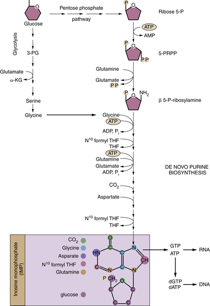
Glucose-Derived Metabolites Contribute to All Classes of Macromolecules

Glycolytic flux provides substrates for biosynthetic reactions that are required for the synthesis of lipids, nucleotides, and proteins (Figure 13-5 ). Glycolytic intermediates can be diverted to the pentose phosphate pathway to produce ribose 5-phosphate, which serves as the foundation for the de novo synthesis of purines and pyrimidines. The oxidative arm of the pentose phosphate pathway also generates NADPH, which can provide reducing equivalents for nucleotide and fatty acid synthesis. Similarly, the glycerol required for phospholipid synthesis is derived from the glycolytic intermediate dihydroxyacetone phosphate. As the major component of mammalian cell membranes, phospholipid synthesis is critical for cell growth. Glycolytic intermediates can also contribute to amino acid synthesis: 3-Phosphoglycerate can be converted to serine and ultimately glycine.
image
Figure 13-5 Glucose metabolism provides critical intermediates for cell growth Glycolytic intermediates provide the substrates for the pentose phosphate pathway (PPP) that generates the ribose 5-phosphate that is critical for nucleotide biosynthesis. The oxidative arm of the PPP, which utilizes glucose 6-phosphate, additionally generates NADPH. Glycerol 3-phosphate, which contributes the glycerol head groups for phospholipid biosynthesis, is formed from the intermediate dihydroxyacetone phosphate. 3-Phosphoglycerate provides the foundation for the serine synthesis pathway, which can further fuel glycine production for protein synthesis. This pathway also contributes to the pool of one-carbon units (CH2-THF) that are used in nucleotide biosynthesis. Glucose-derived citrate provides the acetyl-CoA that represents the fundamental building block for the synthesis of the fatty acids that comprise cellular lipids. Key metabolic enzymes are shown in blue; NADPH required for biosynthetic reactions is shown in green. CH 2 -THF, methylene tetrahydrofolate; GLDC, glycine decarboxylase; PHGDH, phosphoglycerate dehydrogenase; PKM2, pyruvate kinase M2 isoform; PPP, pentose phosphate pathway; THF, tetrahydrofolate.
A series of recent studies documenting tumors whose growth depends on diversion of glycolytic flux to anabolic pathways lends support to the hypothesis that glycolytic intermediates play a key role fueling tumor growth. Phosphoglycerate dehydrogenase (PHGDH), the enzyme that catalyzes the first step of the serine synthesis pathway that branches from glycolysis, is frequently amplified in melanoma and breast cancers, and flux into the serine synthesis pathway can support tumor cell growth. 24,25 Similarly, multiple tumor types display high levels of glycine decarboxylase (GLDC), which generates methylene tetrahydrofolate necessary for pyrimidine synthesis, and GLDC expression can promote cellular transformation and tumorigenesis. 26

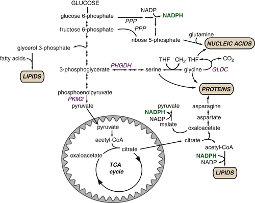
Macromolecular Synthesis, Not ATP, May Be the Major Purpose of Elevated Glycolysis in Cancer Cells

The importance of glycolytic intermediates as biosynthetic building blocks supporting cell proliferation was underscored by the observation that most proliferating cells express the M2 isoform of pyruvate kinase (PK), whereas quiescent cells express the PKM1 isoform. 27 Pyruvate kinase catalyzes the final ATP-producing step of glycolysis, converting phosphoenolpyruvate to pyruvate. Perhaps counterintuitively, PKM2 has less intrinsic activity than PKM1; PKM2 is also inhibited by growth factor signaling. These observations suggest that inhibiting the final step of glycolysis can be beneficial for cell growth. By slowing the final step of glycolysis, PKM2 enables accumulation of upstream glycolytic intermediates that can then be diverted into biosynthetic pathways. These findings indicate that proliferating cells do not undergo glycolysis for the sole purpose of ATP production; rather, they appear to selectively slow pyruvate production in order to maximize the flow of glucose carbons into anabolic pathways.
Expression of PKM2 may have the added benefit of reducing ATP production. Although ATP is required for anabolic reactions, NADPH is quantitatively more important. Biosynthesis of many macromolecules requires more reducing equivalents than energy from ATP. 1 Consequently, if all glucose were oxidized completely, ATP production would far outstrip NADPH production, thus greatly circumscribing potential cell growth. Furthermore, ATP is a potent inhibitor of phosphofructokinase-1, a key enzyme in the regulation of glycolysis, and so ATP produced in excess of biosynthetic demands would have the adverse effect of blocking glycolysis, further reducing NADPH production.
Although cancer cells preferentially reduce pyruvate kinase activity, the final step in glycolysis cannot be abrogated completely, as pyruvate fulfills many important metabolic roles. The pyruvate produced at this step has three fates: It can be transaminated to alanine, reduced to lactate to regenerate NAD+, or imported to the mitochondrion, where it is converted to acetyl-CoA that can enter the TCA cycle. In many tumors, glucose consumption is high enough to support anabolic pathways while also maintaining pyruvate flux into the mitochondria and still resulting in high amounts of lactate secretion. It is likely that reducing pyruvate kinase activity slows glucose flux through glycolysis in order to increase the likelihood that glucose can be diverted into anabolic pathways rather than being completely and rapidly exported as lactate or oxidized in the mitochondria. Given the high rates of lactate produced from glucose (as much as 90% of glucose is converted to lactate and alanine in glioblastoma cells), 28 cancer cells still have appreciable pyruvate consumption. It is likely that despite the high percentage of pyruvate that is secreted as lactate, the rate of glycolysis is so high that sufficient pyruvate remains to fuel the TCA cycle.

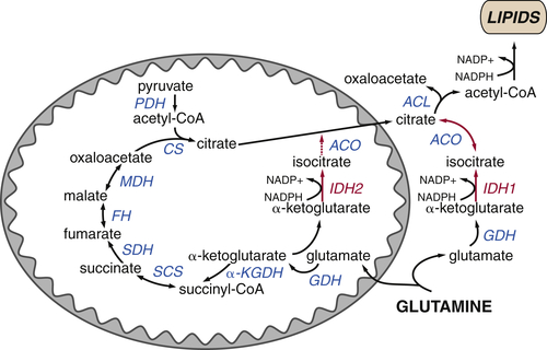
The TCA Cycle as a Biosynthetic Hub

Just as glycolytic intermediates are harnessed to support anabolic pathways, proliferating cells rewire the TCA cycle to prioritize cell growth over ATP generation. In its traditional form, the TCA cycle begins with the condensation of pyruvate-derived acetyl-CoA with oxaloacetate to form citrate (Figure 13-6 ). A series of reactions in the mitochondrial matrix convert citrate back to oxaloacetate, releasing two carbons as two molecules of carbon dioxide and producing three molecules of NADH and one molecule of FADH2. NAD+ and FAD are regenerated when NADH and FADH2 donate their electrons to support oxidative phosphorylation. The cycle can continue by combining the recycled oxaloacetate with a new molecule of acetyl-CoA. In this manner, the oxidation of acetyl-CoA is used to maximize ATP production.
image
Figure 13-6 The tricarboxylic acid (TCA) cycle supports macromolecular biosynthesis Pyruvate derived from glucose enters the mitochondrion and is converted to acetyl-CoA by pyruvate dehydrogenase (PDH), and fatty acids are oxidized to produce acetyl-CoA units within the mitochondrion. Acetyl-CoA condenses with oxaloacetate to form citrate, which proceeds through the TCA cycle in highly oxidative tissues, releasing two carbons as carbon dioxide and regenerating oxaloacetate for further cycling. These reactions maximize ATP production from acetyl-CoA by reducing the electron carriers NADH and FADH2 to fuel the electron transport chain. In proliferating cells, citrate is also exported from the mitochondrion to the cytosol, where it can provide the acetyl-CoA required for the synthesis of fatty acids and cholesterol. Similarly, oxaloacetate can form aspartate and other amino acids, contributing to intracellular amino acid pools for protein synthesis. Both of these reactions deplete TCA cycle intermediates; consequently, cells must replenish TCA cycle metabolites in order to regenerate the oxaloacetate necessary for citrate synthesis. Most cells use glutamine to fulfill this anaplerotic role: glutamine is converted to glutamate and then α-ketoglutarate, preserving TCA cycle flux. Metabolites that fuel the TCA cycle are shown in light blue. Metabolite efflux from the TCA cycle is shown in pink. Enzymes are shown in dark blue, reduced electron carriers in green, and high-energy triphosphates in orange. PDH, pyruvate dehydrogenase; ACL, ATP-citrate lyase; ACO, aconitase; CoA-SH, coenzyme A; CS, citrate synthase; FH, fumarate hydratase; GDH, glutamine dehydrogenase; IDH, isocitrate dehydrogenase; αKGDH, α-ketoglutarate dehydrogenase; MDH, malate dehydrogenase; SCS, succinyl-CoA synthetase; SDH, succinate dehydrogenase.

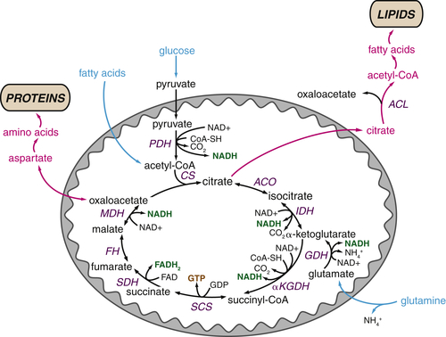
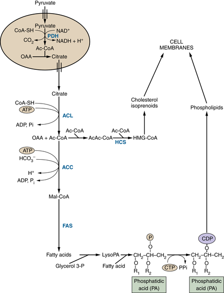
TCA Cycle Metabolites Are Required for Macromolecular Biosynthesis

In proliferating cells, citrate is not only oxidized in the TCA cycle but also can be exported to the cytosol where it is converted back to oxaloacetate and acetyl-CoA by the enzyme ATP citrate lyase (ACL). Cytosolic acetyl-CoA provides the substrate for the synthesis of fatty acids, cholesterol, and prostaglandins (Figure 13-7 ). Through this pathway, glucose acts as the major substrate for de novo lipogenesis; consequently, ACL inhibition blocks cell proliferation and inhibits tumor growth. 2830
Citrate is not the only TCA cycle metabolite that has an important biosynthetic role. Oxaloacetate and α-ketoglutarate can provide the carbon backbone for nonessential amino acids, which are used for protein and nucleic acid synthesis (see Figure 13-6). In this manner, multiple TCA cycle metabolites are diverted to other pathways that support cell growth.

Glutamine Plays Several Roles Supporting the TCA Cycle and Anabolic Metabolism

Although efflux of TCA cycle metabolites supports anabolic reactions, this results in depletion of TCA cycle intermediates from the mitochondrial matrix. Growing cells must draw on alternative sources to maintain the oxaloacetate required for citrate synthesis. Replenishment of TCA cycle intermediates, a process known as anaplerosis, is thus critical to maintain both a constant supply of metabolites for synthesis and appropriate ATP production through oxidative phosphorylation. Most proliferating cancer cells meet their anaplerotic needs through the catabolism of glutamine (see Figure 13-6). Glutamine is converted to the TCA cycle intermediate α-ketoglutarate through a metabolic pathway known as glutaminolysis. Tumor cells take up high levels of glutamine, often in great excess of any other amino acid, and studies using carbon labeling techniques have demonstrated that glutamine is the major source of carbon for the cellular oxaloacetate pool in cancer cell lines. 28
image
Figure 13-7 Glucose-derived acetyl-CoA fuels lipid and sterol synthesis Pyruvate derived from glycolysis can generate citrate in the mitochondrion. This citrate can be exported to the cytosol to provide acetyl-CoA for the synthesis of fatty acids, phospholipids, cholesterol, and isoprenoids, all of which are critical for membrane biogenesis and function. Metabolites are in black; enzymes are in blue. ACC, Acetyl-CoA carboxylase; Ac-CoA, acetyl-CoA; ACL, ATP-citrate lyase; CDP, cytidine diphosphate; CoA-SH, coenzyme A; CTP, cytidine triphosphate; FAS, fatty acid synthetase; glycerol-3-P, glycerol 3-phosphate; HCS, HMG-CoA synthetase; HMG-CoA, 3-hydroxy-3-methylglutaryl CoA; lyso PA, lysophosphatidic acid; Mal-CoA, malonyl-CoA; OAA, oxaloacetate; Pi, inorganic phosphate; R, acyl group on a lipid molecule.
By maintaining flux through the TCA cycle—and therefore delivery of electrons to the electron transport chain—glutamine plays a critical role maintaining mitochondrial bioenergetics in many cancer cells. Consequently, many cancer cell lines are absolutely dependent on glutamine for survival. Addition of cell-permeable TCA cycle analogs can rescue death of glutamine-deprived cells, highlighting the importance of glutamine as an anaplerotic substrate in cancer cells. 31,32
Proliferating cells rely on glutamine to fulfill additional biosynthetic roles. First, glutamine provides an important source of nitrogen for synthesis of nonessential amino acids and nucleotides. Glutamine is required for two independent steps in purine nucleotide synthesis, and oxaloacetate-derived aspartate is required for a third (Figure 13-8 ). Similarly, two steps of pyrimidine synthesis require glutamine. In all cases, glutamine donates nitrogen in the form of an amide group and is converted to glutamic acid, which provides a major source of nitrogen for amino acid synthesis. Transaminases can transfer the amine group from glutamic acid to α-ketoacids, which are themselves derived from the catabolism of glucose or glutamine, producing alanine, serine, aspartate, and ornithine. In turn, these amino acids act as precursors for the synthesis of glycine, cysteine, arginine, and asparagine. Likewise, two enzymatic reactions directly convert glutamate to proline. In this manner, glucose and glutamine contribute to the synthesis of every nonessential amino acid except for tyrosine, which is directly produced from the essential amino acid phenylalanine.
image
Figure 13-8 Nucleotide biosynthesis requires multiple metabolic inputs Nucleotide biosynthesis requires inputs from several metabolic pathways, highlighting why cells must coordinately regulate multiple metabolic pathways during proliferation. The de novo synthesis of purines (shown) and pyrimidines requires glucose, several amino acids, and one-carbon groups from folate metabolism. The origin of individual carbons and nitrogens on inosine monophosphate (IMP), the precursor to GTP and ATP, are color-coded in the purple box. α-KG, α-ketoglutarate; N10 formyl THF, N10 formyl tetrahydrofolate; 3-PG, 3-phosphoglycerate; β 5-P-ribosylamine, β 5-phosphate-ribosylamine; PRPP, 5-phosphoribosyl pyrophosphate; ribose 5-P, ribose 5-phosphate; P, phosphate group.
Intriguingly, cancer cells may convert up to 60% of glutamine carbon into lactic acid, an ostensibly wasteful secretion analogous to the Warburg effect. 28 One possible explanation for this behavior is that the conversion of glutamine to lactate produces NADPH, which is absolutely required for many anabolic reactions. NADPH is produced when malic enzyme converts glutamine-derived malate to pyruvate, which is subsequently reduced to lactate and secreted. In this manner, glutamine may contribute to all three needs of proliferating cells: Glutamine can provide the carbon and nitrogen for most metabolic building blocks, maintain ATP production to support bioenergetics, and generate reducing equivalents required for many anabolic reactions.

TCA Cycle Rearrangements Highlight the Role of TCA Cycle Metabolites as Biosynthetic Precursors

In proliferating cancer cells, the TCA cycle can behave more as a source of anabolic substrates rather than a bona fide cycle. A prime example of how the TCA cycle prioritizes biosynthetic reactions is the reductive carboxylation of α-ketoglutarate to isocitrate. During conditions of hypoxia or mitochondrial dysfunction, NADH accumulates from a combination of increased glycolysis and reduced oxidation of NADH by the electron transport chain. Depletion of oxidized NAD+ poses a bioenergetic challenge for the cell: Both the conversion of glucose-derived pyruvate to acetyl-CoA and the production of oxaloacetate through the enzymes of the TCA cycle require NAD+. Furthermore, during periods of acute hypoxia, pyruvate is converted to lactate at the expense of acetyl-CoA. Both of these events would strongly impair citrate production and thus cell growth. How, then, do hypoxic cells proliferate?
Several groups have demonstrated that under these conditions, the TCA cycle can partially function in reverse (for examples, see Refs. 3335). Normally, isocitrate dehydrogenase (IDH) catalyzes the oxidative decarboxylation of isocitrate to α-ketoglutarate, consuming NAD+ and producing NADH. When NAD+ is limiting, both the mitochondrial and cytosolic isoforms of IDH can function in reverse, carboxylating α-ketoglutarate to isocitrate and producing oxidized NADP+ (Figure 13-9 ). In this manner, cells can use glutamine-derived α-ketoglutarate to generate citrate, providing a mechanism to maintain anabolic reactions even under hypoxic conditions that characterize the tumor microenvironment.

Genetic Mechanisms Driving Cancer Cell Metabolism

Metabolic transformation is a common feature of tumorigenesis, occurring in many tumor types in diverse tissues. It is logical that metabolic transformation and oncogenic transformation would go hand in hand: As cancer cells acquire mutations that enable pathological proliferation, they must also acquire the means to support cell growth. Thus, many of the mutations that promote cancer growth also trigger metabolic reprogramming. This section discusses several common genetic oncogenic events that reprogram metabolism to support tumor growth.
image
Figure 13-9 Reductive carboxylation supports lipid synthesis during hypoxia Citrate levels can be maintained even in cells with defective TCA cycles or during periods of hypoxia by the reductive metabolism of glutamine-derived α-ketoglutarate to citrate. Both mitochondrial and cytosolic isoforms of isocitrate dehydrogenase (IDH2 and IDH1, respectively) can function in reverse, reducing α-ketoglutarate to isocitrate and oxidizing NADPH to NADP+. Isocitrate is reversibly converted to citrate, which can be used to generate acetyl-CoA for lipid synthesis in the cytosol.

Activation of the PI3K Pathway Promotes Metabolic Transformation

In normal cells, extracellular cues regulate the intracellular signaling pathways that tightly coordinate metabolism and proliferation. In many cells, this coordination is accomplished by the phosphatidylinositol-3-kinase (PI3K) pathway, a highly conserved signaling pathway that responds to a variety of extracellular cues. When growth factors bind to their cell surface receptors, receptor tyrosine kinases (RTKs) activate PI3K, which phosphorylates phosphatidylinositol lipids at the plasma membrane. These phosphorylated lipids recruit and activate downstream effectors of the PI3K pathway, triggering an intracellular signaling cascade that promotes cell growth, survival, and metabolism.
Constitutive activation of PI3K provides a growth stimulus and is therefore a major mechanism of tumorigenesis. In normal cells, PI3K activity is tightly guarded by a number of factors, ensuring that pathway activity is set at appropriate levels. RTK activity is strictly controlled by growth factor availability. In addition, negative feedback loops prevent prolonged pathway activation: The lipid phosphatase PTEN dephosphorylates phosphatidylinositol species to dampen PI3K signaling.
Cancers exhibit a variety of mutations to circumvent this regulation and gain cell-autonomous activation of the PI3K pathway. 36 Amplifications or mutations of growth factor receptors occur in a variety of cancers. Common examples are the amplification of the Her2/Neu receptor in breast cancer and epithelial growth factor receptor (EGFR) in lung cancer. Similarly, deletion, downregulation, or loss of function of PTEN increases cancer susceptibility and promotes tumor progression. Several tumor types display activating mutations in the catalytic PI3K subunit, PIK3CA; likewise, amplification of PIK3CA and the major PI3K effector, Akt, are common oncogenic events. All together, genetic activation of the PI3K pathway is one of the most prevalent classes of tumorigenic mutations.
Whereas PI3K signaling influences numerous cellular functions, the serine/threonine kinase Akt and its effector, mechanistic target of rapamycin (mTOR), are the PI3K effectors most commonly implicated in tumorigenesis. Akt coordinates cell growth and survival in a large part by rewiring metabolism to promote nutrient uptake and biosynthetic activities. Certainly, with regard to the regulation of cell metabolism, Akt activation is likely the most important consequence of PI3K pathway activation.
Akt induces many phenotypes consistent with the Warburg effect. Akt promotes the expression and membrane localization of glucose transporters, driving increased glucose uptake. Akt concurrently increases the expression of hexokinase, the enzyme that phosphorylates glucose so that it is retained in the cell and can undergo glycolysis. Akt regulates multiple steps of glycolysis through both gene expression and modulation of enzyme activity. Together, these activities result in a large increase in glycolytic flux in cells with active Akt. Similarly, both Akt and mTOR can trigger expression of lipogenic genes and lipid synthesis. 7,37 mTOR additionally promotes biosynthetic pathways by directly stimulating mRNA translation and ribosome biogenesis. 38 Thus, PI3K signaling is often sufficient to enhance the metabolic activities that support cell growth.

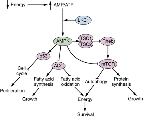
LKB1 and AMPK Are Metabolic Brakes That Circumvent Tumor Growth

While growth factor–stimulated signaling cascades promote cell growth under favorable conditions, cells have sophisticated nutrient sensing systems that serve to block growth when the internal energy supply is limiting. These regulators ensure that, during periods of intracellular nutrient depletion, metabolites are redirected from anabolic pathways and instead used to fuel catabolic pathways that will provide the energy required to survive the period of nutrient limitation. The AMP-activated protein kinase (AMPK) plays a major role coordinating cellular energy status with appropriate metabolic responses.
AMPK directly senses cellular energy levels in the form of the AMP/ATP ratio (Figure 13-10 ). Falling energy levels increase the cellular AMP/ATP ratio, priming AMPK for activation by the liver kinase B1 (LKB1). AMPK phosphorylates multiple targets with the cumulative effect of blocking anabolic reactions and stimulating energy-generating catabolic pathways. For example, AMPK phosphorylates and inhibits acetyl-CoA carboxylase (ACC), with the dual effect of blocking fatty acid synthesis and activating fatty acid oxidation. AMPK also directly inhibits cell growth, both by inducing a p53-dependent cell cycle arrest and by blocking mTOR activity at multiple levels (see Figure 13-10). Through these diverse activities, AMPK functions as a metabolic checkpoint, ensuring that cell growth is halted until bioenergetic conditions are favorable for growth.
This is particularly relevant in the tumor microenvironment, when varied nutrient delivery could occasionally activate such metabolic checkpoints and block growth. Suggestively, LKB1 is classified as a tumor suppressor. Inactivating mutations in LKB1 cause Peutz-Jeghers syndrome, a syndrome characterized by benign gastrointestinal and oral lesions and heightened susceptibility to a variety of cancers. Similarly, inactivating mutations in LKB1 are frequent in lung and ovarian carcinomas. 39 The pathogenic mechanisms of LKB1 loss of function are unclear, as LKB1 targets proteins involved in apoptosis, cell polarity, and cell cycle control in addition to activating AMPK. However, it is possible that failure to curtail cell growth in response to normal environmental cues contributes to the oncogenic potential of LKB1 inhibition. In line with this hypothesis, there is great interest in determining whether activating AMPK through agonists such as metformin could activate metabolic checkpoints to block tumor growth. Given the role of AMPK antagonizing many of the pro-growth functions of the PI3K pathway, such strategies may provide an effective way to harness our normal homeostatic mechanisms to slow cancer growth.
image
Figure 13-10 The LKB1/AMPK pathway coordinates the metabolic response to nutrient deprivation. A fall in cellular energy status, triggered by nutrient deprivation or other stressors, may increase the adenosine monophosphate (AMP):adenosine triphosphate (ATP) ratio. This activates the kinase KLB1 and its target, the AMP-activated protein kinase (AMPK). AMPK phosphorylates a number of targets to coordinately inhibit anabolic processes and activate catabolic, energy-generating pathways to restore cellular energy levels. In particular, AMPK triggers the phosphorylation and activation of p53, inducing cell cycle arrest; phosphorylation and inactivation of acetyl-CoA carboxylase (ACC), which suppresses fatty acid synthesis and promotes fatty acid oxidation, and direct and indirect inhibition of the mechanistic target of rapamycin (mTOR), thereby inhibiting protein synthesis and inducing autophagy. Suppression of growth and proliferation preserve existing energy stores, while fatty acid oxidation and autophagy generate ATP.

The Transcription Factor HIF1 Controls Glycolytic Metabolism

Conditions of low oxygen availability pose a threat to cell survival. Oxygen serves as the terminal electron acceptor of the electron transport chain; as a result, when oxygen is limiting, ATP production through oxidative phosphorylation can be compromised. Excess electrons leak out of the electron transport chain inappropriately, forming reactive oxygen species than can, in high doses, cause great cellular damage. Consequently, cells must rapidly sense and respond to conditions of low oxygen. The cellular response to low oxygen is coordinated by the hypoxia inducible factors (HIF1 and HIF2), transcription factors that are rapidly stabilized and activated during hypoxia. HIF1 and HIF2 promote survival in hypoxia through two mechanisms. First, they induce expression of angiogenic genes, such as vascular endothelial growth factor (VEGF), in order to increase the blood supply to reoxygenate the tissue. Second, they promote metabolic reprogramming in order to maintain ATP supplies while limiting damaging electron loss from the electron transport chain. 40
Many of the activities of HIF1 and HIF2 would benefit tumor cell survival. As discussed earlier, tumors have frequent variations in oxygenation, often resulting in regions of hypoxia. Expression of HIF1 or HIF2 would help a tumor cell adapt and survive in a hypoxic microenvironment. In particular, HIF1 induces multiple metabolic changes that characterize cancer cells. HIF1 increases expression of glucose transporters and glycolytic enzymes, thereby elevating glucose consumption and glycolytic flux.
Many tumors display aberrant HIF1 expression. Most famously, von Hippel–Lindau disease, characterized by hemangioblastomas of the central nervous system, pheochromocytoma, and renal cell carcinomas, is caused by loss-of-function mutations of the von Hippel–Lindau (VHL) ubiquitin ligase that negatively regulates the stability of HIF1 and HIF2. Tumors exhibit high levels of HIF1, even under normoxia, and constitutively high levels of HIF1 targets. Elevated HIF1 is observed in several solid tumors outside of VHL disease, including breast, ovarian, colon, pancreas, and prostate. 41 In several cases, levels of HIF1 correlate with tumor aggressiveness and poor prognosis. Multiple mechanisms result in increased HIF1 expression: Variable oxygenation, ROS production, oncogene activation, and growth factor signaling (including PI3K and mTOR signaling) can all contribute to heightened HIF1 activity.
Although HIF1 increases glycolysis and lactate production, its effects on metabolism are not uniformly pro-tumorigenic. First, hypoxia causes a decrease in protein synthesis rates—up to 50% of the rates in normal cells—and this decline in protein synthesis is mediated at least in part by repression of mTOR by HIF1. 42 Second, while HIF1 promotes glycolysis, it blocks the mitochondrial oxidation of glucose-derived pyruvate. HIF1 induces pyruvate dehydrogenase kinase (PDK), which inhibits PDH, the enzyme responsible for converting pyruvate to acetyl-CoA. HIF1 therefore reduces the amount of acetyl-CoA that is available for lipid synthesis, instead diverting pyruvate to lactate for secretion. 43 Although HIF1 expression is sufficient to drive citrate production through reductive carboxylation of glutamine-derived α-ketoglutarate, it is not clear whether this citrate can fully compensate for reduced conversion of pyruvate to acetyl-CoA. 33 Thus, the effects of HIF1 on tumor metabolism are complex. During conditions of true hypoxia, HIF1 expression promotes metabolic adaptations that support tumor survival and increases angiogenesis to support tumor growth. However, during conditions of adequate oxygenation, persistent HIF1 expression would serve to reduce protein and lipid synthesis and hamper cell proliferation. These disparate effects of HIF1 on tumor growth could explain why HIF1 expression is associated with a tumor-suppressive function under certain circumstances. 44

c-Myc Coordinates Cellular Proliferation and Metabolism

The Myc family of transcription factors (c-Myc, L-Myc, and N-Myc) both stimulates and represses transcription to regulate a vast array of cellular processes, including cell growth and cell cycle progression, and contributes to a variety of human tumors. Myc regulates the expression of an enormous number of genes and consequently can influence processes as diverse as differentiation, vasculogenesis, cell adhesion, cell growth, apoptosis, and DNA damage responses. 45 Although all of these outcomes could contribute significantly to the oncogenic effects of Myc expression, this section focuses on the potential role of Myc in coordinating cell metabolism to support cell proliferation.
Myc heterodimerizes with its binding partner Max to regulate expression of a large number of genes. In particular, c-Myc promotes expression of several cyclins and CDK4 while repressing the cyclin-dependent kinase inhibitors p21 and p27, thus promoting entry into S phase. 45 At the same time, Myc directly reprograms metabolism by increasing expression of genes involved in glycolysis, glutamine metabolism, and mitochondrial biogenesis. 46 c-Myc also targets a number of genes involved in nucleotide biosynthesis, ensuring that cells have the raw material to successfully complete S phase. 47 An extensive survey of Myc targets in a variety of cell lines revealed that genes involved in ribosome biogenesis form a large component of the core Myc signature, further highlighting Myc’s function in regulating biomass accumulation. 48 Together, these findings demonstrate that c-Myc functions to upregulate energy production and biosynthetic processes that are required for successful cell replication, thus directly coordinating proliferative metabolism and cell cycle progression.
Myc is a powerful oncogene whose activity is pathologically enhanced in many human cancers; indeed, MYC is one of the most highly amplified oncogenes across all types of human cancers. 49 In other cases, chromosomal translocations or increased transcription downstream of hyperactive Wnt signaling contribute to oncogenic activation of Myc. 50 Myc has many activities that could contribute to its ability to promote tumor establishment and maintenance. In some cases, the metabolic reprogramming driven by Myc expression provides critical support for tumor growth. Myc-overexpressing cells are extremely sensitive to nutrient depletion. In particular, cells with high levels of Myc are glutamine addicted, using glutamine as a critical source of TCA cycle metabolites to support rapid cell growth. Consequently, removal of glutamine from the extracellular medium triggers apoptosis in Myc-overexpressing cells. Likewise, blocking conversion of glutamine to α-ketoglutarate through inhibition of glutaminase can block cellular transformation and tumor growth. 51,52

Mutations in Metabolic Enzymes Can Influence Tumorigenesis

Although the bulk of cancer-associated metabolic rearrangements are directed by signaling events, cellular signaling pathways are not unidirectional. Metabolites themselves can serve as intracellular cues to regulate signaling proteins and influence gene expression. Activation of AMPK by AMP/ATP and mTOR by amino acid levels are common examples of how metabolites can inform signaling pathways. As with canonical signaling pathways, dysregulated activation of these metabolic signaling pathways can influence cancer susceptibility and tumor growth. Mutations in three TCA-cycle enzymes provide major examples of how inappropriate metabolite accumulation can trigger oncogenic signaling events.
Loss-of-function mutations in two consecutive enzymes of the TCA cycle, succinate dehydrogenase (SDH) and fumarate hydratase (FH), predispose patients to tumor formation. In both cases, the genes behave as classic tumor suppressors, in which mutation in one allele causes a dominant risk of tumor formation, followed by loss of function of the second, wild-type allele, in the tumor itself. SDH has four subunits and an assembly factor, and mutation in any of these five genes predisposes individuals to a variety of paragangliomas and pheochromocytomas, and in some cases, renal cell carcinomas or gastrointestinal stromal tumors. 53 In all cases, the pathogenic mechanisms linking SDH deficiency to tumor formation are attributed to elevated succinate levels, triggering a pseudohypoxic cellular state. In normoxia, HIF1 activity is repressed by the action of a family of prolyl hydroxylases (PHDs) that target the labile subunit of HIF1 (HIF1α) for ubiquitination and degradation by the proteasome. PHDs use oxygen, α-ketoglutarate, and ferrous iron to hydroxylate HIF1α, releasing carbon dioxide, succinate, and ferric iron. Consequently, accumulation of succinate blocks PHD activity, resulting in aberrant stabilization of HIF1α. 54 Enhanced HIF1 activity is likely an important mechanism driving SDH-deficient tumor growth, as the clinical presentations of patients with dominant SDH mutations are similar to several hypoxia syndromes. 53
Fumarate, likewise, is a potent inhibitor of the PHDs. 55,56 However, FH defects are associated with a predisposition to hereditary leiomyomatosis and renal cell carcinoma. 57 Although elevated HIF1 may contribute to the pathogenic mechanism of FH mutations, several additional consequences of elevated intracellular fumarate provide alternative avenues through which FH mutations may increase cancer susceptibility. First, fumarate can directly modify specific cysteine residues in Kelch-like ECH-associated protein 1 (KEAP1), thereby blocking its ability to suppress the Nrf2-mediated antioxidant response pathway. 58 Enhanced antioxidant defenses may, in some contexts, help guard cancer cell survival in the face of high proliferation-induced ROS production. Other studies have demonstrated that FH deficiency triggers a shift toward glycolytic metabolism, accompanied by reduced AMPK activity, lower intracellular iron, and elevated HIF1. 59 Regardless of the mechanism of tumorigenesis, both SDH- and FH-deficient tumors have the striking phenotype of interrupted TCA cycle function. Elevated aerobic glycolysis can increase glucose consumption and alleviate the bioenergetic constraints. In some cases, mitochondria may be able to funnel accumulated metabolites into alternate metabolic pathways, enabling partial mitochondrial NADH generation. 60 Similarly, glutamine-dependent reductive carboxylation can maintain citrate pools in cells with defective oxidative phosphorylation. 35 Given the critical role of the TCA cycle in supporting anaplerotic reactions, it will be important for future studies to clarify how cells with impaired TCA cycle activities can proliferate at pathological rates.
More recently, the discovery of mutations in the cytosolic and mitochondrial forms of isocitrate dehydrogenase (IDH) has provided the strongest evidence that altered metabolism can drive tumorigenesis. Somatic mutations at a defined number of residues on IDH occur at a high frequency in a variety of malignancies, most notably glioblastoma and acute myeloid leukemia. Oncogenic mutations in IDH confer a neomorphic activity: Rather than oxidizing isocitrate to α-ketoglutarate, mutant IDH isoforms reduce α-ketoglutarate, forming 2-hydroxyglutarate (2-HG). 61,62 A series of studies demonstrated that 2-HG, by virtue of its structural similarity to α-ketoglutarate, can act as an inhibitor of multiple α-ketoglutarate–dependent dioxygenases. In particular, 2-HG can inhibit the TET family of enzymes that convert 5-methylcytosine to 5-hydroxymethylcytosine, an intermediate step in the process of DNA demethylation. 6365 Likewise, 2-HG can inhibit various histone demethylases in vitro, and mutant IDH expression correlates with increased H3K9 histone methylation in vivo. 6567 In both cases, hypermethylation profiles (of DNA and/or histones) contribute to the silencing of differentiation-related genes, promoting the accumulation of cells arrested in the course of their differentiation, capable of self-renewal and prone to tumorigenesis. 68
Together, these studies underscore the complex interplay between cellular signaling networks and metabolic pathways. In all cases, oncogenic activity is attributed to altered levels of particular metabolites (fumarate, succinate, or 2-HG), indicating that cells are highly responsive to alterations in metabolic enzymes and highlighting the possibility that metabolites themselves can function as potent signaling molecules.

Clinical Implications of Metabolic Transformation

The ultimate goal of cancer research is to improve cancer detection and treatment. Elucidating the differences between cancer and normal cells will provide a means to identify and attack cancer cells specifically. In this regard, the distinctive metabolic phenotype of cancer cells may provide a key therapeutic window to distinguish pathological proliferation.
The propensity of tumors to take up high levels of glucose is already extensively exploited in the clinic. Positron emission tomography (PET) enables in vivo visualization of radioactive tracers such as [18F]fluorodeoxyglucose (FDG), a glucose analog that can be taken up by cells and phosphorylated by hexokinase but not further metabolized. Cellular accumulation of FDG therefore provides a reliable readout of the glycolytic activity of a cell, and whole-body scanning by PET can reveal regions of abnormally high glucose uptake and retention. Because cancer cells often take up high levels of glucose, FDG-PET provides a robust tool for detecting a variety of tumors, including breast, colorectal, lung, brain, and lymphoma. FDG-PET can also be used to monitor tumor growth and metastases. Importantly, FDG-PET provides a rapid readout of tumor response to therapy. Most therapies that effectively impair tumor growth or survival trigger a rapid decrease in FDG-PET signal, a change that can be observed within a few days after therapy initiation. 69,70 Because cancer cells often have aberrant nutrient uptake, future studies may reveal other radioactive metabolic tracers with tumor-specific uptake.
Tumor dependence on glycolysis provides additional therapeutic avenues. Promising preclinical data exist for the inhibition of multiple steps of glucose uptake and catabolism through glycolysis. One large caveat in the targeting of metabolic enzymes is that cancer cells often have a metabolic profile very similar to rapidly proliferating normal cells. Consequently, blocking enzymes that promote proliferative metabolism may have adverse effects on multiple cell types in vivo, including the rapidly proliferating cells in the intestinal epithelium and bone marrow. Nevertheless, it may be possible to safely target glucose metabolism in patients. As discussed earlier, the rate of entry of pyruvate into the TCA cycle is determined by pyruvate dehydrogenase, which is inhibited by the PDKs. Dichloroacetate (DCA) inhibits the PDKs, thus increasing pyruvate flux into the TCA cycle and reducing lactate production. Although the therapeutic benefit of increasing glucose oxidation is not yet demonstrated in tumors in vivo, DCA is well tolerated in patients, suggesting that it may be possible to target aberrant glucose metabolism in cancer patients. 71
The therapeutic potential of many of the pathways discussed in this chapter are targets of ongoing studies. In animal studies, inhibitors of several steps of glycolysis (phosphofructokinase, 72 lactate dehydrogenase 23,73 ), glutamine anaplerosis (glutaminase, 51 glutamate dehydrogenase 74 ), and fatty acid synthesis (ATP citrate lyase, 29 fatty acid synthase 75,76 ) can inhibit tumor growth. These data suggest that carefully targeted administration of agents that block key nodes of proliferative metabolism may hold therapeutic promise.
Suggestively, some of the oldest and most effective cancer treatments target aspects of cell metabolism. For example, antifolates interfere with folate metabolism and thus disrupt nucleotide biosynthesis and other reactions that require one-carbon units, such as protein methylation. 5-Fluorouracil, a nucleoside analog, inhibits thymidylate synthase and blocks DNA synthesis. These antimetabolites are still effective therapies today, reinforcing the critical role of altered metabolism in the support of tumor growth. Further research into the specific metabolic abnormalities of cancer cells, and how these perturbations foster tumor growth, may uncover pharmacologic targets that will be effective and well tolerated.
References

1. Vander Heiden M.G. , Cantley L.C. , Thomspon C.B. Understanding the Warburg effect: the metabolic requirements of cell proliferation . Science . 2009 ; 324 : 1029 1033 .

2. Schulze A. , Harris A.L. How cancer metabolism is tuned for proliferation and vulnerable to disruption . Nature . 2012 ; 491 : 364 373 .

3. Wallace D.C. Mitochondria and cancer . Nat Rev Cancer . 2012 ; 12 : 685 698 .

4. Wallace D.C. A mitochondrial paradigm of metabolic and degenerative diseases, aging, and cancer: a dawn for evolutionary medicine . Annu Rev Genet . 2005 ; 39 : 359 407 .

5. Wise D.R. , Thompson C.B. Glutamine addiction: a new therapeutic target in cancer . Trends Biochem Sci . 2010 ; 35 : 427 433 .

6. Berg J.M. et al. Biochemistry . New York, NY : W.H. Freeman ; 2002 .

7. Cairns R.A. , Harris I.S. , Mak T.W. Regulation of cancer cell metabolism . Nat Rev Cancer . 2011 ; 11 : 85 95 .

8. Lunt S.Y. , Vander Heiden M.G. Aerobic glycolysis: meeting the metabolic requirements of cell proliferation . Annu Rev Cell Dev Biol . 2011 ; 27 : 441 464 .

9. Ward P.S. , Thompson C.B. Metabolic reprogramming: a cancer hallmark even Warburg did not anticipate . Cancer Cell . 2012 ; 21 : 297 308 .

10. Hanahan D. , Weinberg R.A. Hallmarks of cancer: the next generation . Cell . 2011 ; 144 : 646 674 .

11. Warburg O. On the origin of cancer cells . Science . 1956 ; 123 : 309 314 .

12. Moreno-Sánchez R. , Rodríguez-Enríquez S. , Marín-Hernández A. , Saavedra E. Energy metabolism in tumor cells . FEBS J . 2007 ; 274 : 1393 1418 .

13. Warburg O. On respiratory impairment in cancer cells . Science . 1956 ; 124 : 269 270 .

14. Guppy M. , Greiner E. , Brand K. The role of the Crabtree effect and an endogenous fuel in the energy metabolism of resting and proliferating thymocytes . Eur J Biochem . 1993 ; 212 : 95 99 .

15. MacDonald M.J. et al. High activity of mitochondrial glycerol phosphate dehydrogenase in insulinomas and carcinoid and other tumors of the amine precursor uptake decarboxylation system . Cancer Res . 1990 ; 50 : 7203 7205 .

16. Grivell A.R. , Korpelainen E.I. , Williams C.J. , Berry M.N. Substrate-dependent utilization of the glycerol 3-phosphate or malate/aspartate redox shuttles by Ehrlich ascites cells . Biochem J . 1995 ; 310 ( Pt 2 ) : 665 671 .

17. Mazurek S. , Eigenbrodt E. , Failing K. , Steinberg P. Alterations in the glycolytic and glutaminolytic pathways after malignant transformation of rat liver oval cells . J Cell Physiol . 1999 ; 181 : 136 146 .

18. Chowdhury S.K. , Gemin A. , Singh G. High activity of mitochondrial glycerophosphate dehydrogenase and glycerophosphate-dependent ROS production in prostate cancer cell lines . Biochem Biophys Res Commun . 2005 ; 333 : 1139 1145 .

19. Gatenby R.A. , Gillies R.J. Why do cancers have high aerobic glycolysis? Nat Rev Cancer . 2004 ; 4 : 891 899 .

20. Gatenby R.A. , Gawlinski E.T. , Gmitro A.F. et al. Acid-mediated tumor invasion: a multidisciplinary study . Cancer Res . 2006 ; 66 : 5216 5223 .

21. Robey I.F. , Baggett B.K. , Kirkpatrick N.D. et al. Bicarbonate increases tumor pH and inhibits spontaneous metastases . Cancer Res . 2009 ; 69 : 2260 2268 .

22. Sonveaux P. et al. Targeting lactate-fueled respiration selectively kills hypoxic tumor cells in mice . J Clin Invest . 2008 ; 118 : 3930 3942 .

23. Fantin V.R. , St-Pierre J. , Leder P. Attenuation of LDH-A expression uncovers a link between glycolysis, mitochondrial physiology, and tumor maintenance . Cancer Cell . 2006 ; 9 : 425 434 .

24. Locasale J.W. , Grassian A.R. , Melman T. et al. Phosphoglycerate dehydrogenase diverts glycolytic flux and contributes to oncogenesis . Nat Genet . 2011 ; 43 : 869 874 .

25. Possemato R. , Marks K.M. , Shaul Y.D. et al. Functional genomics reveal that the serine synthesis pathway is essential in breast cancer . Nature . 2011 ; 476 : 346 350 .

26. Zhang W.C. , Shyh-Chang N. , Yang H. et al. Glycine decarboxylase activity drives non-small cell lung cancer tumor-initiating cells and tumorigenesis . Cell . 2012 ; 148 : 259 272 .

27. Mazurek S. , Boschek C.B. , Hugo F. , Eigenbrodt E. Pyruvate kinase type M2 and its role in tumor growth and spreading . Semin Cancer Biol . 2005 ; 15 : 300 308 .

28. DeBerardinis R.J. , Mancuso A. , Daikhin E. et al. Beyond aerobic glycolysis: transformed cells can engage in glutamine metabolism that exceeds the requirement for protein and nucleotide synthesis . Proc Natl Acad Sci U S A . 2007 ; 104 : 19345 19350 .

29. Hatzivassiliou G. , Zhao F. , Bauer D.E. et al. ATP citrate lyase inhibition can suppress tumor cell growth . Cancer Cell . 2005 ; 8 : 311 321 .

30. Bauer D.E. , Hatzivassiliou G. , Zhao F. et al. ATP citrate lyase is an important component of cell growth and transformation . Oncogene . 2005 ; 24 : 6314 6322 .

31. Wise D.R. , DeBerardinis R.J. , Mancuso A. et al. Myc regulates a transcriptional program that stimulates mitochondrial glutaminolysis and leads to glutamine addiction . Proc Natl Acad Sci U S A . 2008 ; 105 : 18782 18787 .

32. Yuneva M. , Zamboni N. , Oefner P. et al. Deficiency in glutamine but not glucose induces MYC-dependent apoptosis in human cells . J Cell Biol . 2007 ; 178 : 93 105 .

33. Wise D.R. , Ward P.S. , Shay J.E. et al. Hypoxia promotes isocitrate dehydrogenase-dependent carboxylation of alpha-ketoglutarate to citrate to support cell growth and viability . Proc Natl Acad Sci U S A . 2011 ; 108 : 19611 19616 .

34. Metallo C.M. , Gameiro P.A. , Bell E.L. et al. Reductive glutamine metabolism by IDH1 mediates lipogenesis under hypoxia . Nature . 2012 ; 481 : 380 384 .

35. Mullen A.R. , Wheaton W.W. , Jin E.S. et al. Reductive carboxylation supports growth in tumour cells with defective mitochondria . Nature . 2012 ; 481 : 385 388 .

36. Engelman J.A. Targeting PI3K signalling in cancer: opportunities, challenges and limitations . Nat Rev Cancer . 2009 ; 9 : 550 562 .

37. Yecies J.L. , Manning B.D. Transcriptional control of cellular metabolism by mTOR signaling . Cancer Res . 2011 ; 71 : 2815 2820 .

38. Laplante M. , Sabatini D.M. mTOR signaling in growth control and disease . Cell . 2012 ; 149 : 274 293 .

39. Shackelford D.B. , Shaw R.J. The LKB1-AMPK pathway: metabolism and growth control in tumour suppression . Nat Rev Cancer . 2009 ; 9 : 563 575 .

40. Semenza G.L. Hypoxia-inducible factors in physiology and medicine . Cell . 2012 ; 148 : 399 408 .

41. Semenza G.L. Defining the role of hypoxia-inducible factor 1 in cancer biology and therapeutics . Oncogene . 2010 ; 29 : 625 634 .

42. Spriggs K.A. , Bushell M. , Willis A.E. Translational regulation of gene expression during conditions of cell stress . Mol Cell . 2010 ; 40 : 228 237 .

43. Lun J.J. , Bui T. , Gruber M. The transcription factor HIF-1alpha plays a critical role in the growth factor-dependent regulation of both aerobic and anaerobic glycolysis . Genes Dev . 2007 ; 21 : 1037 1049 .

44. Shen C. , Beroukhim R. , Schumacher S.E. et al. Genetic and functional studies implicate HIF1alpha as a 14q kidney cancer suppressor gene . Cancer Discov . 2011 ; 1 : 222 235 .

45. Adhikary S. , Eilers M. Transcriptional regulation and transformation by Myc proteins . Nat Rev Mol Cell Biol . 2005 ; 6 : 635 645 .

46. Dang C.V. , Kim J.W. , Gao P. , Yustein J. The interplay between MYC and HIF in cancer . Nat Rev Cancer . 2008 ; 8 : 51 56 .

47. Liu Y.C. , Li F. , Handler J. et al. Global regulation of nucleotide biosynthetic genes by c-Myc . PLoS One . 2008 ; 3 : e2722 .

48. Ji H. , Wu G. , Zhan X. et al. Cell-type independent MYC target genes reveal a primordial signature involved in biomass accumulation . PLoS One . 2011 ; 6 : e26057 .

49. Beroukhim R. , Mermel C.H. , Porter D. et al. The landscape of somatic copy-number alteration across human cancers . Nature . 2010 ; 463 : 899 905 .

50. Dang C.V. MYC on the path to cancer . Cell . 2012 ; 149 : 22 35 .

51. Wang J.B. , Erickson J.W. , Fuji R. et al. Targeting mitochondrial glutaminase activity inhibits oncogenic transformation . Cancer Cell . 2010 ; 18 : 207 219 .

52. Le A. , Lane A.N. , Hamaker M. et al. Glucose-independent glutamine metabolism via TCA cycling for proliferation and survival in B cells . Cell Metab . 2012 ; 15 : 110 121 .

53. Raimundo N. , Baysal B.E. , Shadel G.S. Revisiting the TCA cycle: signaling to tumor formation . Trends Mol Med . 2011 ; 17 : 641 649 .

54. Selak M.A. , Armour S.M. , MacKenzie E.D. et al. Succinate links TCA cycle dysfunction to oncogenesis by inhibiting HIF-alpha prolyl hydroxylase . Cancer Cell . 2005 ; 7 : 77 85 .

55. Iaacs J.S. , Jung Y.J. , Mole D.R. et al. HIF overexpression correlates with biallelic loss of fumarate hydratase in renal cancer: novel role of fumarate in regulation of HIF stability . Cancer Cell . 2005 ; 8 : 143 153 .

56. MacKenzie E.D. et al. Cell-permeating alpha-ketoglutarate derivatives alleviate pseudohypoxia in succinate dehydrogenase-deficient cells . Mol Cell Biol . 2007 ; 27 : 3282 3289 .

57. Tomlinson I.P. , Alam N.A. , Rowan A.J. et al. Germline mutations in FH predispose to dominantly inherited uterine fibroids, skin leiomyomata and papillary renal cell cancer . Nat Genet . 2002 ; 30 : 406 410 .

58. Adam J. , Hatipoglu E. , O’Flaherty L. et al. Renal cyst formation in Fh1-deficient mice is independent of the Hif/Phd pathway: roles for fumarate in KEAP1 succination and Nrf2 signaling . Cancer Cell . 2011 ; 20 : 524 537 .

59. Tong W.H. , Sourbier C. , Kovtunovych G. et al. The glycolytic shift in fumarate-hydratase-deficient kidney cancer lowers AMPK levels, increases anabolic propensities and lowers cellular iron levels . Cancer Cell . 2011 ; 20 : 315 327 .

60. Frezza C. , Zheng L. , Folger O. et al. Haem oxygenase is synthetically lethal with the tumour suppressor fumarate hydratase . Nature . 2011 ; 477 : 225 228 .

61. Dang L. , White D.W. , Gross S. et al. Cancer-associated IDH1 mutations produce 2-hydroxyglutarate . Nature . 2009 ; 462 : 739 744 .

62. Ward P.S. , Patel J. , Wise D.R. et al. The common feature of leukemia-associated IDH1 and IDH2 mutations is a neomorphic enzyme activity converting alpha-ketoglutarate to 2-hydroxyglutarate . Cancer Cell . 2010 ; 17 : 225 234 .

63. Figueroa M.E. , Abdel-Wahab O. , Lu C. et al. Leukemic IDH1 and IDH2 mutations result in a hypermethylation phenotype, disrupt TET2 function, and impair hematopoietic differentiation . Cancer Cell . 2010 ; 18 : 553 567 .

64. Turcan S. , Rohle D. , Goenka A. et al. IDH1 mutation is sufficient to establish the glioma hypermethylator phenotype . Nature . 2012 ; 483 : 479 483 .

65. Xu W. , Yang H. , Liu Y. et al. Oncometabolite 2-hydroxyglutarate is a competitive inhibitor of alpha-ketoglutarate-dependent dioxygenases . Cancer Cell . 2011 ; 19 : 17 30 .

66. Lu C. , Ward P.S. , Kapoor G.S. et al. IDH mutation impairs histone demethylation and results in a block to cell differentiation . Nature . 2012 ; 483 : 474 478 .

67. Chowdhury R. et al. The oncometabolite 2-hydroxyglutarate inhibits histone lysine demethylases . EMBO Rep . 2011 ; 12 : 463 469 .

68. Lu C. , Thompson C.B. Metabolic regulation of epigenetics . Cell Metab . 2012 ; 16 : 9 17 .

69. Engelman J.A. et al. Effective use of PI3K and MEK inhibitors to treat mutant Kras G12D and PIK3CA H1047R murine lung cancers . Nat Med . 2008 ; 14 : 1351 1356 .

70. Young H. et al. Measurement of clinical and subclinical tumour response using [18F]-fluorodeoxyglucose and positron emission tomography: review and 1999 EORTC recommendations. European Organization for Research and Treatment of Cancer (EORTC) PET Study Group . Eur J Cancer . 1999 ; 35 : 1773 1782 .

71. Vander Heiden M.G. Targeting cancer metabolism: a therapeutic window opens . Nat Rev Drug Discov . 2011 ; 10 : 671 684 .

72. Clem B. , Telang S. , Clem A. et al. Small-molecule inhibition of 6-phosphofructo-2-kinase activity suppresses glycolytic flux and tumor growth . Mol Cancer Ther . 2008 ; 7 : 110 120 .

73. Shim H. , Dolde C. , Lewis B.C. et al. c-Myc transactivation of LDH-A: implications for tumor metabolism and growth . Proc Natl Acad Sci U S A . 1997 ; 94 : 6658 6663 .

74. Yang C. , Sudderth J. , Dang T. et al. Glioblastoma cells require glutamate dehydrogenase to survive impairments of glucose metabolism or Akt signaling . Cancer Res . 2009 ; 69 : 7986 7993 .

75. Pizer R.D. , Wood F.D. , Heine H.S. et al. Inhibition of fatty acid synthesis delays disease progression in a xenograft model of ovarian cancer . Cancer Res . 1996 ; 56 : 1189 1193 .

76. Flavin R. , Peluso S. , Nguyen P.L. , Loda M. Fatty acid synthase as a potential therapeutic target in cancer . Future Oncol . 2010 ; 6 : 551 562 .