Tachyarrhythmias

Published on 07/02/2015 by admin

Filed under Anesthesiology

Last modified 22/04/2025

Print this page

rate 1 star rate 2 star rate 3 star rate 4 star rate 5 star
Your rating: none, Average: 0 (0 votes)

This article have been viewed 2622 times

Tachyarrhythmias

Srikanth Hosur, MD and Adebola Adesanya, MD, MPH

Tachyarrhythmias are classified as either narrow complex tachycardias (NCT) or wide complex tachycardias (WCT), based on the width of the QRS complexes. NCTs are further classified as atrioventricular (AV) node-passive or AV node-active tachycardias, based on whether the AV node is involved in the propagation and maintenance of the arrhythmia. AV node-passive tachycardias have a regular rhythm, as in atrial tachycardia or atrial flutter, or an irregular rhythm, as in multifocal atrial tachycardia, atrial flutter with varying conduction, or atrial fibrillation. (AV node reentry tachycardia [AVNRT]) or accessory pathway-dependent tachycardia. The accessory pathway conduction can be orthodromic (using the AV node as forward conduction) and, hence, have a narrow complex, or antidromic (using the accessory pathway as forward conduction and the AV node itself for retrograde conduction) and, hence, have a wide complex.

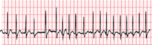
It is important to understand the mechanisms of these arrythmias because AV node-active tachycardias can usually be terminated by maneuvers that prolong AV node conduction, such as vagal maneuvers, or the administration of drugs, such as adenosine (Figure 36-1).

Narrow complex tachycardias

Treatment

NCTs can be treated medically or by cardioversion. The administration of adenosine and vagal maneuvers are helpful only in the termination of AV node-active tachycardias. Although all NCTs can be terminated using synchronized cardioversion, the use of cardioversion should be reserved for patients in whom the arrhythmia is accompanied by hemodynamic instability.

In patients with hemodynamically stable NCTs, diltiazem, 0.25 mg/kg, is preferred over β-adrenergic receptor blocking agents or verapamil because dilitiazem has less negative inotropy. Patients with a low ejection fraction are candidates for amiodarone administered intravenously as a bolus of 150 mg that can be repeated. Procainamide is useful as a second-line agent in this situation.

Adenosine should be given in a central or antecubital vein at a dose of 6 to 12 mg. It is short acting, has a half-life of 12 to 18 sec, and can cause flushing, bronchospasm, and chest pain. Adenosine is antagonized by the administration of methylxanthines, such as aminophylline.

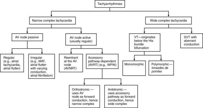
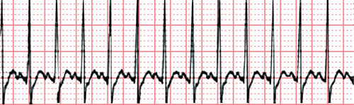
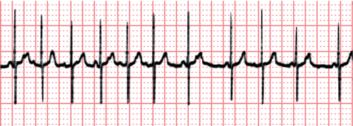
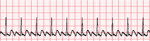
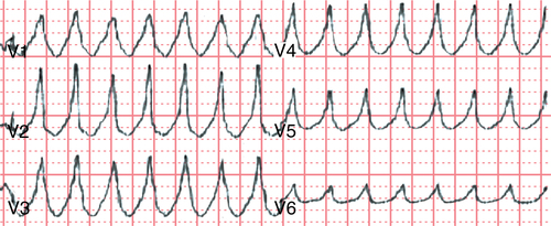
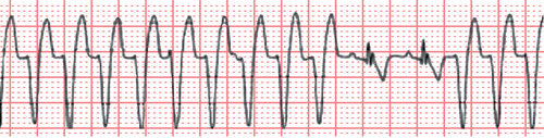
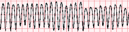
Wide complex tachycardias

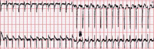
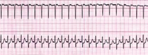
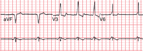
WCTs are defined as arrhythmias with a QRS complex duration longer than 0.12 sec at a rate greater than 100 beats/min. WCTs are presumed to be ventricular tachycardias (VTs) until proved otherwise, although some supraventricular tachycardias (SVTs) can present as WCT (SVT with aberrancy). Differentiating between SVTs with a wide QRS complex and a VT is critical because the treatment is very different (Figures 36-2 to 36-11).

Diagnosis

For the purpose of diagnosis, WCTs can be classified into four categories based on the following:

WCTs associated with drug overdose usually have terminal alterations of the QRS complex with right-axis deviation (RV wave in lead aVR, and S waves in leads I and aVL). The diagnosis can be made by reviewing the patient’s history and electrocardiogram. VT is more likely to occur in patients with coronary artery disease and signs of AV dissociation (cannon A waves). Hemodynamic stability does not rule out a diagnosis of VT.

A diagnosis of VT is likely when, on the 12-lead electrocardiogram, the QRS complex duration is longer than 160 ms, all of the QRS complexes in the precordial leads are positive or negative (positive or negative concordance), fusion beats or capture beats are seen, and AV dissociation is present. Polymorphic VT has beat-to-beat variations in morphologic appearance as a cyclic progressive change in cardiac axis. Polymorphic VT occurring in the setting of a prolonged QT interval is called torsades de pointes. VT that occurs with hyperkalemia is sinusoidal, is preceded by tall T waves, has short QT intervals, has prolonged PR intervals, and has flattened P waves. With tricyclic antidepressant toxicity, VT is characterized by a right-axis pattern with prominent S waves in leads I and aVL and an R wave in lead aVR.

Treatment

Regardless of the cause of WCT, electrical cardioversion with 100 to 200 J (monophasic) or 50 to 100 J (biphasic) is the treatment of choice in patients who are hemodynamically unstable.

The use of β-adrenergic receptor blocking agents, digitalis, and calcium channel blocking agents is recommended for patients with WCTs that are thought to be due to SVT with aberrancy but are contraindicated in patients with WPW syndrome. Procainamide and amiodarone are acceptable alternative choices because they are used to treat WCTs due to VT or SVT. Electrical cardioversion should be performed when the WCT does not respond to antiarrhythmic agents or if the patient is hemodynamically unstable. Torsades de pointes is treated with intravenously administered magnesium; the temporary use of transvenous overdrive pacing should be considered for patients with heart rates of 100 beats/min or greater and who do not respond to magnesium. Isoproterenol (2 μg/min in adults) can be infused with the same heart rate goal (≤100 beats/min) until pacing can be established.

Toxicity of sodium channel blocking agents, such as tricyclic antidepressants and antihistamines, should be treated with induction of alkalosis and diuresis. The administration of sodium bicarbonate infusions should be considered when the QRS intervals exceed 100 ms and the patient has persistent hypotension and arrhythmias. Hyperkalemia is treated with calcium, glucose-insulin infusions, β-agonists, and sodium bicarbonate.