Published on 18/03/2015 by admin
Filed under Dermatology
Last modified 22/04/2025
This article have been viewed 2666 times
Bernice R. Krafchik
Evidence Levels: A Double-blind study B Clinical trial ≥ 20 subjects C Clinical trial < 20 subjects D Series ≥ 5 subjects E Anecdotal case reports
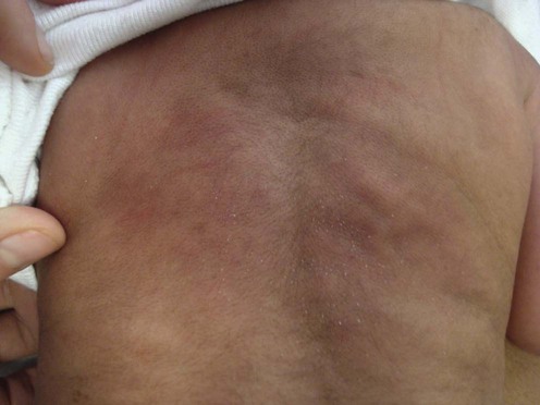
Subcutaneous fat necrosis (SFN) of the newborn is a rare, self-limited skin disease that occurs in the first few weeks of life. It affects areas where fat tissue is found and presents with a typical clinical picture of erythema and induration, commonly over the extremities, back, buttocks, and thighs. Individual lesions vary from a few millimeters to several centimeters and may coalesce. The exact etiopathogenesis is unclear but most infants suffer from some form of perinatal difficulty that includes asphyxia, peripheral hypoxemia, meconium aspiration or trauma; it is also seen in association with maternal diabetes. There have been a number of recent articles describing SFN in infants who have been treated with total body cooling for hypoxic ischemic encephalopathy (HIE). It is unlikely that the HIE is the cause of the SFN as it has also has been described in patients receiving hypothermia for cardiac surgery. Most infants with SFN have been delivered by cesarean section which makes birth trauma an unlikely cause.
The main and important complication seen with SFN is hypercalcemia, which occurred in one-third of patients who were admitted to hospital for SFN. The cause of the hypercalcemia is unknown; one theory postulates that increased calcium absorption occurs as a result of unregulated extrarenal production of 1,25-dihydroxyvitamin D. In cases of severe hypercalcemia seizures and death may ensue, in addition to nephrocalcinosis and hypercalciuria. Other symptoms of hypercalcemia are usually non-specific and consist of fever, vomiting, lethargy, constipation, feeding difficulties, and failure to thrive. Rarely, thrombocytopenia, hypoglycemia, anemia, hypervitaminosis D, and hypertriglyeridemia may also be seen.
SFN resolves spontaneously within 3 to 6 months, leaving normal skin or small pitted scars, and occasionally an absence of fat tissue in the affected areas. Treatment is seldom necessary, except in the rare instances when liquefaction of the fat tissue occurs, or when hypercalcemia or other blood chemistry abnormalities develops.
Biopsy, with its characteristic pathology, is usually not warranted as the clinical picture is typical. Aspiration cytology can be helpful in establishing the diagnosis. It is a simple alternative to a biopsy with lesions that are not clinically diagnostic. MRI scans and ultrasound demonstrate characteristic features and highlight nephrocalcinosis and other areas of calcification if present. In patients with SFN it is important to perform blood sampling looking for abnormal calcium values. Serum calcium levels may be low in normal infants and in patients with SFN during the first 2 weeks of life. Infants with SFN should have calcium values checked weekly, or, if there is any increase in serum calcium, bi-weekly for up to 6 months. If the infant is sick, tests for other abnormal blood indices should be performed.
Treatment regimens are determined by the levels of hypercalcemia, and hypercalciuria. If serum levels are marginally raised, monitoring the serum and urine calcium may be all that is necessary and a spontaneous return to normal levels often occurs. If the increased levels persist or rise further, treatment should be instigated. Mild hypercalcemia is treated by the withdrawal of vitamin D and by using a low-calcium diet. This is best accomplished by breastfeeding, as breast milk is low in both vitamin D and calcium. If this is not possible or helpful, a formula low in calcium with no vitamin D should be considered, and if a reduction in calcium does not occur or the levels continue to rise intravenous saline at 1.5 times the maintenance requirement is given, to promote the renal excretion of calcium. This is augmented with furosemide (a diuretic drug), which further promotes excretion. Other options for non-responders include calcitonin (4–8 IU/kg every 6 to 12 hours), bisphosphonates (pamidronate and others), and oral corticosteroids (hydrocortisone or prednisone). The latter treatments should be instituted with the help of a pediatric endocrinologist. The rare occurrence of liquefaction in the lesions is treated by removal of the liquid with a large-bore needle.
Schubert PT, Razack R, Vermaak A, Jordaan HF. Diagn Cytopathol 2012; 40: 245–7.
The diagnosis of SFN is usually clinically characteristic. An alternative to biopsy, fine needle aspiration is easy to perform and gives as much information as biopsy The typical pathological picture is seen; this includes clumped lobules of fat with opaque cytoplasm to necrotic aspirates with dispersed fat cells with opaque cytoplasm, foamy macrophages, multinucleated giant cells, lymphocytes, and neutrophils, and the characteristic radially oriented, and loose refractile needle-shaped crystals in the cytoplasm of the fat cells lying in the necrotic background.
Vasireddy S, Long SD, Sacheti B, Mayforth RD. Pediatr Radiol 2008; 89: 73–6.
This article reviews the presentation of the MRI findings of SFN and distinguishes these findings from other diseases that cause a similar picture. The authors also emphasize how ultrasound can identify calcification in the kidneys of some patients with SFN and hypercalcemia, and how MRI can discern calcification in other organs.
Nair S, Nair SG, Borade A, Ramakrishnan K. Indian J Pediatr 2009; 76: 1155–7.
This article discusses the management of a patient with SFN and metastatic calcification using saline, furosemide, oral steroids, and bisphosphonates.
Burden AD, Krafchik BR. Pediatr Dermatol 1999; 16: 384–7.
A review of SFN in patients requiring hospitalization. Ten of 11 patients had been delivered by cesarean section for fetal distress, and four of 11 developed hypercalcemia. Patients should be monitored for at least 6 months, even if the lesions of SFN have disappeared.
Strohm B, Hobson A, Brocklehurst P, Edwards AD, Azzopardi D. UK TOBY Cooling Register. Pediatrics 2011; 128: e450–2.
The authors review over 1239 cases of hypoxic ischemic encephalopathy treated with hypothermia. Twelve developed SFN. They discuss that hypothermia is becoming a common mode of treatment for various conditions and that physicians should be aware of the complication of SFN and hyperkalemia in patients treated with this modality.
Biopsy or aspiration cytology for diagnosis
Ultrasound of the kidneys for nephrocalcinosis or nephrolithiasis
MRI of torso if indicated by high calcium levels
Follow serum and urine calcium for 6 months weekly
If hypercalcemia occurs, calcium should be monitored bi-weekly
Monitor calcium/creatinine ratio in the urine
Borgia F, De Pasquale L, Cacace C, Meo P, Guarneri C, Cannavo SP. J Paediatr Child Health 2006; 42: 316–18.
A report on the extracutaneous and cutaneous features of SFN. The associated metabolic complications include hypoglycemia, thrombocytopenia, hypertriglyceridemia, anemia, and hypercalcemia. The delayed onset of hypercalcemia, 1 to 6 months after the development of the skin manifestations, and its frequent non-specific symptoms necessitates prolonged follow-up to prevent toxic effects on the cardiovascular and renal systems, and metastatic calcification.
Singalavanija S, Limponsanurak W, Wannaprasert T. J Med Assoc Thai 2007; 90: 1214–22.
This article discusses seven cases of SFN, all term babies: there were four males and three females. Five (70%) had perinatal asphyxia. The mean age of onset was 14 days (range 3–42 days). The locations of SFN were the back (three cases), shoulder (two), arm (two), buttock (one), and neck (one). Skin biopsy was performed in three cases and was compatible with subcutaneous fat necrosis. Treatment was supportive, with close monitoring of serum calcium. Hypercalcemia was seen in five cases (70%) and three were treated with oral prednisolone. Cutaneous lesions of all cases resolved without sequelae.
Khan N, Licata A, Rogers D. Clin Pediatr (Phila) 2001; 40: 217–19.
Oral bisphosphonates are poorly absorbed. This is the first report of intravenous administration of a bisphosphonate that resulted in fast normalization of the hypercalcemia from SFN. The drug acts by slowing the calcium turnover from bone.
Vijayakumar M, Prahlad N, Nammalwar BR, Shanmughasundharam R. Indian Pediatr 2006; 43: 360–3.
A report of a 28-day-old infant with SFN, hypercalcemia, and nephrocalcinosis who was managed with intravenous saline followed by furosemide, oral prednisolone, potassium citrate, and etidronate.
Alos N, Eugène D, Fillion M, Powell J, Kokta V, Chabot G. Horm Res 2006; 65: 289–94.
This report reviews the various treatments of hypercalcemia in SFN, including hydration, furosemide, and corticosteroids, citing one report only on the use of intravenous bisphosphonates. They describe four newborns with SFN complicated by severe hypercalcemia. All had ionized calcium levels higher than 1.4 mmol/L, associated with high urinary calcium/creatinine ratios and high 1,25-dihydroxyvitamin D levels. Despite treatment with intravenous fluids, a low-calcium diet, and furosemide, calcium levels remained high. Four doses (0.25–0.50 mg/kg/dose) of pamidronate were given. Urinary calcium/creatinine ratios and calcium levels decreased within 48–96 hours. 1,25-Dihydroxyvitamin D levels normalized with resolution of the skin lesions. No persistent nephrocalcinosis was observed. To prevent nephrocalcinosis, pamidronate might be considered as first-line treatment for severe hypercalcemia in SFN.
Lombardi G, Cabano R, Bollani L, Del Forno C, Stronati M. Eur J Pediatr 2009; 168: 625–7.
A case of severe hypercalcemia complicating SCFN in a newborn who was treated with hyperhydration, furosemide, prednisone, and, lastly, pamidronate.
Treatment of Skin Disease Comprehensive Therapeutic Strategies 4e
WhatsApp us


 No treatment for the majority of patients
 If mild hypercalcemia develops with or without symptoms, treatment should be instigated
 Low calcium diet and low vitamin D (either breast milk or formula)
 Intravenous saline
 Furosemide
 Oral prednisone
 Subcutaneous calcitonin
 Pamidronate