[level-membership-for-dermatology-category]
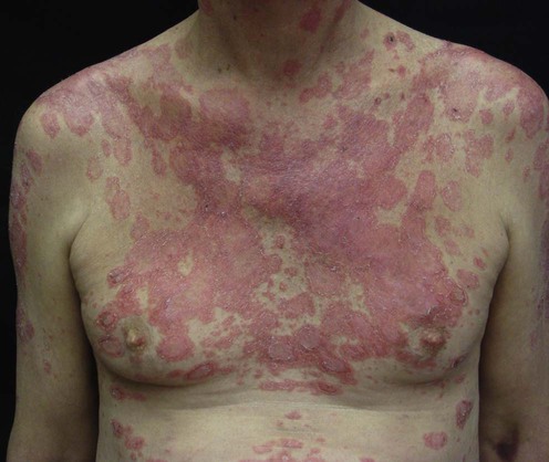
Subacute cutaneous lupus erythematosus
Specific investigations


First-line therapies
 Cosmetics
 Sunscreens and protective clothing
 Corticosteroids
 Antimalarials
 Topical retinoids
 Topical tacrolimus or pimecrolimus
Second-line therapies
 Dapsone
 Gold
Gold Thalidomide
 Retinoids
 Immunosuppressive agents: methotrexate, azathioprine, mycophenolate mofetil
Third-line therapies
 Clofazimine
 Phenytoin
 High-dose intravenous immunoglobulin
 Cytokine therapy
 Rituximab
 Ustekinumab
 Leflunomide
Regression of subacute cutaneous lupus erythematosus in a patient with rheumatoid arthritis treated with a biologic tumor necrosis factor alpha-blocking agent: comment on the article by Pisetsky and the letter from Aringer et al.
Fautrel B, Foltz V, Frances C, Bourgeois P, Rozenberg S. Arthritis Rheum 2002; 46: 1408–9.
[/level-membership-for-dermatology-category][not-level-membership-for-dermatology-category]
Subacute cutaneous lupus erythematosus
Specific investigations
First-line therapies
The association of the two antimalarials chloroquine and quinacrine for treatment-resistent chronic and subacute cutaneous lupus erythematosus.
Feldmann R, Salomon D, Saurat JH. Dermatology 1994; 189: 425–7.
Buy Membership for Dermatology Category to continue reading. Learn more here
[/not-level-membership-for-dermatology-category]
