Published on 19/03/2015 by admin
Filed under Dermatology
Last modified 19/03/2015
This article have been viewed 2563 times
Dimitra Koch and Saleem M. Taibjee
Evidence Levels: A Double-blind study B Clinical trial ≥ 20 subjects C Clinical trial < 20 subjects D Series ≥ 5 subjects E Anecdotal case reports
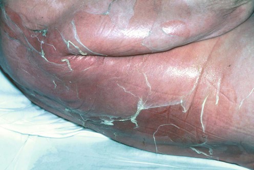
Staphylococcal scalded skin syndrome (SSSS) is a highly contagious blistering condition triggered by exfoliative toxin-producing Staphylococcus aureus, most commonly affecting neonates and children under the age of 6 years. It rarely presents in adults, usually in association with renal failure or immune compromise, and possibly non-steroidal anti-inflammatory drug (NSAID) use. In healthy infants mortality is uncommon, with reported rates up to 3%, contrasting with up to 50–60% in adults with comorbidity.
SSSS is caused by hematogenous spread of exfoliative toxin (ET), mainly ET-A and/or -B produced by S. aureus, most commonly phage group II (types 3A, 3B, 3C, 55, 71), including both methicillin-sensitive and methicillin-resistant (MRSA) strains. ET, a serine protease, specifically cleaves the desmosomal protein desmoglein-1, leading to disruption of keratinocyte adhesion in the superficial epidermis. Increased susceptibility to SSSS in neonates and adults with renal failure, or immunocompromise, is thought to be due to impaired renal clearance of ET or low titers of ET-specific antibodies, respectively.
Clinical features include a prodrome of malaise, irritability, and fever. Skin manifestations develop within 24 to 48 hours. Tender erythema is followed by superficial, flaccid blistering, most pronounced in flexural and periorificial areas. Nikolsky sign is positive. Mucosal surfaces are not involved. With successful treatment SSSS resolves over 10 to 14 days. Bullae rupture to leave denuded skin, followed by desquamation and re-epithelialization without scarring.
Typically, the causative organism cannot be cultured from affected skin, as this is a toxin-mediated disease. The infection is acquired by skin-to-skin contact or from contact with nasal carriers. The source of infection is mainly via nasal or perianal colonization. It may therefore be possible to culture ET-producing S. aureus from extracutaneous sites, most commonly the nasopharynx, but also the rectum, conjunctivae, urinary tract, umbilicus and blood. Polymerase chain reaction (PCR) or random amplified polymorphic DNA analysis may identify the toxin-encoding genes. Histology of affected skin typically shows a superficial, subcorneal blister, absence of epidermal necrosis, and minimal inflammation. Frozen section of the blister roof is an alternative method of confirming the superficial level of cleavage and may facilitate more rapid diagnosis.
The main differential diagnosis is the Stevens-Johnson syndrome–toxic epidermal necrolysis spectrum, in which targetoid skin lesions, mucosal involvement, and deeper level of blistering with full-thickness epidermal necrosis are distinguishing features.
Treatment includes antibiotic therapy in combination with supportive measures addressing electrolyte and fluid balance, temperature regulation, nutrition, analgesia, and skin care. Bland emollients such as 50 : 50 white soft paraffin/liquid paraffin should be applied regularly to reduce friction and insensible fluid losses. Non-adherent dressings are used to cover denuded areas, with avoidance of adhesive tapes directly to skin. Systemic β-lactamase-resistant antibiotics are necessary to eradicate the causative S. aureus strain. Patients with extensive SSSS require intravenous antibiotics and in-hospital care provided by a multidisciplinary team experienced in delivering optimal skin care, e.g., on a burns unit. In view of the growing emergence of resistant ET-producing S. aureus strains, in particular MRSA and fusidic acid-resistant strains, antibiotic choice should be guided by the local microbiology team.
SSSS outbreaks on maternity and neonatal units are reported. Strict infection control measures are advisable, including patient isolation, barrier nursing, and scrupulous hand-washing. Screening programs may identify S. aureus carriers amongst patient contacts including healthcare workers. Colonized individuals require treatment with topical antiseptics, e.g., chlorhexidine, with nasal carriage eradicated by topical antibiotics such as mupirocin.
The choice of antibiotic therapy should be guided by local incidence of multidrug-resistant S. aureus strains and microbiology advice.
Bell FG, Fenton PA. Lancet 1993; 342: 120.
An outbreak of SSSS due to ET-producing S. aureus carriage by a pediatrician.
This was not initially recognized since many cases presented following hospital discharge.
El Helali N, Carbonne A, Naas T, Kerneis S, Fresco O, Giovangrandi YP, et al. J Hosp Infect 2005; 61: 130–8.
An SSSS outbreak affecting 13 neonates in a maternity unit. Temporary removal from duty of a nurse with chronic hand dermatitis confirmed as a carrier of the causative ET-producing S. aureus with infection control measures (patient isolation, barrier-nursing, chlorhexidine handwashing, treatment of carriers with nasal mupirocin, and chlorhexidine showers) controlled the outbreak. All affected neonates were successfully treated with oxacillin.
Healthcare workers are an important potential source of outbreaks.
Treatment of Skin Disease Comprehensive Therapeutic Strategies 4e
WhatsApp us