Spinal anesthesia

Published on 07/02/2015 by admin

Filed under Anesthesiology

Last modified 22/04/2025

Print this page

rate 1 star rate 2 star rate 3 star rate 4 star rate 5 star
Your rating: none, Average: 0 (0 votes)

This article have been viewed 1112 times

Spinal anesthesia

Lopa Misra, DO

The use of spinal anesthesia gained popularity in the United States in the 1940s. However, due to reports of adverse neurologic effects, the number of spinal anesthetics administered declined until a large study demonstrated the relative rarity of such complications, leading to a resurgence in the use of spinal blocks.

When spinal anesthetics are used in appropriately selected patients, their advantages include a decreased incidence of thromboembolism, cardiac morbidity and death, and bleeding, the latter resulting in a decreased need for transfusion. In addition, the use of subarachnoid blocks decreases the incidence of lower extremity vascular graft occlusion and of postoperative pneumonia. The advantages of subarachnoid blocks are probably due to multiple factors, including a decreased hypercoagulable state, increased tissue blood flow (because of vasodilation below the level of the block), increased oxygenation (because of maintenance of normal ventilation), increased peristalsis (related to the need for lower doses of opioids), and decreased stress response (because of the blockade of afferent activity at the spinal cord level as opposed to blockade at the level of the reticular activating center/cortex).

The mechanism of action of spinal anesthesia is due to the effect of local anesthetic agents on the individual nerve roots, effects that depend on the size and myelin content of nerve fibers, concentration of the local anesthetic agent, and duration of contact between the nerve root and the local anesthetic agent. The loss of conductivity of impulses through the fibrils follows a fixed sequence—first, sympathetic and parasympathetic activity are lost, and then sensation in the C, B, and A fibers is lost. C—fibers quit firing first, progressing finally to loss of activity in the Aα fibers, which accounts for the loss of motor function lastly. Heavy myelinated fibers present in motor nerves are most resistant to the effects of local anesthetic agents; hence, loss of activity in these nerves is the last group of nerves to be blocked. Because of the increased sensitivity of autonomic nerve fibers, blockade of these nerves extends for two or more dermatomes above the level of skin anesthesia, and blockade of motor fibers is seen two or more levels below the level of skin anesthesia (Box 122-1, Table 122-1).
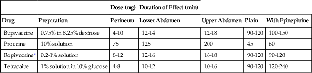
Table 122-1

Dose and Duration of Effect for Local Anesthetic Agents Commonly Administered Intrathecally

    Dose (mg) Duration of Effect (min)
Drug Preparation Perineum Lower Abdomen Upper Abdomen Plain With Epinephrine
Bupivacaine 0.75% in 8.25% dextrose 4-10 12-14 12-18 90-120 100-150
Procaine 10% solution 75 125 200 45 60
Ropivacaine* 0.2-1% solution 8-12 12-16 16-18 90-120 90-120
Tetracaine 1% solution in 10% glucose 4-8 10-12 10-16 90-120 120-240

image

*Intrathecal injection is an off-label use for ropivacaine.

Cardiovascular effects

The loss of sympathetic activity that accompanies a spinal anesthetic results in vasodilation below the level of blockade, decreasing systemic vascular resistance, and the associated venodilation decreases cardiac preload and, therefore, cardiac output. If the level of blockade is sufficiently cephalad, loss of activity occurs in the cardiac accelerator nerves, which is manifested by bradycardia. The net effect of these physiologic changes can manifest as a profound decrease in systemic blood pressure. The most effective approach to the sympathectomy is (1) to provide prophylaxis by administering a bolus of intravenous fluids to the patient before the intrathecal administration of the local anesthetic agent and (2) to have a vagolytic drug and a vasopressor readily available to administer intravenously to treat the bradycardia and hypotension. Risks for the development of bradycardia (<50 beats/min) following a spinal anesthetic include (1) a baseline heart rate below 60 beats/min, (2) the use of β-adrenergic receptor blocking agents prior to presentation for a spinal anesthetic, (3) prolonged PR interval on the electrocardiogram (4) sensory level above T6, (5) age younger than 50 years and (6) American Society of Anesthesiologists I physical status.

Pulmonary effects

Pulmonary effects seen with subarachnoid blocks are usually minimal because the diaphragm is innervated by motor fibers in the phrenic nerves that exit the spinal column at C3-C5. However, if spinal blockade is sufficiently high, in the midthoracic to upper thoracic region, vital capacity decreases because of loss of abdominal muscle activity, but tidal volume remains unchanged.

In patients with severe chronic lung disease, a high thoracic spinal anesthetic may lead to loss of accessory muscle function. Many patients with chronic lung disease depend on the accessory muscles for maintenance of ventilation.

As a general rule all patients should receive supplemental O2 because acute airway closure, atelectasis, and hypoxia have been observed in patients with otherwise normal pulmonary function. The apnea seen with a high spinal block is more commonly due to hypoperfusion of the brainstem because of hypotension and not necessarily from the activity of the local anesthetic agents on the C3 to C5 nerve roots. When a high spinal block is identified, the patient should be placed in the Trendelenburg position to increase cardiac preload, cardiac output, and perfusion of the brainstem; if pulmonary ventilation remains compromised, tracheal intubation, assisted ventilation, or both may be necessary.

Contraindications to spinal anesthesia

Absolute contraindications include patient refusal, coagulation abnormalities, severe hypovolemia, increased intracranial pressure, infection, and severe stenotic aortic and mitral valvular heart disease (Box 122-2). Other relative and controversial contraindications are also listed in Box 122-2.