Serologic Diagnosis of Infectious Diseases

Published on 08/02/2015 by admin

Filed under Basic Science

Last modified 22/04/2025

Print this page

rate 1 star rate 2 star rate 3 star rate 4 star rate 5 star
Your rating: none, Average: 5 (1 votes)

This article have been viewed 5093 times

Serologic Diagnosis of Infectious Diseases

Immunochemical methods are used as diagnostic tools for serodiagnosis of infectious disease. An understanding of how these methods have been adapted for this purpose requires a basic working knowledge of the components and functions of the immune system. Immunology is the study of the components and functions of the immune system. The immune system is the body’s defense mechanism against invading “foreign” antigens. One of the functions of the immune system is distinguishing “self” from “nonself” (i.e., the proteins or antigens from foreign substances). (Chapter 3 presents a more in-depth discussion of the host’s response to foreign substances.) This chapter is intended to provide a brief overview and review of immunology. The complexity and detail required to fully understand immunology and serology are beyond the scope of this text.

Features of the Immune Response

The host, or patient, has physical barriers, such as intact skin and ciliated epithelial cells, and chemical barriers, such as oils produced by the sebaceous glands and lysozyme found in tears and saliva, to prevent infections by foreign organisms. In addition, natural (innate) immunity, which is not specific, activates chemotaxis, the process by which phagocytes are recruited to a site of invasion and engulf organisms entering the host. Acquired active immunity is the specific response of the host to an infecting organism.

The human specific immune responses are simplistically divided into the following two categories: cell-mediated and antibody-mediated.

Cell-mediated immune responses are carried out by special lymphocytes of the T-cell (thymus derived) class. T cells proliferate and differentiate into various effector T cells, including cytotoxic and helper cells. Cytotoxic T lymphocytes specifically attack and kill microorganisms or host cells damaged or infected by pathogens. Helper cells promote the maturation of B cells by producing activator cytokines that induce the B cells to produce antibodies and attach to and kill invading organisms. Although diagnosis of certain diseases may be aided by measuring the cell-mediated immune response to the pathogen, such tests entail skin tests performed by physicians or in vitro cell function assays performed by specially trained immunologists. These tests are usually not within the repertoire of clinical microbiology laboratories.

Antibody-mediated immune responses are produced by specific proteins generated by lymphocytes of the B-cell (bone marrow derived) class. Because these proteins exhibit immunologic function and fold into a globular structure in the active state, they are also referred to as immunoglobulins. Antibodies are either secreted into the blood or lymphatic fluid (and sometimes other body fluids) by activated B lymphocytes (plasma cells), or they remain attached to the surface of the lymphocyte or other cells. Because the cells involved in this category of immune response primarily circulate in the blood, this type of immunity is also called humoral immunity. For purposes of determining whether a patient’s body has produced an antibody against a particular infectious agent, the serum (or occasionally the plasma) is examined for the presence of the antibody. The study of the diagnosis of disease by measuring antibody levels in serum is referred to as serology.

Characteristics of Antibodies

Immunocompetent humans are able to produce antibodies specifically directed against almost all the antigens with which they may come into contact throughout their lifetimes and that the body recognizes as “foreign.” Antigens may be part of the physical structure of a pathogen or a chemical produced and released by the pathogen, such as an exotoxin. One pathogen may contain or produce many different antigens that the host recognizes as foreign. Infection with one agent may cause the production of a number of different antibodies. In addition, some antigenic determinants on a pathogen may not be available for recognition by the host until the pathogen has undergone a physical change. For example, until a pathogenic bacterium has been digested by a human polymorphonuclear (PMN) leukocyte, certain antigens deep in the cell wall are not detected by the host immune system. Once the bacterium has been broken down, these new antigens are released and the specific antibodies can be produced. For this reason, a patient may produce different antibodies at different times during the course of a single disease. The immune response to an antigen also matures with continued exposure, and the antibodies produced become more specific and more avid (able to bind more tightly).

Antibodies function by (1) attaching to the surface of pathogens and making the pathogens more amenable to ingestion by phagocytic cells (opsonizing antibodies); (2) binding to and blocking surface receptors for host cells (neutralizing antibodies); or (3) attaching to the surface of pathogens and contributing to their destruction by the lytic action of complement (complement-fixing antibodies). Routine diagnostic serologic methods are used to measure primarily two antibody classes, IgM and IgG; however, antibodies are categorized into five classes: immunoglobulin G (IgG), immunoglobulin M (IgM), immunoglobulin A (IgA), immunoglobulin D (IgD), and immunoglobulin E (IgE). IgA, also referred to as secretory antibody, is the predominant class of antibody in saliva, tears, and intestinal secretions. IgD is attached to the surface of B cells and is involved in immune regulations. IgE levels increase as a result of infections caused by several parasites or in response to allergic reactions.

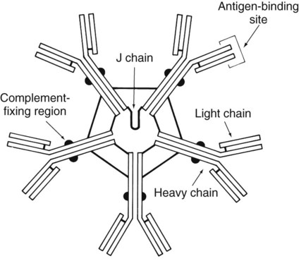
The basic structure of an antibody molecule comprises two mirror images, each composed of two identical protein chains (Figure 10-1). At the terminal ends are the antigen binding sites, or variable regions, which specifically attach to the antigen against which the antibody was produced. Depending on the specificity of the antibody, antigens of some similarity, but not total identity, to the inducing antigen may also be bound; this is called a cross reaction. The complement binding site is found in the center of the molecule in a structure similar for all antibodies of the same class and is referred to as the constant region. IgM is produced as a first response to many antigens, although the levels remain high transiently. Thus, the presence of IgM usually indicates recent or active exposure to an antigen or infection. IgG, on the other hand, may persist long after an infection has run its course.

The IgM antibody type (Figure 10-2) consists of five identical proteins (pentamer), with the basic antibody structures linked at the bases with 10 antigen binding sites on the molecule. IgG consists of one basic antibody molecule (monomer) that has two binding sites (see Figure 10-1). The differences in the size and conformation between these two classes of immunoglobulins result in differences in activities and functions.

Features of the Humoral Immune Response Useful in Diagnostic Testing

Immunocompetent individuals produce both IgM and IgG antibodies in response to most pathogens. In most cases, IgM is produced by a patient after the first exposure to a pathogen and is no longer detectable within a relatively short period. For serologic diagnostic purposes, it is important to note that IgM is unable to cross the placenta. Therefore, any IgM detected in the serum of a newborn must have been produced by the infant and indicates an infection in utero. The larger number of binding sites on IgM molecules provides for more rapid clearance of the offending pathogen, even though each individual antigen binding site may not be the most efficient for binding to the antigen. Over time, the cells producing IgM switch to production of IgG. IgG is the highest circulating antibody in the human body.

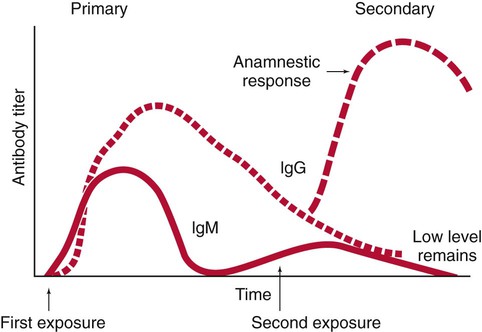
IgG is often more specific for the antigen (i.e., it has higher avidity). IgG has two antigen binding sites, but it can also bind complement. Complement is a complex series of serum proteins that is involved in modulating several functions of the immune system, including cytotoxic cell death, chemotaxis, and opsonization. When IgG is bound to an antigen, the base of the molecule (Fc portion) is exposed in the environment. Structures on this Fc portion attract and bind the cell membranes of phagocytes, increasing the chances of engulfment and destruction of the pathogen by the host cells. A second exposure to the same pathogen induces a faster and greater IgG response and a much lesser IgM response. Several B lymphocytes retain memory of the pathogen, allowing a more rapid response and a higher level of antibody production than the primary exposure or response. This enhanced response is called the anamnestic response. B-cell memory is not perfect. Occasional clones of memory cells can be stimulated through interaction with an antigen that is similar but not identical to the original antigen. Therefore, the anamnestic response may be polyclonal and nonspecific. For example, reinfection with cytomegalovirus may stimulate memory B cells to produce antibody against Epstein-Barr virus (another herpes family virus), which the host encountered previously, in addition to antibody against cytomegalovirus. The relative humoral responses are diagrammatically represented in Figure 10-3.

Interpretation of Serologic Tests

In serology, a change in antibody titer is a central concept for the diagnosis and monitoring of disease progression. The titer of antibody is the reciprocal of the highest dilution of the patient’s serum in which the antibody is still detectable. Patients with large amounts of antibody have high titers, because antibody is still detectable at very high dilutions of serum. Serum for antibody levels should be drawn during the acute phase of the disease (when it is first discovered or suspected) and again during convalescence (usually at least 2 weeks later). These specimens are called acute and convalescent sera. For some infections, such as legionnaires’ disease and hepatitis, titers may not rise until months after the acute infection, or they may never rise. Therefore, changes in titer must be carefully correlated with the patient’s signs and symptoms of the specific disease or suspected infectious agent.

Patients with intact humoral immunity develop increasing amounts of antibody to a pathogen over several weeks. If it is the patient’s first exposure to the pathogenic organism and the specimen has been obtained early enough, no or very low titers of antibody are detected at the onset of disease. In the case of a second exposure, the patient’s serum usually contains measurable antibody during the initial phase of the disease, and the antibody level quickly increases as a result of the anamnestic response. For most pathogens, an increase in the patient’s titer of two doubling dilutions (e.g., from a positive result of 1 : 8 to a positive result of 1 : 32) is considered to be diagnostic of current infection. This is defined as a fourfold rise in titer.

For many infections, accurate results used for diagnosis are achieved when acute and convalescent sera are tested concurrently in the same test system. Variables inherent in the procedures and laboratory error can cause a difference of one doubling (or twofold) dilution in the results obtained from a same sample tested concurrently in different laboratories. Unfortunately, a certain proportion of infected patients never demonstrate a rise in titer, necessitating the use of other diagnostic tests. Because the delay inherent in testing paired acute and convalescent sera results in diagnostic information arriving too late to affect the initial therapy, increasing numbers of early (IgM) serologic testing assays are being commercially evaluated. Moreover, it is sometimes more realistic to see a fourfold fall in titer between acute and convalescent sera when samples are tested concurrently in the same system. This is a result of the sera being collected late in the course of an infection, when antibodies have already begun to decrease.

Serodiagnosis of Infectious Diseases

With most diseases, a spectrum of responses may be seen in infected humans, such that a person may develop antibody from a subclinical infection or after colonization by an agent without actually having symptoms of the disease. In these cases, the presence of antibody in a single serum specimen or a similar titer of antibody in paired sera may merely indicate past contact with the agent and cannot be used to accurately diagnose recent disease. Therefore, in the vast majority of serologic procedures for diagnosis of recent infection, testing of both acute and convalescent sera is the method of choice. Except for detecting the presence of IgM, testing of a single serum may be recommended in certain cases. Mycoplasma pneumoniae and viral influenza B infections are examples in which high titers may indicate recent infection. IgM levels may be diagnostic if the infecting or disease-causing agent is extremely rare, such as rabies or exposure to botulism toxin, and people without disease or prior immunization would have no chance of developing an immune response.

The prevalence of antibody to an etiologic agent of disease in the population correlates with the number of people who have come into contact with the agent, not the number who actually develop disease. For most diseases, only a small proportion of infected individuals actually develop symptoms; others develop protective antibodies without experiencing signs and symptoms of the disease. In a number of circumstances, serum is tested to determine whether a patient is immune; that is, whether the patient has antibody to a particular agent either in response to a past infection or to immunization. These tests can be performed with a single serum sample. The results of the tests must be correlated with the actual immune status of individual patients to determine the level of detectable antibody present, in order to determine whether the individual has developed a true immunity to infection or a secondary reinfection. For example, sensitive tests can detect the presence of very tiny amounts of antibody to the rubella virus. Certain people, however, may still be susceptible to infection with the rubella virus with such small amounts of circulating antibody, and a higher level of antibody may be required to ensure protection from disease.

Alternatively, depending on the etiologic agent, even low levels of antibody may protect a patient from pathologic effects of disease and not prevent a second reinfection. For example, a person previously immunized with killed poliovirus vaccine who becomes infected with pathogenic poliovirus experiences multiplication of the virus in the gut and virus entry into the circulation. Damage to the central nervous system is blocked by humoral antibody in the circulation. As more sensitive testing methods are developed and these types of problems become more common, microbiologists must work closely with clinicians to develop guidelines for interpreting serologic test results in relation to the immune status of individual patients. Moreover, patients may respond to an antigenic stimulus by producing cross-reacting antibodies. These antibodies are nonspecific and may cause misinterpretation of serologic tests.

Table 10-1 provides a brief list of representative serologic tests available for immunodiagnosis of infectious diseases, the specimen required, interpretation of positive and negative test results, and examples of applications of each technique. Because serologic assays are rapidly evolving, this table is not intended to be all-inclusive.

TABLE 10-1

Noninclusive Overview of Tests Available for Serodiagnosis of Infectious Diseases

Test Sera Needed Interpretation Application
IgM Single, acute (collected at onset of illness) Newborn, positive: in utero (congenital) infection
Adult, positive: primary or current infection
Adult, negative: no infection or past infection
Newborn: STORCH* agents; other organisms
Adults: any infectious agent
IgG Acute and convalescent (collected 2-6 weeks after onset) Positive: fourfold rise or fall in titer between acute and convalescent sera tested at the same time in the same test system
Negative: no current infection or past infection, or patient is immunocompromised and cannot mount a humoral antibody response, or convalescent specimen collected before increase in IgG (Lyme disease, Legionella sp.)
Any infectious agent
IgG Single specimen collected between onset and convalescence Adult, positive: adult evidence of infection at some unknown time except in certain cases in which a single high titer is diagnostic (rabies, Legionella, Ehrlichia spp).
Newborn, positive: maternal antibodies that crossed the placenta
Newborn, negative: patient has not been exposed to microorganism or patient has a congenital or acquired immune deficiency or specimen collected before increase in IgG (Lyme disease or Legionella sp.)
Any infectious agent
Immune status evaluation Single specimen collected at any time Positive: previous exposure
Negative: no exposure
Rubella testing for women of childbearing age, syphilis testing may be required in some states to obtain a marriage license, cytomegalovirus testing for transplant donor and recipient

image

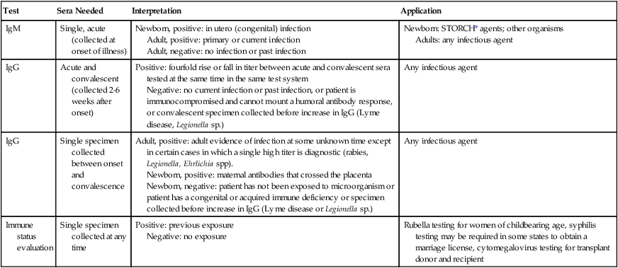
*STORCH, Syphilis, Toxoplasma, rubella virus, cytomegalovirus, herpes simplex virus.

Principles of Serologic Test Methods

Antibodies can be detected in many ways. In some cases, antibodies to an agent may be detected in more than one way, but the different antibody detection tests may not be measuring the same antibody. For this reason, the presence of antibodies to a particular pathogen, as detected by one method, may not correlate with the presence of antibodies to the same agent as detected by another test method. Moreover, different test methodologies have varying degrees of sensitivity in detecting antibodies. However, because IgM is produced at an initial higher level during a patient’s first exposure to an infectious agent, the detection of specific IgM can help the clinician a great deal in establishing a diagnosis. Most of the serologic test methods can be adapted for analysis of IgM.

Separating IgM from IgG for Serologic Testing

IgM testing is especially helpful for diseases that have nonspecific clinical presentations, such as toxoplasmosis, and for conditions that require rapid therapeutic decisions. For example, rubella infection in pregnant women can lead to congenital defects in the unborn fetus, such as cataracts, glaucoma, mental retardation, and deafness. Therefore, pregnant women who are exposed to rubella virus and develop a mild febrile illness can be tested for the presence of anti-rubella IgM. In addition, identification of IgM within the amniotic fluid of a pregnant mother is diagnostic of neonatal infection. Because IgG can readily cross the placenta, newborns carry titers of IgG passed from the mother to the fetus during the first 2 to 3 months of life until the infant produces his or her own antibodies. This is the only form of natural passive immunity. Accurate serologic diagnosis of infection in neonates requires either demonstration of a rise in titer (which takes time to occur) or the detection of specific IgM directed against the putative agent. Because the IgM molecule does not cross the placental barrier, any IgM would have to be of fetal origin and diagnostic of neonatal infection. Agents difficult to culture or those that adult females would be expected to have encountered during their lifetimes, such as Treponema pallidum, cytomegalovirus, herpes virus, Toxoplasma sp., or rubella virus, are organisms that may cause an infection and elevation of fetal IgM. The names of some of these agents have been grouped together with the acronym STORCH (syphilis, Toxoplasma sp., rubella, cytomegalovirus, and herpes). These tests should be ordered separately, depending on the clinical illness of a newborn suspected of having one of these diseases. In many instances, however, infected babies display no clinical signs or symptoms of infection. Furthermore, in many cases serologic tests yield false-positive or false-negative results. Therefore, multiple considerations, including the patient history and the clinical signs and symptoms, must be included in the serodiagnosis of neonatal infection, and in many cases culture is still the most reliable diagnostic method.

Several methods have been developed to measure specific IgM in sera that may also contain IgG. In addition to using a labeled antibody specific for IgM as the marker or the IgM capture sandwich assays, the immunoglobulins can be separated from each other by physical means. Centrifugation through a sucrose gradient, performed at very high speeds, has been used in the past to separate IgM, which has a greater molecular weight than IgG.

Other available IgM separation systems use the presence of certain proteins on the surface of staphylococci (protein A) and streptococci (protein G expressed by group C and G streptococci) that bind the Fc portion of IgG. A simple centrifugation step separates the particles and their bound immunoglobulins from the remaining mixture, which contains the bulk of the IgM. Other methods use antibodies to remove IgM from sera containing both IgG and IgM. An added bonus of IgM separation systems is that rheumatoid factor, IgM antibodies produced by some patients against their own IgG, often binds to the IgG molecules being removed from the serum. Consequently, these IgM antibodies are removed along with the IgG. Rheumatoid factor can cause nonspecific reactions and interfere with the results in a variety of serologic tests.

Methods of Antibody Detection

Direct Whole Pathogen Agglutination Assays

Basic tests for antibody detection measure the antibody produced by a host to determinants on the surface of a bacterial agent in response to infection. Specific antibodies bind to surface antigens of the bacteria in a thick suspension and cause the bacteria to clump in visible aggregates. Such antibodies are called agglutinins, and the test is referred to as bacterial agglutination. Electrostatic and additional chemical interactions influence the formation of aggregates in solutions. Because most bacterial surfaces have a negative charge, they tend to repel each other. Performance of agglutination tests in sterile physiologic saline (0.9% sodium chloride in distilled water), which contains free positive ions, enhances the ability of antibody to cause aggregation of bacteria. Although bacterial agglutination tests can be performed on the surface of both glass slides and in test tubes, tube agglutination tests are often more sensitive, because a longer incubation period can be used, allowing more antigen and antibody to interact. The small volume of liquid used for slide tests requires a rather rapid reading of the result, before the liquid evaporates, causing erroneous results.

Examples of bacterial agglutination tests include assays for antibodies to Francisella tularensis and Brucella spp., which are part of a panel referred to as febrile agglutinin tests. Bacterial agglutination tests are often used to diagnose diseases in which the bacterial agent is difficult to cultivate in vitro. Diseases diagnosed by this technique include tetanus, yersiniosis, leptospirosis, brucellosis, and tularemia. The reagents necessary to perform many of these tests are commercially available, singly or as complete systems. Because most laboratories are able to culture and identify the causative agent, agglutination tests for certain diseases, such as typhoid fever, are seldom used today. Furthermore, the typhoid febrile agglutinin test (called the Widal test) is often positive in patients with infections caused by other bacteria because of cross-reacting antibodies or a previous immunization against typhoid. Appropriate specimens from patients suspected of having typhoid fever should be cultured for the presence of salmonellae.

Whole cells of parasites, including Plasmodium and Leishmania spp., or Toxoplasma gondii, have also been used for direct detection of antibody by agglutination. In addition to using the actual infecting bacteria or parasites as the agglutinating particles, certain bacteria may be agglutinated by antibodies produced against another infectious agent. Many patients infected with one of the rickettsiae produce antibodies capable of nonspecifically agglutinating bacteria of the genus Proteus, specifically Proteus vulgaris. The Weil-Felix test detects these cross-reacting antibodies. Because newer, more specific serologic methods of diagnosing rickettsial disease have become more widely available, the use of the Proteus agglutinating test is no longer offered in many laboratories.

Particle Agglutination Tests

Numerous serologic procedures have been developed to detect antibody via the agglutination of an artificial carrier particle with antigen bound to its surface. As noted in Chapter 9, similar systems using artificial carriers coated with antibodies are commonly used for detection of microbial antigens. Either artificial carriers (e.g., latex particles or treated red blood cells) or biologic carriers (e.g., whole bacterial cells) can carry an antigen on their surface capable of binding with antibody. The size of the carrier enhances the visibility of the agglutination reaction, and the artificial nature of the system allows the antigen bound to the surface to be extremely specific.

The results of particle agglutination tests depend on several factors, including the amount and avidity of antigen conjugated to the carrier, the time of incubation with the patient’s serum (or other source of antibody), and the microenvironment of the interaction (including pH and protein concentration). Commercial tests have been developed as systems, complete with their own diluents, controls, and containers. For accurate results, a serologic test kit should be used as a unit, without modification or mixing from another kit. In addition, tests developed for use with cerebrospinal fluid, for example, should not be used with serum unless the package insert or the technical representative has certified such use.

Treated animal red blood cells have also been used as carriers of antigen for agglutination tests; these tests are called indirect hemagglutination, or passive hemagglutination tests, because it is not the original red blood cell antigens, but rather the passively attached antigens, that are bound by antibody. The most widely used indirect assays include the microhemagglutination test for antibody to T. pallidum (MHA-TP, so called because it is performed in a microtiter plate), the hemagglutination treponemal test for syphilis (HATTS), the passive hemagglutination tests for antibody to extracellular antigens of streptococci, and the rubella indirect hemagglutination tests, all of which are available commercially. Certain reference laboratories, such as the Centers for Disease Control and Prevention (CDC), also perform indirect hemagglutination tests for antibodies to some clostridia, Burkholderia pseudomallei, Bacillus anthracis, Corynebacterium diphtheriae, Leptospira sp., and the agents of several viral and parasitic diseases.

Complete systems for the use of latex or other particle agglutination tests are available commercially for accurate and sensitive detection of antibody to cytomegalovirus, rubella virus, varicella-zoster virus, the heterophile antibody of infectious mononucleosis, teichoic acid antibodies of staphylococci, antistreptococcal antibodies, mycoplasma antibodies, and others. Latex tests for antibodies to Coccidioides, Sporothrix, Echinococcus, and Trichinella spp. are available, although they are not widely used because of the uncommon occurrence of the corresponding infection or its limited geographic distribution. Use of tests for Candida antibodies has not yet shown results reliable enough for accurate diagnosis of disease.

Flocculation Tests

In contrast to the aggregates formed when particulate antigens bind to specific antibody, the interaction of soluble antigen with antibody may result in the formation of a precipitate, a concentration of fine particles, usually visible only because the precipitated product is forced to remain in a defined space within a matrix. Variations of precipitation and flocculation are widely used for serologic studies.

In flocculation tests the precipitin end product forms macroscopically or microscopically visible clumps. The Venereal Disease Research Laboratory test, known as the VDRL, is the most widely used flocculation test. Patients infected with pathogenic treponemes, most commonly T. pallidum, the agent of syphilis, form an antibody-like protein called reagin that binds to the test antigen, cardiolipin-lecithin–coated cholesterol particles, causing the particles to flocculate. Reagin is not a specific antibody directed against T. pallidum antigens, therefore the test is highly sensitive but not highly specific; however, it is a good screening test, detecting more than 99% of cases of secondary syphilis.

The VDRL is the single most useful test available for testing cerebrospinal fluid in cases of suspected neurosyphilis, although it may be falsely positive in the absence of disease. Performance of the VDRL test requires scrupulously clean glassware and attention to detail, including numerous daily quality control checks. In addition, the reagents must be prepared fresh immediately before the test is performed, and patients’ sera must be inactivated (complement inactivation) by heating for 30 minutes at 56°C before testing. Because of this complexity, the VDRL has been replaced in many laboratories by a qualitatively comparable test, the rapid plasma reagin (RPR) test.

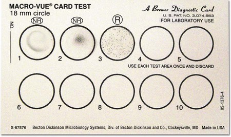
The RPR test is commercially available as a complete system containing positive and negative controls, the reaction card, and the prepared antigen suspension. The antigen, cardiolipin-lecithin–coated cholesterol with choline chloride, also contains charcoal particles to allow for macroscopically visible flocculation. Sera can be tested without heating, and the reaction takes place on the surface of a specially treated cardboard card, which is then discarded (Figure 10-4). The RPR test is not recommended for testing of cerebrospinal fluid. All procedures are standardized and clearly described in product inserts, and these procedures should be strictly followed. Overall, the RPR appears to be a more specific screening test for syphilis than the VDRL, and it is not as technically complex. Several modifications have been made, such as the use of dyes to enhance visualization of results and the use of automated techniques.

Conditions and infections other than syphilis can cause a patient’s serum to yield a positive result in the VDRL or RPR test; these are referred to as biologic false-positive tests. Autoimmune diseases, such as systemic lupus erythematosus and rheumatic fever, in addition to infectious mononucleosis, hepatitis, pregnancy, and old age have been known to cause false-positive reactions. The results of screening tests should always be considered presumptive until confirmed with a specific treponemal test.

Immunodiffusion Assays

The Ouchterlony double immunodiffusion assay, which closely resembles the precipitation test, is used to detect antibodies directed against fungal cell components (see Chapter 9). Whole-cell extracts or other antigens of the suspected fungus are placed in wells in an agarose plate, and the patient’s serum and a positive control serum are placed in adjoining wells. If the patient has produced specific antibody against the fungus, precipitin lines become visible in the agarose between the homologous (identical) antigen and antibody wells; the patient’s sample identity, with similar lines from the control serum, helps confirm the results. The type and thickness of the precipitin bands may have both prognostic and diagnostic value. Antibodies against the pathogenic fungi Histoplasma, Blastomyces, Coccidioides, and Paracoccidioides spp., as well as some opportunistic fungi, are routinely detected by immunodiffusion. The test usually requires at least 48 hours, but additional time may be required for the bands to become visible.

Hemagglutination Inhibition Assays

Many human viruses can bind to surface structures on red blood cells from different species. For example, rubella virus particles can bind to human type O, goose, or chicken erythrocytes and cause agglutination of the red blood cells. Influenza and parainfluenza viruses agglutinate guinea pig, chicken, or human O erythrocytes; many arboviruses agglutinate goose red blood cells; adenoviruses agglutinate rat or rhesus monkey cells; mumps virus binds red blood cells of monkeys; and herpes virus and cytomegalovirus agglutinate sheep red blood cells. Serologic tests for the presence of antibodies to these viruses exploit the agglutinating properties of the virus particles. Patients’ sera that have been treated with kaolin or heparin-magnesium chloride (to remove nonspecific inhibitors of red cell agglutination and nonspecific agglutinins of the red cells) are added to a system containing the suspected virus. If antibodies to the virus are present, they form complexes and block the binding sites on the viral surfaces. When the proper red cells are added to the solution, all of the virus particles are bound by antibody, preventing the virus from agglutinating the red cells. Thus, the patient’s serum is positive for hemagglutination-inhibiting antibodies. As for most serologic procedures, a fourfold increase in the titer is considered diagnostic. The hemagglutination inhibition tests for most agents are performed at reference laboratories. Rubella antibodies, however, are often detected with this method in routine diagnostic laboratories. Several commercial rubella hemagglutination inhibition test systems are available.

Neutralization Assays

Antibody that inhibits the infectivity of a virus by blocking the host cell receptor site is called a neutralizing antibody. The test serum is mixed with a suspension of infectious viral particles of the same type as the virus suspected in a patient’s infection. A control suspension of viruses is mixed with normal serum. The viral suspensions are then inoculated into a cell culture system that supports growth of the virus. The control cells display evidence of viral infection. If the patient’s serum contains antibody to the virus, that antibody binds the viral particles and prevents them from invading the cells in culture; the antibody has neutralized the “infectivity” of the virus. These tests are technically demanding and time-consuming and are performed in reference laboratories.

Antibodies to bacterial toxins and other extracellular products that display measurable activities can be tested in a similar fashion. The ability of a patient’s serum to neutralize the erythrocyte-lysing capability of streptolysin O, an extracellular enzyme produced by Streptococcus pyogenes during infection, has been used for many years as a test for identifying a previous streptococcal infection. After pharyngitis with streptolysin O–producing strains, most patients show a high titer of the antibody to streptolysin O (i.e., antistreptolysin O [ASO] antibody). Streptococci also produce the enzyme deoxyribonuclease B (DNase B) during infections of the throat, skin, or other tissue. A neutralization test that prevents activity of this enzyme, the anti–DNase B test, has also been used extensively as an indicator of recent or previous streptococcal disease. However, the use of particle agglutination tests (latex or indirect hemagglutination) for the presence of antibody to many of the streptococcal enzymes has replaced the use of these neutralization tests in many laboratories.

Complement Fixation Assays

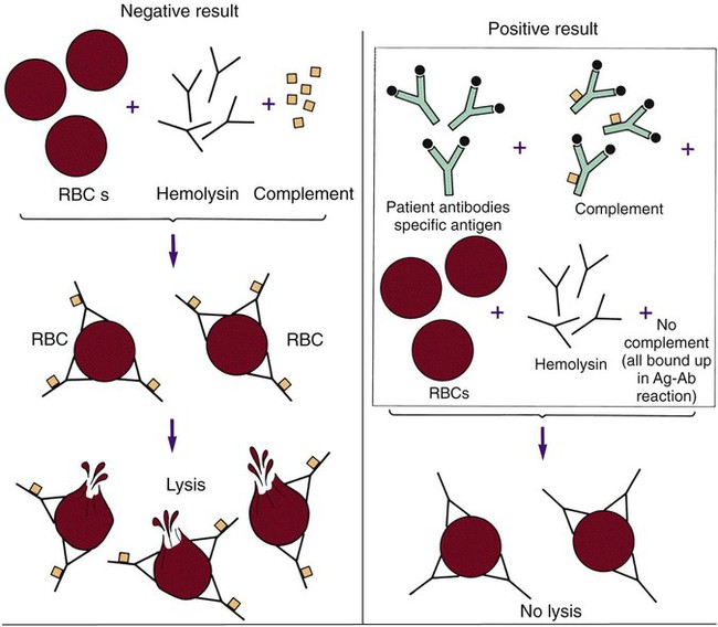
One of the classic methods of demonstrating the presence of antibody in a patient’s serum is the complement fixation (CF) test. This test consists of two separate systems. The first (the test system) consists of the antigen suspected of causing the patient’s disease and the patient’s serum. The second (the indicator system) consists of a combination of sheep red blood cells, complement-fixing antibody (IgG) raised against the sheep red blood cells in another animal, and an exogenous source of complement (usually guinea pig serum). When these three components are mixed together in optimum concentrations, the anti-sheep erythrocyte antibody binds to the surface of the red blood cells, and the complement then binds to the antigen-antibody complex, ultimately causing lysis (bursting) of the red blood cells. For this reason the anti-sheep red blood cell antibody is also called hemolysin. For the CF test, these two systems are tested in sequence (Figure 10-5). The patient’s serum is first added to the putative antigen; then the limiting amount of complement is added to the solution. If the patient’s serum contains antibody to the antigen, the resulting antigen-antibody complexes bind all the complement added. In the next step, the sheep red blood cells and the hemolysin (indicator system) are added. The patient’s complement is available to bind to the sheep cell–hemolysin complexes and cause lysis if the complement has not been bound by a complex formed with antibody from the patient’s serum. A positive result, meaning the patient has complement-fixing antibodies, is revealed by failure of the red blood cells to lyse in the final test system. Lysis of the indicator cells indicates lack of antibody and a negative CF test result.

Although this test requires many manipulations, takes at least 48 hours to complete both stages, and often yields nonspecific results, it has been used for many years to detect many types of antibodies, particularly antiviral and antifungal antibodies. Many new systems have gradually been introduced to replace the CF test, because they demonstrate improved recovery of pathogens or their products and provide more sensitive and less demanding procedures for detecting antibodies, such as particle agglutination, indirect fluorescent antibody tests, and enzyme-linked immunosorbent assay (ELISA). CF tests are performed chiefly for diagnosis of unusual infections and are done primarily in laboratories.

Enzyme-Linked Immunosorbent Assays

ELISA tests available for the detection of antibodies to infectious agents are sensitive and specific. As described in depth in Chapter 9, the presence of a specific antibody is detected by the ability of a second antibody, conjugated to a colored or fluorescent marker, to bind to the target antibody, which is bound to its homologous antigen. (Various enzyme-substrate systems, including the use of avidin-biotin to bind marker substances, are also discussed in Chapter 9.) The antigen to which the antibodies bind, if antibodies are present in the patient’s sera, is either attached to the inside of the wells of a microtiter plate, adherent to a filter matrix, or bound to the surface of beads or plastic paddles. Advantages of ELISA tests include ease of performance on many serum samples at the same time and easy detection of the colored or fluorescent end products with appropriate instrumentation, removing the element of subjectivity inherent in so many serologic procedures. Disadvantages include the need for special equipment, the fairly long reaction times (often hours instead of minutes for particle agglutination tests), the relative end point of the test (which relies on measuring the amount of a visible end product that is not dependent on the original antigen-antibody reaction itself but on a second enzymatic reaction, compared to a directly quantitative result), and the requirement for batch processing to ensure cost effectiveness.

Commercial microdilution or solid-phase matrix systems are available to detect antibody specific for hepatitis virus antigens, herpes simplex viruses 1 and 2, respiratory syncytial virus (RSV), cytomegalovirus, human immunodeficiency virus (HIV), rubella virus (both IgG and IgM), mycoplasmas, chlamydiae, Borrelia burgdorferi, Entamoeba histolytica, and many other agents.

The introduction of membrane-bound ELISA components has improved sensitivity and ease of use dramatically. Slot-blot and dot-blot assays force the target antigen through a membrane filter, causing it to become affixed in the shape of the hole (a dot or a slot). Several antigens can be placed on one membrane. When test (patient) serum is layered onto the membrane, specific antibodies, if present, bind to the corresponding dot or slot of antigen. Addition of a labeled second antibody and subsequent development of the label allows visual detection of the presence of antibodies based on the pattern of antigen sites. Cassette-based membrane-bound ELISA assays, designed for testing a single serum, can be performed rapidly (often within 10 minutes). Commercial kits to detect antibodies to Helicobacter pylori, Taxoplasma gondii, and some other infectious agents are available.

Antibody capture ELISAs are particularly valuable for detecting IgM in the presence of IgG. Anti-IgM antibodies are fixed to the solid phase; therefore, only IgM antibodies, if present in the patient’s serum, are bound. In a second step, specific antigen is added in a sandwich format and a second antigen-specific labeled antibody is added. Toxoplasmosis, rubella, and other infections are diagnosed using this technology, typically in research settings.

Indirect Fluorescent Antibody Tests and Other Immunomicroscopic Methods

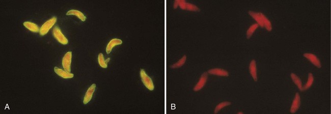
Indirect fluorescent antibody determination (IFA) is a widely applied method of detecting diverse antibodies (see Chapter 9). For these types of tests, the antigen against which the patient makes antibody (e.g., whole Toxoplasma organisms or virus-infected tissue culture cells) is affixed to the surface of a microscope slide. The patient’s serum is diluted and placed on the slide, covering the area in which antigen was placed. If present in the serum, antibody binds to the specific antigen. Unbound antibody is then removed by washing the slide. In the second stage of the procedure, a conjugate of antihuman globulin directed specifically against IgG or IgM and a fluorescent dye (e.g., fluorescein) is placed on the slide. This labeled marker for human antibody binds to the antibody already bound to the antigen on the slide and serves as a detector, indicating binding of the antibody to the antigen when viewed under a fluorescence microscope (Figure 10-6). Commercially available test kits include slides coated with the antigen, positive and negative control sera, diluent for the patients’ sera, and the properly diluted conjugate. As with other commercial products, IFA systems should be used as units, without modification of the manufacturer’s instructions. Commercially available IFA tests include those for antibodies to Legionella species, B. burgdorferi, T. gondii, varicella-zoster virus, cytomegalovirus, Epstein-Barr virus capsid antigen, early antigen and nuclear antigen, herpes simplex viruses types 1 and 2, rubella virus, M. pneumoniae, T. pallidum (the fluorescent treponemal antibody absorption test [FTA-ABS]), and several rickettsiae. Most of these tests, if performed properly, give extremely specific and sensitive results. Proper interpretation of IFA tests requires experienced and technically competent technologists. These tests can be performed rapidly and are cost effective.

Radioimmunoassays

Radioimmunoassay (RIA) is an automated method of detecting antibodies that usually is performed in the chemistry section of the laboratory rather than in the serology section. RIA tests were originally used to detect antibody to hepatitis B viral antigens. Radioactively labeled antibody competes with the patient’s unlabeled antibody for binding sites on a known amount of antigen. A reduction in radioactivity of the antigen–patient antibody complex, compared with the radioactive counts in a control test with no antibody, is used to quantitate the amount of patient antibody bound to the antigen. The development of new marker substances, such as ELISA systems, chemiluminescence, and fluorescence, resulted in the production of diagnostic tests as sensitive as RIA without the hazards associated with the use and disposal of radioactive reagents.

Fluorescent Immunoassays

Fluorescent immunoassays (FIA) were developed in response to the inconveniences associated with RIA (i.e., radioactive substances and expensive scintillation counters). These tests, which use fluorescent dyes or molecules as markers instead of radioactive labels, are based on the same principle as RIA. The primary difference is that the competitive antibody in RIA systems is labeled with a radioisotope, and in FIA the antigen is labeled with a compound that fluoresces under the appropriate light emission source. Binding of patient antibody to a fluorescent-labeled antigen can reduce or quench the fluorescence, or binding can cause fluorescence by allowing conformational change in a fluorescent molecule. Measurement of fluorescence is a direct measurement of antigen-antibody binding and is not dependent on a second marker, as in ELISA tests. Systems are commercially available to measure antibody developed against numerous infectious agents, as well as against self-antigens (autoimmune antibodies).

Western Blot Immunoassays

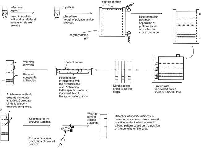
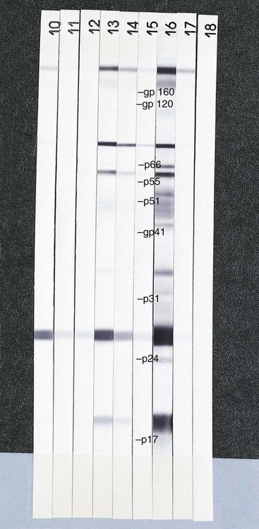
Requirements for the detection of very specific antibodies have driven the development of the Western blot immunoassay (Figure 10-7). The method is based on the electrophoretic separation of major proteins of an infectious agent in a two-dimensional agarose (first dimension) and acrylamide (second dimension) matrix. A suspension of the organism is mechanically or chemically disrupted, and the solubilized antigen suspension is placed at one end of a polyacrylamide (polymer) gel. Under the influence of an electrical current, the proteins migrate through the gel. Most bacteria or viruses contain several major proteins that can be recognized based on their position in the gel after electrophoresis. Smaller proteins travel faster and migrate farther in the lanes of the gel. The protein bands are transferred from the gel to a nitrocellulose or other type of thin membrane, and the membrane is treated to immobilize the proteins. The membrane is then cut into many thin strips, each carrying the pattern of protein bands. When patient serum is layered over the strip, antibodies bind to each of the protein components represented by a band on the strip. The pattern of antibodies present can be used to determine whether the patient has a current infection or is immune to the agent (Figure 10-8). Antibodies against microbes with numerous cross-reacting antibodies, such as T. pallidum, B. burgdorferi, herpes simplex virus types 1 and 2, and HIV, are identified more specifically using this technology than a single method that is used to identify a single antibody type. For example, the CDC defines an ELISA or immunofluorescence assay as a first-line test for Lyme disease antibody, but positive or equivocal results must be confirmed by a Western blot test.