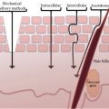
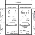
FIGURE 19.1 Pathomechanism of sensitive skin.
AD and ACD are both common skin diseases having an immune pathogenesis (9). AD is a common skin condition, characterized by a complex, heterogeneous pathogenesis, including skin barrier dysfunctions and significant pruritus. Recently, the skin barrier dysfunction induced by the FLG mutation has been demonstrated in AD. The barrier dysfunction shifts Th1 to Th2 response. Th2 cells produce IL-31, which provokes pruritus, and Th2 cytokines decrease filaggrin expressions by keratinocytes. These findings suggest that the evolution of a Th2 response lead to pruritus and barrier dysfunctions in patients with AD (10,11).
There is some similarity between sensitive skin and AD. It is likely that the same factors that play a role in the development of sensitive skin are also operative in AD. The most common and an important factor is skin-barrier dysfunction. There may also be differences among neural thresholds. Some patients have an abnormal response to mediators such as histamine. In some, exposure to histamine generates a significant wheal, whereas in others, there is only an insignificant response or none at all. This differing response can be due to their diverse neural threshold.
Atopic Diathesis and Sensitive Skin
It was thought that patients with AD were unable or less likely to develop contact dermatitis. In various studies, patients with AD stimulated with strong allergens failed to develop sensitization at rates similar to patients without AD. However, recent literature evidence from the United States and Europe has shown that patients with AD have similar if not higher rates of positive patch test results to common contact allergens, than in patients without AD (12).
There are studies supporting the link between sensitive skin and an atopic diathesis (13). It was first suggested by Thiers that features of atopic diathesis could explain sensitive skin syndrome (SSS) (14).
In a survey from the United Kingdom, only 49% of persons with sensitive skin had atopic diathesis concurrently. Hence, all cases with sensitive skin cannot be attributed to underlying atopic diathesis (15).
Clinical Features
The most practical, functional clinical description of sensitive skin is the one that reacts to cosmetic products. That is the hallmark feature of sensitive skin. It is mainly diagnosed by the subjective perceptions of the patient or from patient observation.
The usual complaints with which the patients approach the dermatologists are “I applied that moisturizer and it irritated me so much” or “I cannot find a sunscreen that feels good” or “Everything I apply to my skin burns and stings,” and so on. These are the clues toward the diagnosis of sensitive skin. Demonstrable clinical signs are rare or none in these patients. Only identifiable clinical features may be less hydrated and less supple skin (1). Subjective, sensorial signs, such as stinging, burning, and itching sensations on an apparently normal-looking skin are the clinching points for the diagnosis of sensitive skin (16). The face is the main target of cosmetic use and, hence, the main body part affected. However, sensitive skin may as well involve other locations, such as the hands or scalp (5). In contrast, all the eczematous dermatoses have specific clinical signs and symptoms, specific distribution or body site predilection, and well-defined diagnostic criteria.
People with sensitive skin may identify and mention various aggravating factors for their condition; these include environmental factors such as cold, wind, sun, heat, humid weather, pollution, and exposure to water during showers and in swimming pools (17). Such triggering factors may be part of various eczematous disorders, such as AD, and may even be misdiagnosed as rosacea.
Diagnosis
The diagnosis of sensitive skin is challenging, as there is no consensual definition and way of objective assessment of the condition. The diagnosis mostly relies upon the way subjects report about their feeling of sensations; assessment can also be undertaken by using a questionnaire, prepared by the clinician based upon the existing knowledge on sensitive skin. A provocative test lactic acid stinging test (LAST) can be performed with patients’ consent in order to reproduce the symptoms, which can be considered as indirect evidence of the condition (16). A thorough clinical examination to eliminate obvious as well as more subtle contact eczemas, AD, etc., is mandatory in all patients with sensitive skin (2).
In vivo and in vitro tests for evaluation of sensitive skin are mentioned in Table 19.1. Hematological investigations such as absolute eosinophil count and serum immunoglobulin E level help in the diagnosis of AD. Patch tests and photopatch tests are diagnostic of CD and photocontact dermatitis (18). Some in vivo tests similar to patch tests are in use for the detection of sensitive skin. One of these is the repeat insult patch test, where the product to be tested is applied on the patient’s skin multiple times per week and evaluated for irritation or sensitization. Another such test is the chamber scarification test, where an area of skin is scratched to cause little damage, and then the product is applied to evaluate the reaction.
The main pathogenesis of sensitive skin is impaired barrier function. Physiological changes associated with sensitive skin can be detected when the patient is minimally symptomatic. Noninvasive biophysical instruments can be used for such objective detection of sensitive skin. Such techniques are detection of transepidermal water loss (TEWL), corneometry, skin capacitance, and estimation of skin hydration and skin pH. These tests should be undertaken either at baseline or after a provocative test to identify the biophysical variations linked to sensitive skin (6).
TABLE 19.1
In Vivo Tests for Evaluation of Sensitive Skin (6)
|
In Vivo Test |
Method/Evaluation |
|
Patch test |
Performed with raw ingredients or finished products. |
|
Repeat insult patch test |
Ten patches are applied to the same site at 48–72 hour intervals for 3–4 week periods; after 2 weeks of rest, test site is rechallenged and graded. |
|
Chamber scarification test |
Volar aspect of the forearm is scarified, and the test product is applied under a Finn chamber repetitively for 3 days and evaluated up to 1 week after patch removal. |
|
Cumulative irritancy test |
Patch test is applied to the same test site for 10–21 days and evaluated for irritant reaction. |
Source: Draelos, ZD, Am J Contact Dermat, 8, 67–78, 1997.
Management
Management of sensitive skin due to cosmetics involves simple procedures with a step-by-step elimination, avoidance, and introduction of cosmetics (1). The skin should be prepared and strengthened with barrier repair creams. Good moisturizers should be applied to dry skin and, moreover, to dampen the skin immediately after bathing to improve hydration and enhance the skin barrier. Water-based products are better avoided as these may result in dryness. Water-based products also generally contain more preservatives, which may be the source of irritants or allergens.
Harsh cleansers and soaps should be avoided, and synthetic detergents should be used sparingly when needed. The natural acid mantle can be preserved by using products that are at least pH neutral, ideally a little bit acidic, by avoiding alkaline soaps and moisturizing the skin to strengthen the barrier. If the acid mantle turns alkaline due to use of harsh cleansers or creams, the barrier is damaged very quickly and it promotes Staphylococcal growth (19).
Even in AD, it is important to moisturize the skin effectively to correct or repair the associated barrier dysfunction. The same principles of managing disrupted barrier apply in both AD and sensitive skin. Whereas other eczematous dermatoses are managed by the standard disease-specific protocols/guidelines designed and updated for these disorders regularly.
REFERENCES
1. Inamadar AC, Palit A. Sensitive skin: An overview. Indian J Dermatol Venereol Leprol 2013; 79:9–16.
2. Lev-Tov H, Maibach HI. The sensitive skin syndrome. Indian J Dermatol 2012; 57:419–423.
3. Zug KA, McKay M. Eczematous dermatitis: A practical review. Am Fam Physician 1996; 54:1243–1250.
4. Loffler H. Contact allergy and sensitive skin. In: Berardesca E, Maibach HL, Fluhr JW, Eds. Sensitive Skin Syndromes. New York: CRC Press; 2006. pp. 225–230.
5. Saint-Martory C, Roguedas-Contios AM, Sibaud V, Degouy A, Schmitt AM, Misery L. Sensitive skin is not limited to the face. Br J Dermatol 2008; 158:130–133.
6. Draelos ZD. Sensitive skin: Perceptions, evaluation and treatment. Am J Contact Dermat 1997; 8:67–78.
7. Ale IS, Maibach HI. Irritant contact dermatitis. Rev Environ Health 2014; 29:195–206.
8. de Jongh CM, Khrenova L, Verberk MM, Calkoen F, van Dijk FJ, Voss H et al. Loss-of-function polymorphisms in the filaggrin gene are associated with an increased susceptibility to chronic irritant contact dermatitis: A case-control study. Br J Dermatol 2008; 159:621–627.
9. Thyssen JP, McFadden JP, Kimber I. The multiple factors affecting the association between atopic dermatitis and contact sensitization. Allergy 2014; 69:28–36.
10. Yokozeki H. The research for atopic dermatitis: Up to date. Nihon Rinsho 2014; 72:1503–1509 [Article in Japanese].
11. Kabashima K. New concept of the pathogenesis of atopic dermatitis: Interplay among the barrier, allergy, and pruritus as a trinity. J Dermatol Sci 2013; 70:3–11.
12. Aquino M, Fonacier L. The role of contact dermatitis in patients with atopic dermatitis. Allergy Clin Immunol Pract 2014; 2:382–387.
13. Kamide R, Misery L, Perez-Cullell N, Sibaud V, Taïeb C. Sensitive skin evaluation in the Japanese population. J Dermatol 2013; 40:177–181.
14. Thiers H. Peau sensible. In: Thiers H, editor. Les Cosmétiques. second ed. Paris: Masson; 1986. pp. 266–268.
15. Willis CM, Shaw S, de Lacharrière O et al. Sensitive skin: An epidemiological study. Br J Dermatol 2001; 145:258–263.
16. Jourdain R, Amaral F, Inamadar AC, Godse K. Sensitive skin. In: Srinivas C, Verschoore M, Eds. Basic Science for Modern Cosmetic Dermatology. New Delhi: Jaypee Brothers Medical Publishers; 2015. pp. 127–137.
17. Farage M. Perceptions of sensitive skin: Changes in perceived severity and associations with environmental causes. Contact Dermat. 2008; 59:226–232.
18. Ali SM, Yosipovitch G. Skin pH: From basic science to basic skin care. Acta Derm Venereol 2013; 93:261–267.
19. Ale IS, Maibach HA. Diagnostic approach in allergic and irritant contact dermatitis. Expert Rev Clin Immunol 2010; 6:291–310.


