Published on 16/03/2015 by admin
Filed under Dermatology
Last modified 22/04/2025
This article have been viewed 3554 times
Agustin Martin-Clavijo and John Berth-Jones
Evidence Levels: A Double-blind study B Clinical trial ≥ 20 subjects C Clinical trial < 20 subjects D Series ≥ 5 subjects E Anecdotal case reports
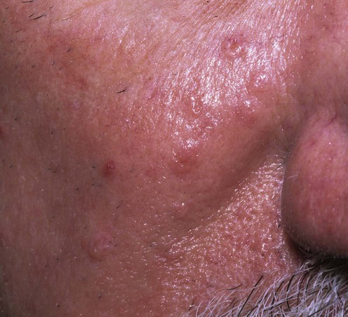
Sebaceous hyperplasia is a common benign condition. Incidence increases with age, but it may occur at any time and even at birth. It presents as single or multiple soft yellow papules, often with central umbilication, typically measuring 1–4 mm, mainly on the face (commonly the nose, cheeks, and forehead). Less commonly lesions are seen in other areas such as the chest, areola, mouth, and genitalia. Its frequency is increased in immunocompromised patients, especially after transplantation in patients on cyclosporine and corticosteroids.
This is a benign condition with no potential for malignant transformation. Lesions are asymptomatic and therefore need treatment only for cosmetic reasons. The differential diagnosis includes rhinophyma, nevus sebaceous, basal cell carcinoma, dermal nevus, plane warts, lupus miliaris disseminatus faciei, and syringoma.
We normally use cautery (electrodesiccation) or cryotherapy first-line. It can be helpful to treat some test lesions to assess patient satisfaction before treating the rest. Other treatments include surgical excision, photodynamic therapy, laser, isotretinoin, and chemical peels. It is important to stress to the patient the risk of scarring with many of these techniques.
No specific investigations are usually needed, as the diagnosis is clinical.
If the diagnosis is uncertain, histology will show enlargement of individual glands with increased numbers of fully mature lobules with no atypia or dysplasia.
Perez-Espana L, Prats I, Sanz A, Mayor M. Nefrologia 2003; 23: 179–80.
The authors looked at 163 renal transplant patients, of whom 25.9% had sebaceous hyperplasia. This was greatest in patients on cyclosporine. However, other immunosuppressants (azathioprine, mycophenolate mofetil, and tacrolimus) showed no significant increase in the incidence of sebaceous hyperplasia.
Bader RS, Scarborough DA. J Am Acad Dermatol 2000; 42: 127–8.
The authors describe the technique for intralesional electrodesiccation. They used it on more than 30 patients with no recurrences after 7 months.
American Academy of Dermatology Committee on Guidelines of Care. J Am Acad Dermatol 1994; 31: 648–53.
The authors include sebaceous hyperplasia as a condition treatable with cryotherapy.
McDonald SK, Goh MS, Chong AH. Australas J Dermatol 2011; 52: 227–30.
Treatment was effective and well tolerated.
Grimalt R, Ferrando J, Mascaro JM. J Am Acad Dermatol 1997; 37: 996–8.
Three closely related patients with premature familial sebaceous hyperplasia were treated with isotretinoin 1 mg/kg/day for 6 weeks. Response was maintained with isotretinoin 20 mg on alternate days in one case and isotretinoin gel 0.05% in the others. Follow-up was for 5 months.
Grekin RC, Ellis CN. Cutis 1987; 34: 90–2.
Two patients were treated with isotretinoin. Both had a good response but relapsed after the treatment was withdrawn, needing maintenance treatment. The first patient received 20 mg daily with a maintenance dose of 10 mg on alternate days; the second received 40 mg on alternate days with a maintenance dose of 40 mg twice a week.
Retinoids are also sometimes used topically, although there is scant published experience of this.
Horio T, Horio O, Miyaichi-Hashimoto H, Ohnuki M, Isei T. Br J Dermatol 2003; 148: 1270–90.
The authors treated one patient with photodynamic therapy. The response was good, with no scarring or hyperpigmentation at 12 months follow-up.
Gold MH, Bradshaw VL, Boring MM, Bridges TM, Biron JA, Lewis TL. J Drugs Dermatol 2004; 3: S6–9.
Twelve patients were randomized to topical aminolevulinic acid and either blue light or intense pulsed light. They had four treatments at 1-month intervals. Both treatment arms had more than 50% reduction in the number of the lesions without recurrence at 12 weeks follow-up.
Alster TS, Tanzi EL. J Drugs Dermatol 2003; 2: 501–4.
Ten patients were treated with topical 5-ALA followed 1 hour later by the 595 nm pulsed dye laser. They were compared with untreated lesions and laser-only treated lesions. Laser photodynamic therapy yielded the best results; 70% of lesions had total clearing after one treatment, the remaining 30% after two treatments.
No D, McLaren M, Chotzen V, Kilmer SL. Dermatol Surg 2004; 30: 382–4.
The authors treated 10 patients. Both objective and subjective improvement was noted in the majority of patients, with very few side effects.
Aghassi D, Gonzalez E, Anderson RR, Rajadhyaksha M, Gonzalez S. J Am Acad Dermatol 2000; 43: 49–53.
The authors treated 29 lesions on seven different patients with pulsed dye laser. There was improvement in 93% of lesions and complete clearance in 28%. No scarring or hyperpigmentation was noted. However, 28% of the lesions reappeared and 7% returned to their original size.
Landthaler M, Haina D, Waidelich W, Braun-Falco O. J Dermatol Surg Oncol 1984; 10: 456–61.
The authors report on 477 patients with different cutaneous lesions treated with an argon laser, including sebaceous hyperplasia.
Walther T, Hohenleutner U, Landthaler M. Dtsch Med Wochenschr 1998; 123: 798–800.
This article reports the treatment of a patient with CO2 laser. The lesions cleared without scarring.
Riedel F, Bergler W, Baker-Schreyer A, Stein E, Hormann K. HNO 1999; 47: 101–6.
This article erports on 216 patients with different facial lesions. The authors report good-to-excellent results with sebaceous hyperplasia.
Rosian R, Goslen JB, Brodell RT. J Dermatol Surg Oncol 1991; 17: 876–9.
The authors treated 67 lesions in 20 patients: 66 cleared after one application of 100% bichloroacetic acid, with minimal scarring.
Occasionally, sebaceous hyperplasia is treated surgically. This has the advantage of providing tissue for histology when there is any doubt about the diagnosis. Shave excision or curettage are usually sufficient both to obtain histological confirmation and to achieve removal with minimal scarring.
Treatment of Skin Disease Comprehensive Therapeutic Strategies 4e
WhatsApp us
 Conservative management/cosmetic camouflage
 Electrodesiccation/cautery
 Cryotherapy
 Isotretinoin
 Photodynamic therapy
 Topical retinoids
 Topical aminolevulinic acid and pulsed dye laser
 Diode laser
 Pulsed dye laser
 Argon laser
 Carbon dioxide laser
 Erbium : YAG laser
 Bichloroacetic acid
 Surgery/curettage