[level-membership-for-basic-science-category]
Scope of Microbiology
After reading this chapter, the student will be able to:
• Describe the achievements of scientists in the early years of microbiology
• Discuss the different types of microscopes and their use
• Explain the theory of spontaneous generation
• Discuss the germ theory of disease and its significance
• Name and describe Koch’s postulates
• Discuss the origin and evolution of microorganisms
• Describe the differences between prokaryotes and eukaryotes
• Explain the role of taxonomy
• Describe the different ways of microbial transmission
• Name and briefly describe the different uses of microorganisms in everyday life
Origins of Microbiology and Microscopy
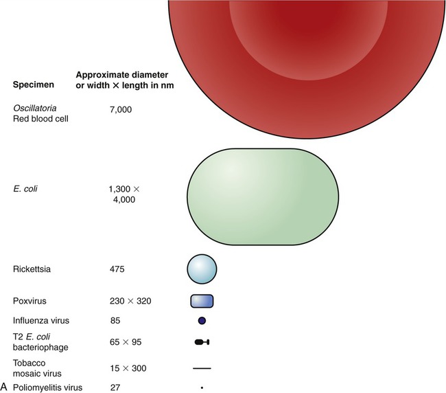
Microbiology is the study of microorganisms, using a variety of techniques for purposes of visualization, identification, and study of their function. The science of microbiology originated with the invention and development of the microscope. Microscopy allowed humans to magnify objects and microorganisms not detectable by the naked eye. Technological advances then have led to the improvement of microscopes, which became an essential investigative tool for biology in general and for the study of cells, tissues, and microorganisms (Figure 1.1) in particular.
Microscopy and Its Founding Fathers
Table 1.1 lists some significant events in the history of microbiology.
TABLE 1.1
Significant Events in Microbiology
| Name | Year | Event |
| Zaccharias Janssen | 1590 | Invention of the first compound microscope |
| Robert Hooke | 1660 | Explores living and nonliving matter with a compound microscope |
| Francesco Redi | 1668 | Experiments to disprove spontaneous generation |
| Antony van Leeuwenhoek | 1676 | Observes bacteria and protozoan “animalcules” with simple microscope |
| Francesco Redi | 1688 | Published experiments on spontaneous generation of maggots |
| Lazzaro Spallanzani | 1776 | Conducts further experiments to disprove spontaneous generation |
| Edward Jenner | 1796 | Introduction of smallpox vaccination |
| Ignaz Semmelweis | 1847–1850 | First use of antiseptics to reduce hand-borne disease |
| Louis Pasteur | 1857 | Proves that fermentation is caused by microorganisms; introduces pasteurization |
| Louis Pasteur | 1861 | Completes experiments that show without doubt that spontaneous generation does not occur |
| Joseph Lister | 1867 | Antiseptic surgery—begins the trend toward modern aseptic techniques |
| Robert Koch | 1876–1877 | Studies anthrax in cattle and implicates Bacillus anthracis as causative agent |
| Louis Pasteur | 1881 | Develops anthrax vaccine for animals |
| Robert Koch | 1882 | Identifies causative agent of tuberculosis |
| Robert Koch | 1884 | Describes his postulates |
Types of Microscopes
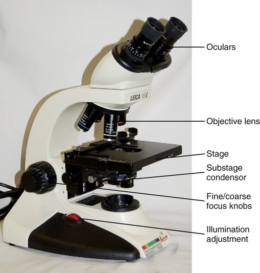
All light microscopes use visible light to illuminate, and optical lenses to observe, enlarged images of specimens. They are classified as either simple or compound. A simple light microscope, such as van Leeuwenhoek’s, has a single magnifying lens and a visible light source, and can magnify objects approximately ×266. A compound light microscope also uses visible light, usually provided by an electric source, but uses multiple lenses for magnification. The lens or lenses close to the eye are called ocular lenses and are located in the headpiece of the microscope. The lenses closer to the specimen are located in the body of the microscope and are referred to as objective lenses. Each lens has its own magnifying power, and the final magnification of a compound microscope is the product of the enlarging power of the ocular lens multiplied by the power of the objective lens. Most often the ocular lenses, either single (monocular) or in pairs (binocular), magnify by a power of 10 (×10). The objective lenses are mounted on a revolving nosepiece and usually magnify ×4 or ×5, ×10, ×40, and ×100. In general, compound microscopes can magnify an object up to 1000 times (i.e., an ocular lens with a magnification of ×10 times the objective lens with a power of ×100 = ×1000). The specimens for compound light microscopy can either be visualized as whole (i.e., bacteria and other microorganisms) or are specially prepared for viewing with a given type of microscope. After specific dehydration procedures larger specimens are cut into 1.0- to 10-µm sections. Both smear preparations, for single cells, and sections are usually stained for better visual images (see Chapter 4, Microbiological Laboratory Techniques). Photographs taken through a microscope are referred to as photomicrographs or micrographs.
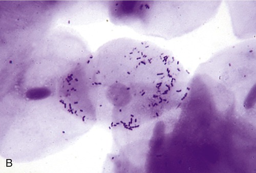
Bright-field Microscopes
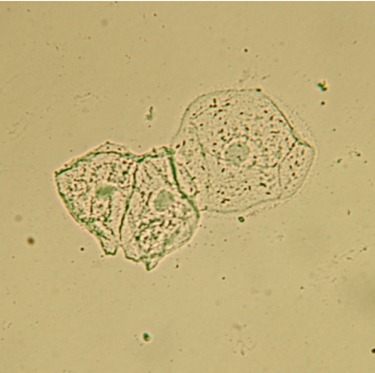
The bright-field microscope, a type of compound microscope, can be used to examine small specimens and some of their details. The microscope exhibits a background brighter than the observed specimen and is dependent on altering the light path (refraction) only. For this reason most specimens require staining for optimal observation. Bright-field microscopes are most commonly used to observe sectioned and stained tissues, organs, and microorganisms (Figure 1.2).
Dark-field Microscopes
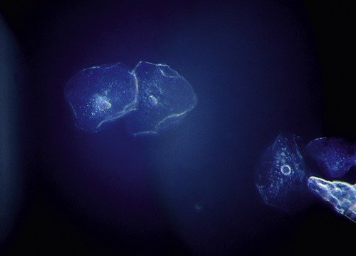
Unfixed, unstained specimens such as living organisms (i.e., bacteria) can be observed with a dark-field microscope. Because of its design, the background of the microscope slide is dark, whereas the organism itself is bright (Figure 1.3). Some bacteria that are difficult to stain, including spirochetes (see Chapter 6, Bacteria and Archaea), can best be observed by dark-field microscopy. Bacterial capsules are also good candidates to be observed with dark-field microscopes.
Fluorescence Microscopes
If a specimen can emit light (fluoresce) of one color when illuminated by ultraviolet radiation then fluorescence microscopy may be the method of choice. Fluorescence microscopes are used to visualize specimens that contain natural fluorescent substances such as chlorophyll or those stained with a fluorescent dye such as fluorescein, auramine, or rhodamine B (Figure 1.5). Fluorescence microscopy is an important and widely used tool in the diagnosis of infectious disease and in studies in microbial ecology. Fluorescence techniques are applied to identify specific antibodies, which are proteins produced in response to antigens (see Chapter 20, The Immune System). By attaching a fluorescent dye to these protein molecules, labeled antibodies are created that can be visualized, monitored, and studied.
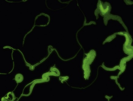
The fluorescence microscope uses an ultraviolet lamp to illuminate specimens stained with fluorescent stain. This is a fluorescence microscopy photo of a modified strain of Pseudomonas. (Approximate magnification: ×1000.) Courtesy Dr. Larry Halverson, Iowa State University.
Transmission Electron Microscopes
In a transmission electron microscope the electron beam travels through an ultrathin sectioned specimen (approximately 100 nm in thickness) and provides a two-dimensional image of the cell or other object. The resolving power of a TEM is approximately 0.002 µm, which is 100 times greater than can be achieved with a light microscope. The usual magnification of a TEM ranges from 500,000 to 1,000,000. Pictures taken of images created with an electron microscope are called electron micrographs (Figure 1.6).

Scanning Electron Microscopes
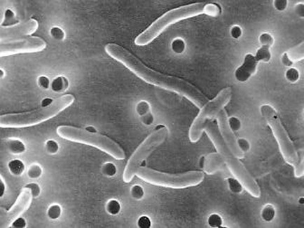
A scanning electron microscope also provides images of high resolution, but in contrast to the TEM, the SEM does not require ultrathin sections. It scans the surface of an object, producing a three-dimensional image (Figure 1.7). Moreover, the SEM has a large depth of field that allows the surface areas of large samples to be in focus at the same time. The usual magnification that can be achieved with an SEM ranges from ×10 to ×100,000.
Spontaneous Generation
Louis Pasteur (1822–1895), a French chemist, finally ended the controversy. In 1861 he designed and conducted a definitive experiment in which he boiled meat broth in a flask and then heated the neck of the flask until it could be bent it into an S shape (swan-necked flask). Thus air could enter the flask but microorganisms trapped in the S-shaped neck were unable to reach the broth. There was no evidence of microbial growth. However, when he tilted the flask so that the broth could reach the organisms in the neck, the broth gradually became cloudy due to microbial growth (Figure 1.8).
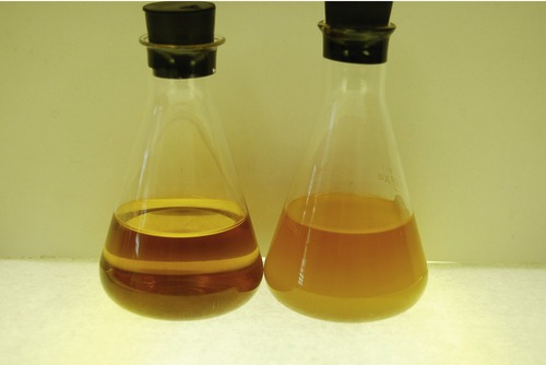
The more bacteria in a container of broth, the cloudier or more turbid it is. The clear flask on the left is uninoculated and contains no bacteria. The turbid (cloudy) flask on the right contains a culture that has grown for 18 hours and contains an enormous number of bacteria.
In 1877 John Tyndall (1820–1893) demonstrated that microorganisms exist in dust. In a set of experiments similar to Pasteur’s, but in the absence of dust, the broths remained sterile even when exposed to air. In addition, Tyndall provided evidence of the existence of heat-resistant bacteria. Ferdinand Cohn (1828–1898), independent of Tyndall’s studies, also described heat-resistant bacterial endospores (see Chapter 3, Cell Structure and Function).
Pasteurization
In addition to his other findings Louis Pasteur invented the process we know as pasteurization (Box 1.1). The Emperor Napoleon III asked Pasteur to investigate the spoiling of wine that caused a negative impact in the production of wine, damaging the French wine industry. Pasteur discovered that wine spoilage (wine disease) is caused by microorganisms. He “pasteurized” the wine by heating it to 55° C for several minutes, which killed enough microorganisms to prevent the wine from spoiling. Pasteurization does not kill all microorganisms, but reduces the number of viable organisms so they are less likely to cause spoilage or disease. Sterilization (see Chapter 4, Microbiological Laboratory Techniques), on the other hand, kills all microorganisms including their endospores.
Germ Theory of Disease
The germ theory of disease proposed by Louis Pasteur and Robert Koch (1843–1910) is based on the existence of infectious microorganisms. Although Pasteur was convinced that microbes caused disease in humans he was never able to link a specific microbe with a particular disease. Koch’s investigations focused on anthrax, an infectious disease that seriously affects animal herds and humans brought in contact with the microorganism. Koch also discovered that anthrax produced endospores that persist after the death of the exposed animals. Moreover, he proved that these spores can survive and later develop into the active anthrax microbe and infect other animals. This established that a specific organism caused a specific disease. After many years of experimentation he developed what we now know as Koch’s postulates (Box 1.2), which set forth the conditions that should identify an organism as the specific cause of a specific disease.
Origin and Evolution of Microorganisms
Origin
• In earliest times the earth was dominated by volcanoes, a lifeless ocean, and a very turbulent atmosphere.
• Intensive chemical activity occurred and the ocean received organic matter from land, atmosphere, and meteorites.
• Several elements formed key molecules such as sugars, amino acids, and nucleotides, all of which are building blocks for living organisms (see Chapter 2, Chemistry of Life).
Evolution
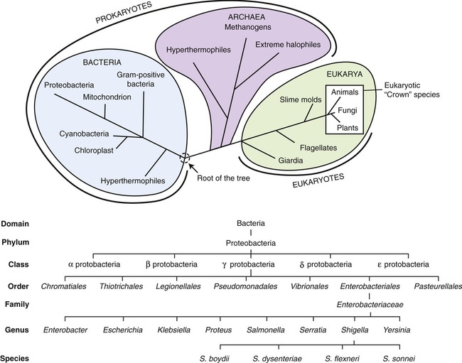
For the first three quarters of evolutionary history the only organisms on earth were microscopic and mostly unicellular. Fossils of prokaryotes go back to 3.5 to 4 billion years followed by eukaryotic life approximately 2.2 billion years ago. Eukaryotic cells are larger and more complex than prokaryotic cells and scientists believe that eukaryotes have evolved from prokaryotic symbiotic communities. It is estimated that there are about 5 to 100 million species of organisms living on earth today. The evolutionary relationship between organisms is the subject of phylogeny. Today, the phylogenetic relationship between organisms can be determined by nucleic acid and nucleotide sequencing. Results from ribosomal RNA (rRNA) sequencing identify three evolutionarily distinct cell lines that are classified as bacteria, archaea, and eukarya. These three distinct lineages, based on the origin of cell lines, are referred to as domains (Figure 1.9).
Classification of Microorganisms
To better understand the different types of microorganisms (microbes), they are grouped or classified in various ways. Microbes are diverse and in terms of numbers, most of the diversity of life on earth is represented by microbes. Further detailed description and discussion of the individual groups of microorganisms is provided in Chapter 6 (Bacteria and Archaea), Chapter 7 (Viruses), and Chapter 8 (Eukaryotic Microorganisms).
Prokaryotes versus Eukaryotes
Differentiation between prokaryotes (pro, before; karyon, nucleus) and eukaryotes (eu, good or with; karyon, nucleus) was greatly advanced as more sophisticated microscopes became available, allowing scientists to identify membrane-bound structures in unicellular organisms. Significantly, scientists discovered that some unicellular organisms contained membrane-bound organelles such as a nucleus, whereas others did not. The cells without membrane-bound organelles and therefore without a nucleus were classified as prokaryotes and the ones with membrane-bound organelles and a nucleus were designated eukaryotes. All bacterial cells are prokaryotic, whereas algae, fungi, and protozoans are eukaryotes (see Chapter 3, Cell Structure and Function).
Bacteria, Archaea, and Eukaryotes
Archaea are a group of single-celled microorganisms, similar to bacteria because they are also prokaryotes, but evolutionarily different. They are characterized by their preference to live in extreme environments including hot springs, glaciers, or highly salty environments (see Chapter 6, Bacteria and Archaea).
Eukaryotic microorganisms include algae, fungi, and protozoans (see Chapter 8, Eukaryotic Microorganisms). Algae are a large and diverse group of simple organisms, ranging from unicellular to multicellular forms, containing chlorophyll that is needed for photosynthesis. They are common in aquatic bodies (both freshwater and saltwater) and in all types of soil, but have limited medical significance. Fungi include yeasts, molds, and mushrooms, which are not microbes. Their energy sources are organic compounds found in soil and water. Fungi play a major role in the breakdown of dead organic matter in a variety of environments. However, some fungi are pathogenic and can cause disease in humans and animals. Protozoans are colorless, mobile organisms feeding on other organisms for their energy source. In general, protozoans are free-living microorganisms and several of them are also pathogenic to humans and animals.
Viruses
Viruses are noncellular, submicroscopic particles (visible only by electron microscopy) and consist of nucleic acid surrounded by a protein coat. They are metabolically inert, incapable of carrying out biosynthetic functions, and they are dependent on host machinery for their multiplication. Viruses are classified according to the type of nucleic acid (RNA or DNA) they contain. Many viruses are pathogenic and can be classified on the basis of their host as animal viruses, plant viruses, and bacterial viruses (see Chapter 7, Viruses).
Taxonomy
• Classification is the assignment of organisms into taxa based on similarities.
• Nomenclature deals with the rules for naming organisms.
• Identification is the process of specifying, identifying, and recording the traits of organisms.
The current system of taxonomy began with a Swedish botanist, Carolus Linnaeus (1707–1778). He provided a system that standardized the naming and classification of organisms on the basis of common characteristics. Linnaeus’s system groups similar species into genera, genera sharing common features into families, similar families into orders, orders into classes, classes into phyla, and phyla into kingdoms (see Figure 1.9). In addition, in 1990, Carl Woese (1928–) and his colleagues introduced a three-domain system of taxonomy (archaea, bacteria and eukarya) based on genetic rather than morphological similarities (see Figure 1.9).
The taxonomic resource for bacteria is the Bergey’s Manual of Systematic Bacteriology, the second edition of which is being published in five volumes. In addition to the number of keys for bacterial identification, newer versions of the manual also contain some molecular sequencing information for various bacterial groups. An outline of Bergey’s Manual can be found in Appendix A of this book.
Microorganisms in Health and Disease
Microbes in the Environment
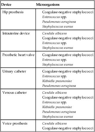
Microorganisms in nature are often organized into complex communities of different organisms called biofilms. A biofilm consists of surface-associated microbial cells enclosed in an extracellular polymeric substance matrix. Antony van Leeuwenhoek observed and described layers of microorganisms on tooth surfaces, representing a microbial biofilm (see Chapter 3, Cell Structure and Function). However, detailed, high-magnification observations of microbial biofilms did not occur until electron microscopy became available. Biofilms are found on a variety of surfaces such as medical devices (Table 1.2), industrial water system piping, natural aquatic systems, foods, and also on living tissues. An established biofilm structure provides a perfect environment for the exchange of genetic material between different organisms in the biofilm community.
TABLE 1.2
Microorganisms Associated With Biofilms on Medical Devices
| Device | Microorganisms |
| Hip prosthesis |
Ecological interactions between organisms in a community, including mutualism, commensalism, synergism, and parasitism, are classified according to the degree of benefit or harm they pose to one another. In mutualism both organisms benefit, in commensalism the waste product of a microbe provides useful nutrients for another organism, in synergism two organisms are dependent on each other to break down a nutrient that neither breaks down alone, and in parasitism one organism benefits and the other is harmed (see Chapter 9, Infection and Disease).
Normal Flora
Blood, lymph, cerebrospinal fluid, and internal organs are sterile. Before birth the entire healthy body is free of microbes. However, starting at birth microorganisms contaminate some of these previously sterile environments; for example, bacteria and other microorganisms colonize the mucous membranes of the upper respiratory tract, the digestive tract, and the surface of the skin. Microorganisms regularly found at any anatomical site in healthy humans and not causing infection or disease are referred to as the normal flora. The normal flora provides protection against pathogens by preventing attachment to the host tissue and by competing for the same nutrients. If successful, the pathogens cannot flourish or colonize and therefore are unable to reach sufficient numbers to cause infection (see Chapter 9, Infection and Disease).
Pathogens
The human body is in continual contact with microorganisms and if a particular microbe is pathogenic, colonizes, and is present in sufficient numbers, this interaction can lead to disease. Diseases caused by communicable microorganisms are referred to as infectious diseases. Control of these diseases can be achieved by application of the aseptic technique, the development and use of antimicrobial drugs, and immunization. These practices have drastically reduced the death rate from infectious diseases in the United States. However, death from infectious diseases in developing countries remains extremely high, due in large part to inadequate healthcare and poor sanitation. Infectious diseases may be transmitted by direct or indirect contact and can be acute, subacute, or chronic. Within communities they can be epidemic, pandemic, endemic, or sporadic (see Chapter 9, Infection and Disease).
• Washing hands with hot, soapy water before food preparation, after using the bathroom, or changing diapers
• Keeping raw meat, poultry, seafood, and their juices away from prepared, ready-to-eat foods
• Cooking foods thoroughly at high enough temperatures to kill harmful bacteria and their toxins
• Refrigerating foods within 2 hours of cooking. Cold temperatures will slow bacterial growth and multiplication
• Cleaning sufficiently all surfaces on which food is to be prepared
• Do not drink, swim, bathe, or play in floodwaters.
• Keep children away from floodwaters.
• Do not expose cuts or open wounds to floodwater.
• Monitor the quality of water supplies frequently.
• Keep the water clean and avoid using contaminated water.
• Drink water that has been purified by boiling or treatment with chlorine.
• Rinse fruits and vegetables with clean water.
• Avoid swallowing water from lakes, rivers, or swimming pools.
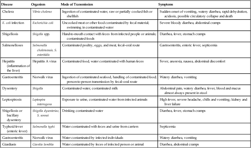
HEALTHCARE APPLICATION
Foodborne Diseases and Waterborne Diseases
| Disease | Organism | Mode of Transmission | Symptoms |
| Cholera | Vibrio cholerae | Ingestion of contaminated water, raw or partially cooked fish or shellfish | Sudden onset of vomiting, watery diarrhea, rapid dehydration, acidosis, possible circulatory collapse and death |
| E. coli infection | Escherichia coli | Uncooked meat or other food contaminated by fecal material; swimming in contaminated water | Severe bloody diarrhea, abdominal cramps |
| Shigellosis | Shigella spp. | Hand-to-mouth contact with feces from infected people or animals; contaminated foods | Diarrhea, fever, stomach cramps |
| Salmonelloses | Salmonella choleraesuis, S. enteritidis | Contaminated poultry, eggs, and meat, fecal–oral route | Gastroenteritis, enteric fever, septicemia |
| Hepatitis (inflammation of the liver) | Hepatitis A virus | Contaminated food, water contaminated with human feces | Fever, anorexia, nausea, abdominal discomfort |
| Gastroenteritis | Norwalk virus | Ingestion of contaminated seafood, handling of contaminated food; person-to-person transmission by fecal–oral route | Watery diarrhea, vomiting |
| Dysentery | Shigella | Contaminated water, contaminated milk | Abdominal pain, watery diarrhea, fever, blood and mucus almost always present in stool |
| Leptospirosis | Leptospira interrogans | Exposure to urine, contaminated water from infected animals | High fever, severe headache, chills and vomiting; kidney and liver failure |
| Shigellosis or bacillary dysentery | Shigella dysenteriae, S. sonnei | Drinking contaminated water | Diarrhea, fever, stomach cramps |
| Typhoid fever (enteric fever) | Salmonella typhi | Water contaminated with feces and urine from carriers | Septicemia |
| Gastroenteritis | Norwalk virus | Water contaminated by infected individuals | Watery diarrhea, vomiting |
| Giardiasis | Giardia lamblia | Water contaminated by feces of infected person or animal | Diarrhea, abdominal cramps |
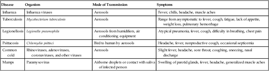
Airborne diseases are transmitted from infected people by coughing, sneezing, or talking. Pathogens are in small mucous saliva particles suspended as aerosols. Movements and directions of air currents play an important role in the spread of airborne respiratory diseases such as tuberculosis, legionellosis, and influenza. The public healthcare issues related to travel and airborne diseases are also discussed in Chapter 18 (Emerging Infectious Diseases).
HEALTHCARE APPLICATION
Airborne Diseases
| Disease | Organism | Mode of Transmission | Symptoms |
| Influenza | Influenza viruses | Aerosols | Fever, chills, headache, muscle aches |
| Tuberculosis | Mycobacterium tuberculosis | Aerosols | Range from asymptomatic to fever, cough, fatigue, lack of appetite, weight loss, pulmonary hemorrhage |
| Legionellosis | Legionella pneumophila | Aerosols from humidifiers, air conditioning equipment | Atypical pneumonia, fever, cough, difficulty in breathing, chest pain |
| Psittacosis | Chlamydia psittaci | Bird to human by aerosols | Headache, fever, nonproductive cough, occasional septicemia |
| Common cold | Rhinoviruses, adenoviruses, coronaviruses, and other viruses | Aerosols | Slight fever, headache, sore throat, coughing, sneezing, nasal discharge |
| Mumps | Paramyxovirus | Airborne droplets or contact with saliva of infected person | Swelling of parotid glands, fever, headache, generalized muscle aches |
Applied Microbiology
Microorganisms in Food Production
Many nonpathogenic microorganisms occur naturally in food, are beneficial, and are used as starter cultures to produce foods such as vinegar, sauerkraut, pickles, fermented milks, yogurt, cheese, and bread (Box 1.3). For example, vinegar is made from foods containing starch or smaller sugars. The production requires a two-way fermentation process that begins with apple juice or other raw materials, to which the yeast Saccharomyces cerevisiae is added to speed up the fermentation process. In the second stage, cultures of acetic acid bacteria such as Acetobacter aceti are added to this alcoholic liquid, converting the alcohol into acetic acid. In the United States most vinegar is produced from apples and is named apple cider vinegar.
Microbes and the Production of Pharmaceutical Agents
Louis Pasteur observed the inhibition of microorganisms by products formed by other microbes. This phenomenon is called antibiosis. The discovery of the penicillin-producing mold Penicillium by Alexander Fleming in the 1920s started the successful search for other antibiotic-producing microorganisms. Today, with the help of genetic engineering and recombinant DNA technology, not only antibiotics and semisynthetic antibiotics but also various hormones and other drugs are available on the market (see Chapter 22, Antimicrobial Drugs).
Microbes in Agriculture
Agricultural microbiology focuses on the relationships between microbes and domesticated plants and animals. Farmers use microbes and their products in a variety of ways, particularly for crop management via recombinant DNA biotechnology (see Chapter 25, Biotechnology). Plant microbiology involves the management of plant disease, soil fertility, and nutritional interactions. For example, the availability of nitrogen in the soil is essential for the growth of crops and bacteria involved in the nitrogen cycle of the soil are essential (see Soil Microbiology in Chapter 24, Microorganisms in the Environment and Environmental Safety). On the animal side, agricultural microbiology deals with the management and prevention of infectious diseases in farm animals as well as other associations animals have with microorganisms.
Summary
• The invention of microscopes made it possible to observe details of organisms and microorganisms that are invisible to the naked eye.
• Different types of microscopes developed over the years have been and continue to be specialized to perform different functions.
• Spontaneous generation was disproved and the germ theory of disease developed. This promoted vaccination and the use of aseptic techniques in surgery among other hygienic practices to reduce infection.
• The first life on earth is dated at 3.5 to 4 billion years ago, and is believed to be represented by prokaryotes, which are still found in every environment on earth today. Through evolution, eukaryotic life appeared approximately 2.2 billion years ago.
• Microorganisms include bacteria, viruses, prions, algae, fungi, and protozoans.
• Taxonomy is the classification, nomenclature, and identification of living organisms. Classification starts with domain, followed by kingdom, phylum, class, order, family, and genus.
• Microbes occur in every environment, may build relationships with other organisms, and often form biofilms. Biofilms represent problems for many industries including healthcare.
• Microorganisms are routinely found in and on humans without causing an infection or disease. These organisms are part of the normal flora.
• Transmission of infectious diseases can be airborne, waterborne, foodborne, or through direct contact.
• Microorganisms play a major role in the production of food, alcoholic beverages, and pharmaceuticals. They are also used in water treatment, agriculture, bioremediation, forensics, and as fuel cells.
Review Questions
1. One type of microscope that provides a three-dimensional image of a specimen is a:
2. One type of microscope capable of observing living microorganisms is the:
3. Which scientist is most responsible for ending the controversy about spontaneous generation?
4. Fossils of prokaryotes go back __________ billion years.
5. Which of the following is not a microorganism?
6. The correct order of the taxonomic category is:
a. Species, domain, phylum, kingdom, order, division, class, genus
b. Domain, kingdom, phylum, class, family, order, genus, species
c. Domain, kingdom, phylum, class, order, family, genus, species
d. Kingdom, domain, phylum, order, class, family, genus, species
7. Complex communities of microorganisms on surfaces are called:
8. A relationship between organisms in which the waste product of one provides nutrients for another is called:
9. Which of the following sites of the human body does not have a normal flora?
10. Which of the following industries use(s) microorganisms?
11. All bacteria are __________ cells.
12. Cells that contain a nucleus are __________ cells.
13. The taxonomic resource for bacteria is the __________.
14. The proteins implicated in spongiform encephalopathy are __________.
15. The cleanup of different industrial waste is referred to as __________.
16. Name and briefly describe the different types of microscopes.
17. Describe Koch’s postulates.
18. Compare and contrast prokaryotic and eukaryotic cells.
[/level-membership-for-basic-science-category][not-level-membership-for-basic-science-category]
Scope of Microbiology
After reading this chapter, the student will be able to:
• Describe the achievements of scientists in the early years of microbiology
• Discuss the different types of microscopes and their use
• Explain the theory of spontaneous generation
• Discuss the germ theory of disease and its significance
• Name and describe Koch’s postulates
• Discuss the origin and evolution of microorganisms
• Describe the differences between prokaryotes and eukaryotes
• Explain the role of taxonomy
• Describe the different ways of microbial transmission
• Name and briefly describe the different uses of microorganisms in everyday life
Origins of Microbiology and Microscopy
Microbiology is the study of microorganisms, using a variety of techniques for purposes of visualization, identification, and study of their function. The science of microbiology originated with the invention and development of the microscope. Microscopy allowed humans to magnify objects and microorganisms not detectable by the naked eye. Technological advances then have led to the improvement of microscopes, which became an essential investigative tool for biology in general and for the study of cells, tissues, and microorganisms (Figure 1.1) in particular.
Microscopy and Its Founding Fathers
Table 1.1 lists some significant events in the history of microbiology.
TABLE 1.1
Significant Events in Microbiology
| Name | Year | Event |
| Zaccharias Janssen | 1590 | Invention of the first compound microscope |
| Robert Hooke | 1660 | Explores living and nonliving matter with a compound microscope |
| Francesco Redi | 1668 | Experiments to disprove spontaneous generation |
| Antony van Leeuwenhoek | 1676 | Observes bacteria and protozoan “animalcules” with simple microscope |
| Francesco Redi | 1688 | Published experiments on spontaneous generation of maggots |
| Lazzaro Spallanzani | 1776 | Conducts further experiments to disprove spontaneous generation |
| Edward Jenner | 1796 | Introduction of smallpox vaccination |
| Ignaz Semmelweis | 1847–1850 | First use of antiseptics to reduce hand-borne disease |
| Louis Pasteur | 1857 | Proves that fermentation is caused by microorganisms; introduces pasteurization |
| Louis Pasteur | 1861 | Completes experiments that show without doubt that spontaneous generation does not occur |
| Joseph Lister | 1867 | Antiseptic surgery—begins the trend toward modern aseptic techniques |
| Robert Koch | 1876–1877 | Studies anthrax in cattle and implicates Bacillus anthracis as causative agent |
| Louis Pasteur | 1881 | Develops anthrax vaccine for animals |
| Robert Koch | 1882 | Identifies causative agent of tuberculosis |
| Robert Koch | 1884 | Describes his postulates |
Types of Microscopes
All light microscopes use visible light to illuminate, and optical lenses to observe, enlarged images of specimens. They are classified as either simple or compound. A simple light microscope, such as van Leeuwenhoek’s, has a single magnifying lens and a visible light source, and can magnify objects approximately ×266. A compound light microscope also uses visible light, usually provided by an electric source, but uses multiple lenses for magnification. The lens or lenses close to the eye are called ocular lenses and are located in the headpiece of the microscope. The lenses closer to the specimen are located in the body of the microscope and are referred to as objective lenses. Each lens has its own magnifying power, and the final magnification of a compound microscope is the product of the enlarging power of the ocular lens multiplied by the power of the objective lens. Most often the ocular lenses, either single (monocular) or in pairs (binocular), magnify by a power of 10 (×10). The objective lenses are mounted on a revolving nosepiece and usually magnify ×4 or ×5, ×10, ×40, and ×100. In general, compound microscopes can magnify an object up to 1000 times (i.e., an ocular lens with a magnification of ×10 times the objective lens with a power of ×100 = ×1000). The specimens for compound light microscopy can either be visualized as whole (i.e., bacteria and other microorganisms) or are specially prepared for viewing with a given type of microscope. After specific dehydration procedures larger specimens are cut into 1.0- to 10-µm sections. Both smear preparations, for single cells, and sections are usually stained for better visual images (see Chapter 4, Microbiological Laboratory Techniques). Photographs taken through a microscope are referred to as photomicrographs or micrographs.
Bright-field Microscopes
The bright-field microscope, a type of compound microscope, can be used to examine small specimens and some of their details. The microscope exhibits a background brighter than the observed specimen and is dependent on altering the light path (refraction) only. For this reason most specimens require staining for optimal observation. Bright-field microscopes are most commonly used to observe sectioned and stained tissues, organs, and microorganisms (Figure 1.2).
Dark-field Microscopes
Unfixed, unstained specimens such as living organisms (i.e., bacteria) can be observed with a dark-field microscope. Because of its design, the background of the microscope slide is dark, whereas the organism itself is bright (Figure 1.3). Some bacteria that are difficult to stain, including spirochetes (see Chapter 6, Bacteria and Archaea), can best be observed by dark-field microscopy. Bacterial capsules are also good candidates to be observed with dark-field microscopes.
Fluorescence Microscopes
If a specimen can emit light (fluoresce) of one color when illuminated by ultraviolet radiation then fluorescence microscopy may be the method of choice. Fluorescence microscopes are used to visualize specimens that contain natural fluorescent substances such as chlorophyll or those stained with a fluorescent dye such as fluorescein, auramine, or rhodamine B (Figure 1.5). Fluorescence microscopy is an important and widely used tool in the diagnosis of infectious disease and in studies in microbial ecology. Fluorescence techniques are applied to identify specific antibodies, which are proteins produced in response to antigens (see Chapter 20, The Immune System). By attaching a fluorescent dye to these protein molecules, labeled antibodies are created that can be visualized, monitored, and studied.
The fluorescence microscope uses an ultraviolet lamp to illuminate specimens stained with fluorescent stain. This is a fluorescence microscopy photo of a modified strain of Pseudomonas. (Approximate magnification: ×1000.) Courtesy Dr. Larry Halverson, Iowa State University.
Transmission Electron Microscopes
In a transmission electron microscope the electron beam travels through an ultrathin sectioned specimen (approximately 100 nm in thickness) and provides a two-dimensional image of the cell or other object. The resolving power of a TEM is approximately 0.002 µm, which is 100 times greater than can be achieved with a light microscope. The usual magnification of a TEM ranges from 500,000 to 1,000,000. Pictures taken of images created with an electron microscope are called electron micrographs (Figure 1.6).
Scanning Electron Microscopes
A scanning electron microscope also provides images of high resolution, but in contrast to the TEM, the SEM does not require ultrathin sections. It scans the surface of an object, producing a three-dimensional image (Figure 1.7). Moreover, the SEM has a large depth of field that allows the surface areas of large samples to be in focus at the same time. The usual magnification that can be achieved with an SEM ranges from ×10 to ×100,000.
