Published on 16/03/2015 by admin
Filed under Dermatology
Last modified 22/04/2025
This article have been viewed 2118 times
John Berth-Jones
Evidence Levels: A Double-blind study B Clinical trial ≥ 20 subjects C Clinical trial < 20 subjects D Series ≥ 5 subjects E Anecdotal case reports
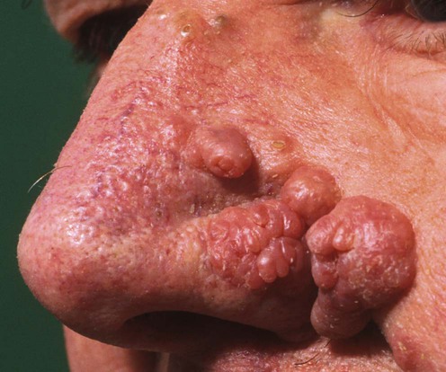
Phymas, of which rhinophyma is much the most common, are localized swellings of facial soft tissues due to a variable combination of fibrosis, sebaceous hyperplasia, and lymphedema. They occur on the nose (rhinophyma) and, less often the ears, forehead, or chin. They are seen much more frequently in males than in females. Rhinophyma may develop in patients with a long history of rosacea, when it is often regarded as a complication or ‘end stage’ of the disease. However, rhinophyma is also seen in patients who have no history of rosacea. Occasionally rhinophyma is complicated by the development of a malignancy.
Phymas require physical ablation or removal, usually by surgery. Remodeling is most often achieved simply by paring off the excess tissue with a scalpel. Other techniques that can be useful in the hands of those with the necessary expertise include electrosurgery, excision/vaporization with argon, CO2, Nd : YAG or Er : YAG lasers, and cryotherapy. Ionizing radiation has been used in cases with coexisting malignancy. Systemic isotretinoin can significantly reduce the bulk of rhinophyma, although it does not restore normal skin contours. It is possible, but not established, that treatment of rosacea may inhibit the development of rhinophyma.
Biopsy is occasionally indicated to exclude malignancy
Lutz ME, Otley CC. Dermatol Surg 2001; 27: 201–2.
Rhinophyma can be complicated by the development of a malignancy, which can be difficult to recognize.
Curnier A, Choudhary S. Ann Plast Surg 2002; 49: 211–14.
The authors report pleasing results in six patients treated by tangential excision for debulking, the use of scissors for sculpting, and mild dermabrasion for final contouring.
Clark DP, Hanke CW. J Am Acad Dermatol 1990; 22: 831–7.
This treatment was inexpensive and associated with few complications, and gave good or excellent cosmetic results in 13 cases.
Rex J, Ribera M, Bielsa I, Paradelo C, Ferrándiz C. Dermatol Surg 2002; 28: 347–9.
Eight male patients were treated using radiofrequency electrosurgery to remove thin layers of tissue until the nose shape was recreated. All patients achieved acceptable cosmetic results.
Halsbergen-Henning JP, van Gemert MJ. Lasers Surg Med 1983; 2: 211–15.
Thirteen cases were treated. This laser is believed to work by selectively coagulating capillaries causing redness of the nose and which feed the hypertrophic regions, as well as by direct coagulation shrinkage of the hypertrophic connective tissue. The result was a smooth and more natural appearance of the nose without redness. Pustulosis was also reduced.
Greenbaum SS, Krull EA, Watnick K. J Am Acad Dermatol 1988; 18: 363–8.
The results from the CO2 laser and electrosurgery were compared in three patients by treating one side of the nose by each method. There was little to choose between them in terms of results, but electrosurgery was more cost-effective.
Madan V, Ferguson JE, August PJ. Br J Dermatol 2009; 161: 814–18.
Exuberant sebaceous tissue was ablated using the Sharplan 40C CO2 laser under local anesthesia. The laser was used in a continuous mode to debulk the larger rhinophymas, and in a resurfacing mode (Silk Touch scanner; Sharplan, 4–7 mm spot at 20–40 W) or continuous mode (10–20 W using a defocused 2–3 mm beam) to reshape the nasal contours. Results were classified as good to excellent in 118 and poor in six patients.
El Azhary RA, Roenigk RK, Wang TD. Mayo Clin Proc 1991; 66: 899–905.
A review of 30 patients treated with the CO2 laser and followed up for 1 to 4 years. Milder cases were treated with laser vaporization, more severe cases with CO2 laser excision and then vaporization. Dilated pores developed in many patients. Leukoderma, unilateral alar lift, and mild hypertrophic scarring developed in single cases.
Wenig BL, Weingarten RT. Laryngoscope 1993; 103: 101–6.
Orenstein A, Haik J, Tamir J, Winkler E, Frand J, Zilinsky I, et al. Lasers Surg Med 2001; 29: 230–5.
Fincher EF, Gladstone HB. Arch Facial Plast Surg 2004; 6: 267–71.
In each of these reports six cases of rhinophyma were treated with satisfactory outcomes. The Er : YAG laser provides both controlled ablation of tissue and hemostasis.
Sonnex TS, Dawber RPR. Clin Exp Dermatol 1986; 11: 284–8.
Five cases were treated using two freeze–thaw cycles, each freeze lasting 30 seconds after the ice field was established, with a 4-minute intervening thaw. A pethidine and diazepam premedication was used. In three cases small residual prominent areas responded to further treatment after 2 months. The final result was satisfactory in each case, with no scarring.
Irvine C, Kumar P, Marks R. In: Marks R, Plewig G, eds. Acne and Related Disorders. London: Martin Dunitz, 1989; 311–15.
A study in which nine men with rhinophyma were treated with isotretinoin 1 mg/kg daily for up to 18 weeks. Isotretinoin reduced the volume of rhinophyma (assessed objectively using molds of the noses) by 9–23%, but did not restore normal skin contours in advanced cases.
The authors report good results in five cases and consider this their treatment of choice.
Kaushik V, Tahery J, Malik TH, Jones PH. J Laryngol Otol 2003; 117: 551–2.
This is a report of a single case treated using this approach. The microdebrider is a powered rotary shaving device. FloSeal™ is a hemostatic mixture of thrombin and gelatin applied topically after the surgery. The use of these adjuncts allowed precise sculpting and immediate hemostasis.
Eisen RF, Katz AE, Bohigian RK, Grande DJ. Arch Dermatol 1986; 122: 307–9.
The Shaw scalpel is a device in which a scalpel blade can be heated. A rhinophyma was treated using a temperature of 150°C to achieve hemostasis while paring. Contours were then refined using a CO2 laser and light dermabrasion.
Plenk HP. Plast Reconstruct Surg 1995; 95: 559–62.
Two patients with basal cell carcinoma complicating rhinophyma had complete control of both conditions by radiotherapy using orthovoltage X-radiation. The authors suggest that this modality might be useful for rhinophyma alone.
Skala M, Delaney G, Towell V, Vladica N. Australas J Dermatol 2005; 46: 88–9.
A rhinophyma was successfully treated with 90 kV photons to a total dose of 40 Gy in 20 daily fractions.
Taghizadeh R, Mackay SP, Gilbert PM. J Plast Reconstruct Aesthet Surg 2008; 61: 330–3.
Six patients were treated successfully.
The Versajet Hydrosurgery System is a novel surgical device which uses a high-velocity jet of water for excision and vacuum aspiration of water and debris.
Treatment of Skin Disease Comprehensive Therapeutic Strategies 4e
WhatsApp us

 Surgical paring
 Electrosurgery
 Argon laser
 CO2 laser
 Nd : YAG laser
 Er : YAG laser
 Cryotherapy
 Isotretinoin
 Microdebrider
 Shaw scalpel
 Radiotherapy
 Hydrosurgery