Positive-pressure mechanical ventilation

Published on 13/02/2015 by admin

Filed under Anesthesiology

Last modified 22/04/2025

Print this page

rate 1 star rate 2 star rate 3 star rate 4 star rate 5 star
Your rating: none, Average: 1 (1 votes)

This article have been viewed 2020 times

Positive-pressure mechanical ventilation

Edmund Carton, MD

Many advances have occurred in intraoperative mechanical ventilation (MV) in the past 5 decades. Multiple modes of ventilatory support can now be delivered by microprocessor-driven mechanical ventilators. Closed-loop technology can provide protective ventilatory strategies with low total gas flows. N2O, He, or air may be added to O2, and the inspired gas and inhaled anesthetic agents are delivered by electronic flowmeters. Contemporary airway circuit technology allows efficient CO2 absorption and improved scavenging. There are also alarm systems for multiple parameters including end-tidal carbon dioxide (ETCO2), tidal volume (VT), respiratory rate, minute ventilation, fraction of inspired O2 (FIO2), and airway pressure to ensure adequate O2 delivery and ventilation.

In addition to these industry-derived improvements, our understanding of intraoperative changes in respiratory mechanics has also improved so that appropriate MV settings can be used in patients with airflow obstruction or lung or chest wall abnormalities. Recent concern about the risk of developing acute lung injury in previously healthy patients after intraoperative MV is also discussed in this chapter. A list of the abbreviations for terms commonly used in respiratory physiology is provided in Box 223-1.

Respiratory mechanics

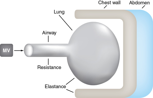
Two independent patient-related forces have to be overcome to achieve lung inflation by positive-pressure MV (Figure 223-1): (1) resistance to the flow of gases within the airway and (2) compliance and elastance of the lung and chest wall, respectively. The latter concept can be difficult for the anesthesia provider to understand. In an inflated lung, the elastin within the lung is stretched. If you then remove a lung from the thoracic cavity and if the airway is open, the lung will collapse. This does not happen within the thoracic cavity because the parietal pleura is adherent to the thoracic cavity. In an intubated paralyzed patient at end expiration, or when the lungs are at their functional residual capacity, positive pressure must be applied to expand the lungs to deliver a breath. The amount of pressure (plateau pressure [Pplat]) applied to expand the lungs to a known volume is a measure of the compliance of the lung (Compliance = ΔV/ΔP). For example, 5 cm of H2O pressure in a patient with a VT of 500 would equal a compliance of 100. In a patient with a diseased lung (e.g., with severe acute respiratory distress syndrome), the compliance would typically be less than 20.

The reciprocal of compliance, elastance, is defined as the ΔP/ΔV and is more commonly used to describe the pressure-volume relationships within the thoracic cavity. Unlike the lung, which collapses if opened, the chest wall or cavity would expand if opened.

Taken together the resistive and elastic properties (also referred to as compliance) of the lung and chest wall make up the patient-related impedance during any mode of positive-pressure inspiration. During each MV inflation, a dynamic interaction occurs between these patient-related variables (airway, lung, and chest wall) and MV settings (VT, airway pressure, and inspiratory flow). Airway pressure is equal to the sum of the pressure required to overcome the resistive component of the respiratory system (influenced by airway resistance and inspiratory flow) plus the pressure required to overcome the elastic component of the respiratory system (influenced by lung compliance, chest wall elastance, and VT):

< ?xml:namespace prefix = "mml" />Airway pressure = (Resistance × Flow) + (Stiffness × VT)

image

Mode of mechanical ventilation

In contemporary mechanical ventilators, microprocessor-controlled valves rapidly adjust airflow and pressure during inspiration to achieve the desired lung inflation. Conventional intraoperative MV usually provides complete ventilatory support (control ventilation) when the patient is expected to have no spontaneous ventilatory effort. The common modes of intraoperative ventilatory support include volume control or vv control at respiratory rates less than 60 breaths/min.

Volume control mechanical ventilation

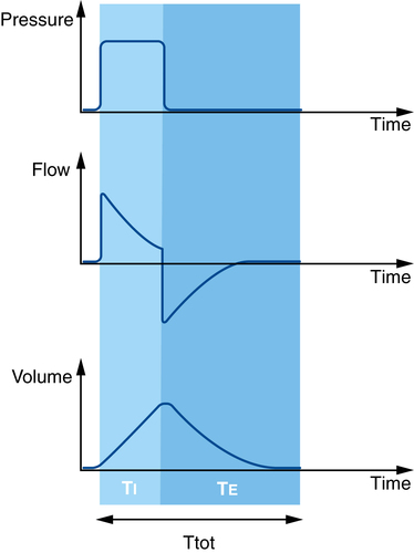
During volume control mode, VT and TI (or respiratory rate and inspiratory: expiratory [I:E] ratio) are set by the clinician and inspiratory flow remains constant (Figure 223-2). In volume control MV, the resistive impedance (transmission of the VT through the airway) and the elastic impedance (compliance of the lung or chest wall) both contribute to the peak airway pressure (Ppeak).

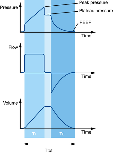
A short end-inspiratory pause may be included in the respiratory cycle in volume control MV so that, after the VT has been fully delivered and just prior to exhalation, airflow is reduced to zero for a fixed proportion of TI. The airway pressure recorded during this end-inspiratory interval is called the plateau (Pplat), pause, or end-inspiratory hold pressure (see Figure 223-2). In many anesthesia ventilators, the default setting in volume control mode may include no end-inspiratory pause so that only Ppeak is recorded. However, it is always possible (and desirable) to include an end-inspiratory pause when using volume control MV so that both Ppeak and Pplat are recorded.

In contrast to Ppeak, which is influenced by both resistive and elastic components, Pplat is influenced only by compliance factors, as there is no resistive contribution during zero-flow conditions. When we use volume control MV and include an end-inspiratory pause, Pplat provides a more accurate estimate of the maximal distending pressure inside the alveolar sac than does Ppeak.

Pressure control mechanical ventilation

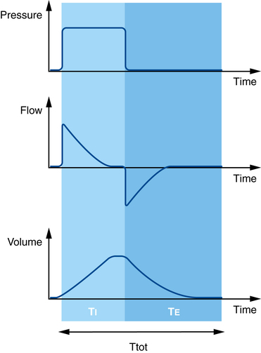
In pressure control MV, inspiratory pressure and TI (or respiratory rate and I:E ratio) are set by the clinician and remain constant, but the delivered VT and inspiratory flow will vary during inspiration (Figure 223-3). In pressure control MV, in which inspiratory pressure remains constant for the duration of TI, no Ppeak or end-inspiratory Pplat is registered (as occurs in volume control mode when an end-inspiratory pause is added into the respiratory cycle). Therefore, the relationship between Ppeak and Pplat that is informative in volume control MV cannot be used in pressure control MV.

Intraoperative mechanical ventilation: Which mode should we use?

No particular mode of ventilation (e.g., volume or pressure control mode) has been shown to be superior for any adult patient subgroup during intraoperative MV. It is more important to appreciate the limitations and the appropriate application of both volume and pressure control mode during intraoperative MV. Traditionally, volume control MV has been used intraoperatively in adult patients to maintain a secure minimum level of alveolar ventilation regardless of temporary changes in airway resistance or respiratory system compliance or elastance. When using volume control MV, an end-inspiratory pause should be routinely included in the respiratory cycle so that the distinction between increased airway resistance and decreased compliance can be observed. Pressure control MV could equally be used in adult patients as long as appropriate adjustments are made to MV settings with intraoperative changes in respiratory system mechanics.

Contemporary anesthetic machines now provide modes of ventilatory support similar to those used in critical care ventilators. Use of assisted (rather than control) modes of ventilatory support during a surgical procedure depends on the intraoperative conditions, patient-related factors (there is no difference between control mode and assisted mode if the patient has received a neuromuscular blocking agent and has a train-of-four of zero and therefore cannot initiate any respiratory effort), the airway device used, and the preference of the individual clinician involved. In pressure support mode, the ventilator delivers a constant inspiratory pressure until the ventilator cycles into expiration. The trigger to change from inspiration to expiration is when the inspiratory flow declines to a set proportion of peak inspiratory flow (Figure 223-4).

The VT and inspiratory flow will vary with each breath, depending on the patient effort and the prevailing respiratory system impedance. The patient must initiate all pressure support breaths and a mode of mandatory minute ventilation should always be used if the triggering of the pressure support breaths is unreliable for any reason.

Transpulmonary pressure in volume or pressure control mechanical ventilation

The transpulmonary pressure (also called transmural pressure), the maximal distending pressure for the alveolar sac, is the difference between maximal intraalveolar and extraalveolar pressure. In either volume or pressure control MV, transpulmonary pressures greater than 30 cm H2O are associated with alveolar overdistention. The extraalveolar pressure is not usually measured in clinical practice but can be clinically estimated by the pleural pressure or by measuring esophageal balloon pressure.

During volume control MV, the maximal intraalveolar pressure is indicated by Pplat and the extraalveolar pressure in normal patients is considered close to atmospheric pressure so that transpulmonary pressure is reflected by Pplat alone. When extraalveolar pressure is thought to be elevated (e.g., pneumoperitoneum) a higher Ppeak and Pplat will be recorded, but the transpulmonary pressure (Pplat – pleural pressure) and the delivered VT will remain unchanged.

In pressure control MV, the maximal intraalveolar pressure is indicated by the inspiratory pressure set by the clinician. When extraalveolar pressure is close to atmospheric pressure, as in patients with normal respiratory dynamics, the transpulmonary pressure is reflected by the set inspiratory pressure alone. If pleural pressure is elevated (e.g., pneumoperitoneum), the transpulmonary pressure (constant set inspiratory pressure – pleural pressure) will be decreased, leading to a reduction in inspiratory flow and VT.

Exhalation

The MV settings discussed thus far define only the inspiratory phase of ventilation. With anesthesia and neuromuscular blockade (no expiratory muscle activity), exhalation after a mechanical ventilator-delivered VT occurs passively and varies exponentially with time. Exhalation is dependent on the time constant of the patient’s respiratory system (resistance and elastance or compliance) and the VT/TE settings on the ventilator. During one time constant, approximately two thirds of the expired volume is exhaled. In patients with increased airway resistance and decreased elastance (e.g., emphysema) the time constant for exhalation will be prolonged.

Dynamic hyperinflation (also referred to as intrinsic or auto-positive end-expiratory pressure) occurs during MV, particularly in patients with airflow obstruction, when complete exhalation has not been achieved prior to delivery of the next mechanical inflation. The high intrathoracic pressure generated by dynamic hyperinflation has physiologic consequences (decreased venous return and cardiac output, increased risk of barotrauma, displacement of the diaphragm and inspiratory muscles from their optimal functional position) similar to those of high levels of externally applied positive end-expiratory pressure (PEEP).

At the end of expiration, airway pressure returns to atmospheric pressure or to a defined PEEP set by the clinician. In patients with hypoxic respiratory failure, PEEP helps to recruit collapsed or flooded lung units and improve oxygenation. In mechanically ventilated patients with chronic obstructive lung disease, PEEP reduces small-airway collapse during expiration in mechanically ventilated patients with emphysema, allowing more complete exhalation, and reduces the work of breathing in spontaneously breathing patients.

Clinical application of intraoperative mechanical ventilation

Even in previously healthy patients, anesthesia, neuromuscular blocking agents, and MV may lead to atelectasis with an increased venous admixture and dead space ventilation. The normal homogeneous pattern of ventilation/perfusion matching is disrupted, because of the regional differences in lung compliance due to atelectasis. In addition, tidal ventilation delivered by the MV as a uniform wave of positive airway pressure will be distributed primarily to the higher-compliance nonatelectic lung units.

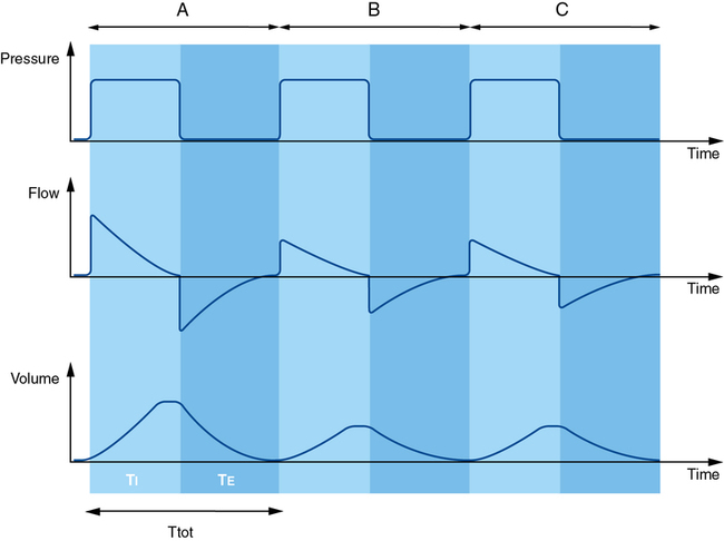
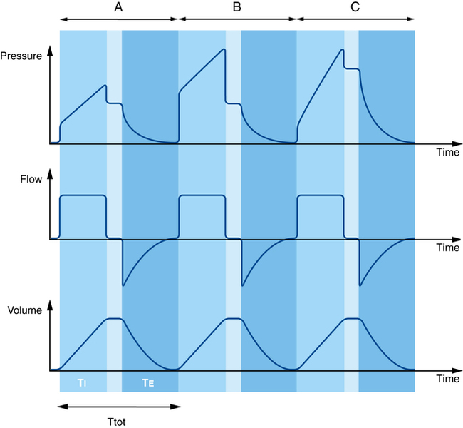
In volume control MV, an increase in respiratory system impedance (either increased resistance or decreased compliance) will lead to the set VT being delivered during the set TI but at the expense of higher airway pressures. Importantly, an increase in airway resistance or decrease in lung compliance can be differentiated by observing the associated changes in Ppeak and Pplat. An increase in airway resistance is reflected by an increase in Ppeak with a relatively unchanged Pplat (Figure 223-5, B) so that the difference between Ppeak and Pplat is increased. In volume control MV, a decrease in lung compliance will increase both Ppeak and Pplat so that the difference between Ppeak and Pplat remains unchanged (Figure 223-5, C).

In pressure control MV (constant inspiratory pressure and TI), any increase in airway resistance or decrease in lung compliance will lead to a decrease in inspiratory flow and delivered VT (Figure 223-6). In contrast to volume control MV, in pressure control MV, there is no way of identifying which factor (airway resistance or lung compliance) is responsible for the decrease in alveolar ventilation.

Mechanical ventilation in patients with airway obstruction

During volume control MV in patients with airflow obstruction, Ppeak and the difference between Ppeak and Pplat can be increased, but the delivered VT remains constant, and there is no change in the transpulmonary pressure. During pressure control MV in patients with airflow obstruction, the achieved VT can be reduced, and, if so, an increase in the set inspiratory pressure or TI should be considered.

During any mode of MV, if an increase in airway resistance is suspected (increase Ppeak but relatively unchanged Pplat during volume control MV or a decrease in achieved VT during pressure control MV), an immediate examination (including passage of a suction catheter) of the patency and position of the tracheal tube or for the presence of bronchospasm should be performed.

Regardless of the mode of MV in patients with airflow obstruction, the expiratory time constant will be prolonged because of the increased airway resistance. The incomplete exhalation prior to delivery of the next scheduled MV breath leads to breathstacking or dynamic hyperinflation. An important MV goal in patients with airflow obstruction is to maintain the lowest end-expiratory volume.

Ventilator adjustments designed to minimize dynamic hyperinflation include reducing VT and extending TE. Extending TE is primarily achieved by decreasing the respiratory rate. Additional measures include increasing inspiratory flow or decreasing the I:E ratio, which will inevitably increase inspiratory pressures and achieve only a modest prolongation of TE. These changes in MV settings will lead to a reduction in minute ventilation, but the resultant respiratory acidosis is generally well tolerated.

Mechanical ventilation in patients with decreased respiratory compliance

During volume control MV in patients with decreased compliance of the lung, both Ppeak and Pplat are elevated, and the difference between them remains unaltered. The set VT is delivered unchanged at the expense of higher airway pressures. During pressure control MV, the achieved VT and inspiratory flow are reduced despite the constant inspiratory pressure during the set TI.

An unusual feature of intraoperative MV is that there may be transient but significant changes in chest wall or abdominal elastance during the course of the operation (e.g., head-down position, insufflation of CO2 into the peritoneum or hemithorax for laparoscopic procedures). These intraoperative events are associated with significant changes in pleural (and extraalveolar) pressure.

During volume control MV, the increased Ppeak and Pplat (and unchanged gap between them) associated with an abrupt increase in pleural pressure is not associated with an increased risk of barotrauma because the transpulmonary pressure ( Pplat – pleural pressure) is unchanged and the set VT is not reduced. When the pleural pressure is decreased to normal (e.g., deflation of pneumoperitoneum), the Pplat and pleural pressure will return to their original settings with no change in the delivered VT.

During pressure control MV, an abrupt increase in pleural pressure (e.g., pneumoperitoneum) will lead to a reduction in the achieved VT but an unchanged airway pressure tracing on the monitor. The decrease in VT will be indicated by an increase in ETCO2, and a decrease in VT and minute ventilation. Under these circumstances, it is appropriate to increase the inspiratory pressure or TI to restore minute ventilation to its prior level. Again, this increase in inspired pressure will not be associated with barotrauma because the transpulmonary pressure (inspired pressure – pleural pressure) remains unchanged. When the pleural pressure is returned to normal, the inspired pressure should also be reduced to achieve an appropriate VT.

Intraoperative mechanical ventilation and acute lung injury

MV has been used to ensure satisfactory gas exchange during general anesthesia for decades. In 1963, Bendixen and associates advocated using a high VT (>10 mL/kg predicted body weight [PBW]) during intraoperative MV to reduce atelectasis, and this practice has continued almost unchanged until recently. Over the same time period, robust evidence has been accumulated for the existence of ventilator-induced lung injury. In the critical care setting, it has now become widely accepted to use protective lung ventilation settings in patients with acute respiratory distress syndrome.

In the past 10 years, the risk of intraoperative MV settings inducing or predisposing previously healthy patients to similar lung injury has been hotly debated. Multiple studies in previously healthy surgical patients have demonstrated that, compared with high VT (12-15 mL/kg PBW) MV, low VT (6-8 mL/kg PBW) MV is associated with the generation of lower levels of proinflammatory biomarkers. Perioperative patients at increased risk of developing acute lung injury (sepsis, shock states, multiple trauma and blood transfusion, patients undergoing high-risk operations) should be identified, and recent reviews support using protective ventilation settings in these patients during intraoperative MV. Measures commonly employed include low VT (6-8 mL/kg PBW), maintaining Pplat less than 30 cm H2O if pleural pressure is normal, use of PEEP and recruitment maneuvers, reduced FIO2, and permissive hypercapnia.

In previously healthy patients at low risk of developing acute lung injury and having elective surgical procedures with a short period (<5 h) of MV, there is little evidence to guide optimal intraoperative MV settings. Some authors suggest that it is reasonable to apply protective MV settings in all patients to avoid sensitizing the lung to any further potential injury. In the face of the very many patients having uneventful intraoperative MV, other authors have questioned whether protective MV settings are necessary or indeed feasible for all patients.