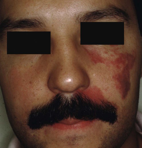
Port wine stain (‘nevus flammeus’)
Specific investigations


First-line therapies
 Pulsed dye laser
Facial port wine stains in childhood: prediction of the rate of improvement as a function of the age of the patient, size and location of the port wine stain and the number of treatments with the pulsed dye (585 nm) laser.
Nguyen CM, Yohn JJ, Huff C, Weston WL, Morelli JG. Br J Dermatol 1998; 138: 821–5.
Pain relief measures and cooling devices
Second-line therapies
 Intense pulsed light source
 Alexandrite 755 nm laser
 Neodymium:yttrium–aluminum–garnet (Nd:YAG) laser
Third-line therapies
 Potassium titanyl phosphate laser
 Imiquimod (as anti-angiogenic therapy)
 Photodynamic therapy
 Rapamycin (as anti-angiogenic therapy)
